Filter-Aided Sample Preparation before Mass Spec Analysis: An Evaluation of FASP and eFASP

12271ma_800px
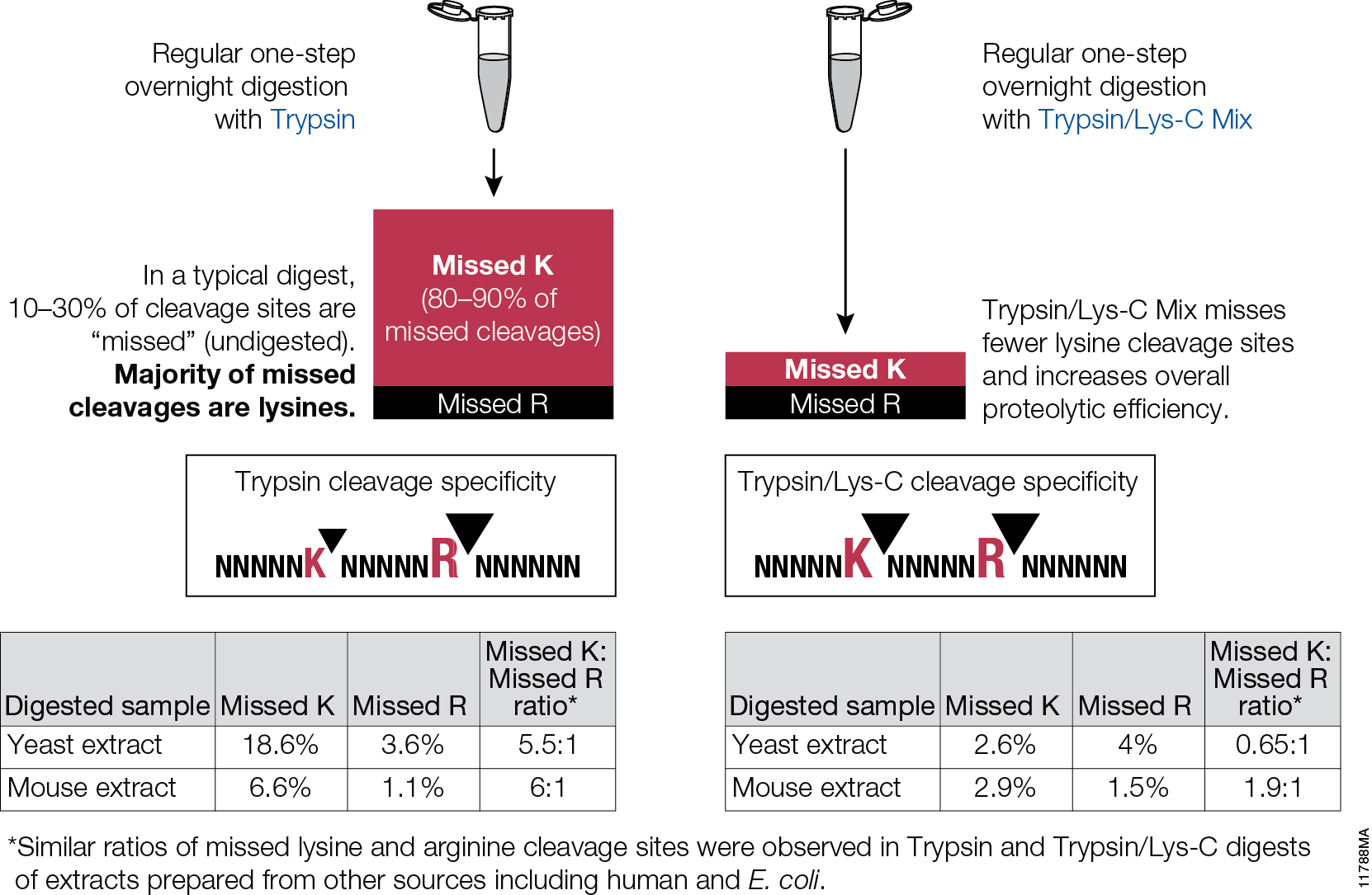
Filter-aided sample preparation (FASP) method is used for the on-filter digestion of proteins prior to mass-spectrometry-based analyses (1,2). FASP was designed for the removal of detergents, and chaotropes that were used for sample preparation. In addition, FASP removes components such as salts, nucleic acids and lipids. Akylation of reduced cysteine residues is also carried out on filter, after which protein is proteolyzed by use of trypsin on filter in the optimal buffer of the enzyme. Subsequent elution and desalting of the peptide-rich solution then provides a sample ready for LC–MS/MS analysis.

Erde et al. (3) described an enhanced FASP (eFASP) workflow that included 0.2% DCA in the exchange, alkylation, and digestion buffers,thus enhancing trypsin proteolysis, resulting in increases cytosolic and membrane protein representation. DCA has been reported (4) to improve the efficiency of the denaturation, solubilization, and tryptic digestion of proteins, particularly proteolytically resistant myoglobin and integral membrane proteins, thereby enhancing the efficiency of their identification with regard to the number of identified proteins and unique peptides.

In a recent publication (5) traditional FASP and eFASP were re-evaluated by ultra-high-performance liquid chromatography coupled to a quadrupole mass filter Orbitrap analyzer (Q Exactive). The results indicate that at the protein level, both methods extracted essentially the same number of hydrophobic transmembrane containing proteins as well as proteins associated with the cytoplasm or the cytoplasmic and outer membranes.

The LC–MS/MS results indicate that FASP and eFASP showed no significant differences at the protein level. However, because of the slight differences in selectivity at the physicochemical level of peptides, these methods can be seen to be somewhat complementary for analyses of complex peptide mixtures.

  1. Manza, L. L. et al. (2005) Sample preparation and digestion for proteomic analyses using spin filters Proteomics  5, 1742–74.
  2. Wiśniewski, J. R. et al. (2009) Universal sample preparation method for proteome analysis Nat. Methods 6, 359–62.
  3. Erde, J. et al. (2014) Enhanced FASP (eFASP) to increase proteomic coverage and sample recovery for quantitative proteome experiments. J. Proteome Res. 13, 1885–95.
  4. Lin, Y. et al. (2008) Sodium-deoxycholate-assisted tryptic digestion and identification of proteolytically resistant proteins Anal. Biochem.  377, 259–66.
  5. Nel. A. et al. (2015) Comparative Reevaluation of FASP and Enhanced FASP methods by LC-MS/MS/ J Proteome Res. 14, 1637–42.

Improved Characterization and Quantification of Complex Cell Surface N-Glycans

MSextractcropped

N-Glycosylation is a common protein post-translational modification occurring on asparagine residues of the consensus sequence asparagine-X-serine/threonine, where X may be any amino acid except proline. Protein N-glycosylation takes place in the endoplasmic reticulum (ER) as well as in the Golgi apparatus.

Approximately half of all proteins typically expressed in a cell undergo this modification, which entails the covalent addition of sugar moieties to specific amino acids. There are many potential functions of glycosylation. For instance, physical properties include: folding, trafficking, packing, stabilization and protease protection. N-glycans present at the cell surface are directly involved in cell−cell or cell−protein interactions that trigger various biological responses.

The standard method used to profile the N-glycosylation pattern of cells is glycoprotein isolation followed by denaturation and/or tryptic digestion of the glycoproteins and an enzymatic release of the N-glycans using PNGase F followed by analysis mass spec. This method has been reported to yield high levels of high-mannose N-glycans that stem from both membrane proteins as well as proteins from the ER.(1,2)

Are you looking for proteases to use in your research?
Explore our portfolio of proteases today.

For those researchers interested in characterizing only cell surface glycans (i.e.,  complex N-glycans)  a recent reference has developed a model system using HEK-292 cells that demonstrates a reproducible, sensitive, and fast method to profile surface N-glycosylation from living cells (3). The method involves standard centrifugation followed by enzymatic release of cell surface N-glycans. When compared to the standard methods the detection and quantification of complex-type N-glycans by increased their relative amount from 14 to 85%.

  1. North, S. J. et al. (2012) Glycomic analysis of human mast cells, eosinophils and basophils. Glycobiology. 2012, 22, 12–22.
  2. Reinke, S. O. et al. (2011) Analysis of cell surface N-glycosylation of the human embryonic
    kidney 293T cell line. J. Carbohydr. Chem.  30, 218–232.
  3. Hamouda, H. et al. (2014) Rapid Analysis of Cell Surface N‑Glycosylation from Living Cells Using Mass Spectrometry. J of Proteome Res. 13, 6144–51.

Mass Spectrometry Application: Antibody Quantitation for Preclinical PK studies

Isoform_Antibodies_LinkedInTherapeutic monoclonal antibodies (mAbs) represent the majority of therapeutics biologics now on the market, with more than 20 mAbs approved as drugs (1–3). During preclinical development of therapeutic antibodies, multiple variants of each antibody are assessed for pharmacokinetic (PK) characteristics across model systems such as rodents, beagles and  primates. Ligand-binding assays (LBA) are the standard technology used to perform the PK studies for mAb candidates (4). Ligand-binding assays (LBAs) are methods used  to detect and measure a macromolecular interaction between a ligand and a binding molecule. In LBAs, a therapeutic monoclonal antibody is considered to be the ligand, or analyte of interest, while the binding molecule is usually a target protein.

LBAs have certain well-documented limitations (5). Specific assay reagents are often not available early in a program. Interferences from endogenous proteins, antidrug antibodies, and soluble target ligands are potential complicating factors.

Liquid chromatography coupled to tandem mass spectrometry (LC–MS/MS)-based methods represent a viable and complementary addition to LBA for mAb quantification in biological matrixes. LC–MS/MS provides specificity, sensitivity, and multiplexing capability.

A recent reference (6) illustrates an automated method to perform LC–MS/MS-based quantitation, with IgG1 conserved peptides, a heavy isotope labeled mAb internal standard,and anti-human Fc enrichment. The method was applied to the pharmacokinetic study of a mAb dosed in cynomolgus monkey, and the results were compared with the immunoassay data. The interesting finding of the difference between ELISA and LC–MRM-MS data indicated that those two methods can provide complementary information regarding the drug’s PK profile.

Literature Cited

  1. Mao, T. et al. (2013) Top-Down Structural Analysis of an Intact Monoclonal Antibody by Electron Capture Dissociation-Fourier Transform Ion Cyclotron Resonance-Mass Spectrometry. Anal.Chem. 85, 4239–46.
  2. Weiner, L. M. et al. (2010) Monoclonal antibodies: versatile platforms for cancer immunotherapy. Nat. Rev. Immunol. 10, 317–27.
  3. Nelson, A. et al. (2010) Development trends for human monoclonal antibody therapeutics. Nat. Rev. Drug Discovery. 9, 767–74.
  4. DeSilva, B. et al. (2003) Recommendations for the Bioanalytical Method Validation of Ligand-Binding Assays to Support Pharmacokinetic Assessments of MacromoleculesPharm. Res. 20, 1885–00.
  5. Ezan, E.et al. (2009) Critical comparison of MS and immunoassays for the bioanalysis of therapeutic antibodiesBioanalysis 1, 1375–88.
  6. Zhang, Q. et al. (2014) Generic Automated Method for Liquid Chromatography–Multiple Reaction Monitoring Mass Spectrometry Based Monoclonal Antibody Quantitation for Preclinical Pharmacokinetic Studies. Anal.Chem. 86, 8776–84.

Use of Cell-Free Technology to Evaluate Nuclease (TALEN) Activity on Target DNA

ImageSource=RCSB PDB; StructureID=1qpf; DOI=http://dx.doi.org/10.2210/pdb1qpf/pdb;

Transcriptional activator-like effector nucleases (TALENs) have rapidly become a technique of choice for precision genome engineering. TALENs are custom-designed nucleases that consist of a modular DNA-binding domain fused to a monomeric, C-terminal FokI nuclease domain (1). TALENs work in pairs and are designed to recognize and bind to tandem-oriented sequences in genomic DNA, separated by a short spacer (15–30 bp). TALEN binding causes dimerization and activation of the FokI nuclease domains, which results in cleavage of the DNA within the spacer region. Small insertions or deletions (indels) are frequently introduced at this site, as the result of errors made during DNA repair by nonhomologous end-joining (NHEJ). These indels can be up to several hundred base pairs in length and result in frameshift mutations that lead to the production of truncated or nonfunctional proteins.

Successful use of TALENs for inducing targeted mutations has been reported in many conventional models, for example: mice, Xenopus and D. melanogaster. TALENs are also reported to be functional in a variety of other invertebrate arthropods, including mosquitos,silkworm and cricket. A recent publication (2) illustrates the use of TALEN technology for the genetic manipulation in P. dumerilii (marine ragworm).

Continue reading “Use of Cell-Free Technology to Evaluate Nuclease (TALEN) Activity on Target DNA”

Shedding Light on Protein:Protein Interactions with NanoBRET™ Technique

NanoBRET™ TechnologyIf you are trying to investigate protein:protein interactions inside cells, you know how important physiologically relevant results are. If you overload your cells with fusion constructs, your protein interactions may not actually reflect what is going on in the cell, and if your BRET energy donor and acceptor do not have sufficiently separated spectra, you can pick up a fair amount of noise in your experiment. Using the new superbright NanoLuc® Luciferase, and the HaloTag® Technology, we have developed a sensitive BRET system to help you take a better look specific protein interactions that interest you. Promega research scientist, Danette Daniels, describes the system in the Chalk Talk below:

Mass Spec-Compatible Proteome Reference Material

MSextractcropped

The complexity of biological samples places high demand on mass spec analytical capability. Adequate monitoring of instrument performance for proteomics studies requires equally complex reference material such as whole-cell extracts. However, whole-cell extracts available commercially are developed for general research (e.g., enzymatic or Western blot analysis) and contain detergents and salts that interfere with reverse phase liquid chromatography and mass spectrometry. Even after clean up, the extracts need to be digested, requiring time, labor and experience to generate samples for use in mass spectrometry. To address the need for complex protein material, we have developed whole-cell protein extracts from yeast and human cells. The yeast extract offers the convenience of a relatively small and well annotated proteome, whereas the human extract provides a complex proteome with large dynamic range. The human extract also serves as reference material for studies targeting the human proteome.

The extracts are free of compounds that interfere with reverse phase liquid chromatography-mass spectrometry (LC-MS), and have been reduced with DTT and alkylated with iodoacetamide then digested with Trypsin/Lys-C Mix and lyophilized. These digested extracts (tryptic peptides) can be readily reconstituted in trifluoroacetic acid (TFA) or formic acid and injected into an instrument. The same human and yeast whole-cell extracts also are provided in an intact (undigested) form for users who would like to develop an independent method for preparing protein mass spectrometry samples. For convenience, the intact extracts are provided as a frozen solution.

Consistent extract protein composition is ensured by tight control over cell culture conditions and manufacturing process. Lot-to-lot consistency of extracts is monitored by various protein and peptide qualitative and quantitation methods, including LC-MS. (Quality control results are provided upon request.) Our manufacturing process assures compatibility with reverse phase liquid chromatography and mass spectrometry, minimal nonspecific protein fragmentation, nonbiological post-translational modifi cations and,for digested extracts, minimal undigested peptides. The extracts are optimized for a high number of peptide and protein identifications in mass spectrometry analysis.

Trypsin/Lys-C Mix: Alternative for standard trypsin protein digestions

Trypsin/Lys-C Mix, Mass Spec Grade, is a mixture of Trypsin Gold, Mass Spectrometry Grade, and rLys-C, Mass Spec Grade. The Trypsin/Lys-C Mix is designed to improve digestion of proteins or protein mixtures in solution.It is a little known fact that trypsin cleaves at lysine residues with lesser efficiency than at arginine residues. Inefficient proteolysis at lysine residues is the major cause of missed (undigested) cleavages in trypsin digests.

11788MA


Supplementing trypsin with Lys-C enables cleavage at lysines with excepetional efficiency and specificity. Following the conventional trypsin digestion protocol (i.e., overnight incubation at nondenaturing conditions, reduction,alkylation, 25:1 protein:protease ratio [w/w], mix and incubate overnight at 37°C.) Replacing trypsin with Trypsin/Lys-C Mix in this conventional protocol leads to multiple benefits for protein analysis including more accurate mass spectrometry-based protein quantitation and improved protein mass spectrometry analytical reproducibility.

Are you looking for proteases to use in your research?
Explore our portfolio of proteases today.

Proteinase K: An Enzyme for Everyone

protein expression purification and analysis

We recently posted a blog about Proteinase K, a serine protease that exhibits broad cleavage activity produced by the fungus Tritirachium album Limber. It cleaves peptide bonds adjacent to the carboxylic group of aliphatic and aromatic amino acids and is useful for general digestion of protein in biological samples. In that previous blog we focused on its use to remove RNase and DNase activities. However, the stability of Proteinase K in urea and SDS and its ability to digest native proteins make it useful for a variety of applications. Here we provide a brief list of peer-reviewed citations that demonstrate the use of proteinase K in DNA and RNA purification, protein digestion in FFPE tissue samples, chromatin precipitation assays, and proteinase K protection assays:

Continue reading “Proteinase K: An Enzyme for Everyone”

Protein:protein Interactions: GST Pulldowns

trypsinGST pull-downs

Pull-down assays probe interactions between a protein of interest that is expressed as a fusion protein (e.g., bait) and the potential interacting partners (prey). In a pull-down assay one protein partner is expressed as a fusion protein (e.g., bait protein) in E. coli and then immobilized using an affinity ligand specific for the fusion tag. The immobilized bait protein can then be incubated with the prey protein. The source of the prey protein can be either from a cell-based or cell-free expression system. After a series of wash steps the entire complex can be eluted from the affinity support using SDS-PAGE loading buffer or by competitive analyte elution, then evaluated by SDS-PAGE.

Continue reading “Protein:protein Interactions: GST Pulldowns”

PNGase F, a Novel Endoglycosidase

11123MA

PNGase F (Cat.# V4831) is a recombinant glycosidase cloned from Elizabethkingia meningoseptica and overexpressed in E. coli, with a molecular weight of 36kD.

PNGase F catalyzes the cleavage of N-linked oligosaccharides between the innermost GlcNAc and asparagine residues of high mannose, hybrid, and
complex oligosaccharides from N-linked glycoproteins. PNGase F will not remove oligosaccharides containing alpha-(1,3)-linked core fucose,
commonly found on plant glycoproteins.

Applications
Determining whether a protein is in fact glycosylated is the initial step in glycoprotein analysis. Polyacrylamide gel electrophoresis in the
presence of sodium dodecyl sulfate (SDS-PAGE) has become the method of choice as the final step prior to mass spec analysis. Glycosylated proteins often migrate as diffused bands by SDS-PAGE. A marked decrease in band width and change in migration position after treatment with PNGase F is considered evidence of N-linked glycosylation.

Gel based data are often correlated with information obtained from mass spec analysis. Asn-linked type glycans can be cleaved enzymatically by PNGase F yielding intact oligosaccharides and a slightly modified protein in which Asn residues at the site of de-N-glycosylation are converted to Asp, by converting the previously carbohydrate-linked asparagine into an aspartic acid, a monoisotopic mass shift of 0.9840Da is observed. The deglycosylated peptides are then analyzed by tandem mass spectrometry (MS/MS), and software algorithms are used to correlate the experimental fragmentation spectra with theoretical tandem mass spectra generated from peptides in a protein database.