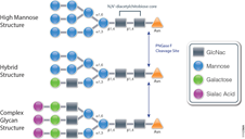
Enhancing Proteomics Data Using Arg-C Protease

Arg-C (clostripain), Sequencing Grade (Cat.# V1881), is a specific endoproteinase isolated from the soil bacterium Clostridium histolyticum. It preferentially cleaves at the C-terminal side of arginine (R) residues. Unlike trypsin, Arg-C efficiently cleaves arginine sites followed by proline (P). This difference is important because every twentieth arginine is followed by proline. To illustrate this benefit, Arg-C was evaluated for protein analysis in two different experiments. In the first experiment, we studied the use of Arg-C for proteomic analysis. Yeast provides an excellent model proteome because its genome is well annotated. Yeast extract was digested in two parallel reactions, using trypsin in the first reaction and Arg-C in the second, using a conventional protocol consistent with LC-MS/MS analysis. As expected the trypsin digestion resulted in a high number of peptide and protein identifications (Figure 1). However, many peptides remained elusive. The parallel Arg-C digestion complemented the trypsin digestion by recovering an additional 2,653 peptides and providing a 37.4% increase in the number of identified peptides. Digesting with Arg-C also resulted in an increase in the number of identified proteins. In fact, 138 new proteins were identified in Arg-C digest compared to the parallel trypsin digest, offering a 13.4% increase in the overall number of identified proteins.

Figure 1. Side-by-side analysis of trypsin-digested and Arg-C digested yeast proteins.

Are you looking for proteases to use in your research?
Explore our portfolio of proteases today.

In a second experiment, the ability of Arg-C to analyze individual proteins was analyzed, selecting human histone H4 as a model protein. Like other histones, this protein is heavily modified post translational modifications (PTMs) that alter histone structure and regulate interaction with transcription factors. As a result, histone PTMs are implicated in gene regulation and associated with multiple disorders. Technical challenges, however, impede histone PTM analysis. Histone PTMs are complex and some, such as acetylation and methylation, prevent trypsin digestion, as shown by our data. In this experiment, trypsin digestion of histone H4 identified several PTMs (Figure 2). However, certain PTMs were missing. By digesting histone H4 with Arg-C, we were able to identify the missing PTMs including mono-, dimethylated and acetylated lysine and arginine residues. We speculate that the PTMs in human histone H4, which modified arginine and lysine residues, rendered trypsin unsuitable for preparing the corresponding histone regions for mass spectrometry. The problem was rectified by replacing trypsin with Arg-C.

Figure 2. Identification of histone h4 PTMs after Arg-C digestion.

Endo H Application: Monitoring Protein Trafficking

Endo H (Endo-ß-N-acetylglucosaminidase H) is a 29,000 dalton protein with optimal activity between pH 5 and 6. In contrast to PNGase F, which cleaves all N-linked glycans at the site of attachment to Asparagine (Asn), (with the exception of those with fucose attached to the core GlcNac moieties), Endo H hydrolyses the bond connecting the two GlcNac groups that comprise the chitobiose core (see Figure 1.). In addition, Endo H cleaves high mannose and hybrid glycans, whereas complex glycans (those with more than 4 different sugar types per glycan chain, including the GlcNac groups) are resistant to hydrolysis.

The unique specificty of Endo H and PNGase F can be used to monitor protein trafficking. Basic N-Glycosylation occurs in the endoplasmic reticulum. Proteins in this stage are sensitive to Endo H digestion. If proteins have entered the Golgi body where additional modifications occur to the glycan, they are resistant to Endo H digestion.

The following references illustrate this application:

Protease K Protection Assay: Cell Free Expression Application

Microsomal vesicles are used to study cotranslational and initial posttranslational processing of proteins. Processing events such as signal peptide cleavage, membrane insertion, translocation and core glycosylation can be examined by the transcription/translation of the appropriate DNA in the TNT® Lysate Systems when used with microsomal membranes.

The most general assay for translocation makes use of the protection afforded the translocated domain by the lipid bilayer of the microsomal membrane. In this assay protein domains are judged to be translocated if they are observed to be protected from exogenously added protease. To confirm that protection is due to the lipid bilayer addition of 0.1% non-ionic detergent (such as Triton® X-100) solubilizes the membrane and restores susceptibility to the protease.

Are you looking for proteases to use in your research?
Explore our portfolio of proteases today.

Many proteases have proven useful for monitoring translocation in this fashion including Protease K or Trypsin.

The following are examples illustrating this application:

  1. Minn, I. et al. (2009) SUN-1 and ZYG-12, mediators of centrosome-nucleus attachment, are a functional SUN/KASH pair in Caenorhabditis elegans. Mol. Biol. Cell. 20, 4586–95.
  2. Padhan, K. et al. (2007) Severe acute respiratory syndrome coronavirus Orf3a protein interacts with caveolin. J.Gen.Virol. 88, 3067–77.
  3. Tews, B.A. et al. (2007) The pestivirus glycoprotein Erns is anchored in plane in the membrane via an amphipathic helix. J.Biol.Chem. 282, 32730–41.
  4. Pidasheva, S. et al. (2005) Impaired cotranslational processing of the calcium-sensing receptor due to signal peptide missense mutations in familial hypocalciuric hypercalcemia. Hum. Mol. Gen. 14, 1679–90.
  5. Smith, D. et al. (2002) Exogenous peptides delivered by ricin require processing by signal peptidase for transporter associated with antigen processing-independent MHC class I-restricted presentation. J. Immun. 169, 99–107.

6X His Protein Pulldowns: An Alternative to GST

ResearchBlogging.orgPull-down assays probe interactions between a protein of interest that is expressed as fusion protein (e.g.,
(e.g., bait) and the potential interacting partners (prey).

In a pull-down assay one protein partner is expressed as a fusion protein (e.g., bait protein) in E. coli and then immobilized using an affinity ligand specific for the fusion tag. The immobilized
bait protein can then be incubated with the prey protein. The source of the prey protein depends on whether the experiment is designed to confirm an interaction or to identify new interactions. After a series of wash steps, the entire complex can be eluted from the affinity support using SDS-PAGE loading buffer or by competitive analyte elution, then evaluated by SDS-PAGE.

Successful interactions can be detected by Western blotting with specific antibodies to both the prey and bait proteins, or measurement of radioactivity from a [35S] prey protein. bait) and potential interacting partners (prey).

The most commonly used method to generate a bait protein is expression as a fusion protein contain a GST (glutathione-S transferase) tag in E. coli. This is followed by immobilization on particles that contain reduced glutathione, which binds to the GST tag of the fusion protein. The primary advantage of a GST tag is that it can increase the solubility of insoluble or semi-soluble proteins expressed in E. coli.

Among fusion tags, His-tag is the most widely used and has several advantages including: 1) It’s small in size, which renders it less immunogenically active, and often it does not need to be removed from the purified protein for downstream applications; 2) There are a large number of commercial vectors available for expressing His-tagged proteins; 3) The tag may be placed at either the N or C terminus; 4) The interaction of the His-tag does not depend on the tag structure, making it possible to purify otherwise insoluble proteins using denaturing conditions. Continue reading “6X His Protein Pulldowns: An Alternative to GST”

Use of Multiple Proteases for Improved Protein Digestion

One of the approaches to identify proteins by mass spectrometry includes the separation of proteins by gel electrophoresis or liquid chromatography. Subsequently the proteins are cleaved with sequence-specific endoproteases. Following digestion the generated peptides are investigated by determination of molecular masses or specific sequence. For protein identification the experimentally obtained masses/sequences are compared with theoretical masses/sequences compiled in various databases.

Trypsin is the favored enzyme for this application, for the following reasons: A) the peptides contain a basic residue (Arg or Lys) on the C terminus and thus are good candidates for collision induced activation (CAD) in tandem experiments (low charge states and high mass-to-charge ratios); B) it is relatively Inexpensive; and C) optimal digestion conditions have been well characterized.

An inherent limitation of trypsin is the size of the peptides that it generates. For most organisms > 50% of tryptic peptides are less than 6 amino acids, too small for mass spectrometry based sequencing.

Are you looking for proteases to use in your research?
Explore our portfolio of proteases today.

One recent publication examined the use of multiple proteases (trypsin, LysC, ArgC , AspN and GluC) in combination with either CAD or electron-based fragmentation (ETD) to improve protein identification (1). Their results indicated a significant improvement from a single protease digestion (trypsin), which yielded 27,822 unique peptides corresponding to 3313 proteins. In contrast using a combination of proteases with either CAD or ETD fragmentation methods yielded 92,095 unique peptides mapping to 3908 proteins.

Swaney DL, Wenger CD, & Coon JJ (2010). Value of using multiple proteases for large-scale mass spectrometry-based proteomics. Journal of proteome research, 9 (3), 1323-9 PMID: 20113005

Trypsin: Innovative Applications

3D model of protein and protease cleavage

Tryptic digestion of samples and subsequent analysis by mass spectrometry is a popular technique for the identification of proteins typically those related to interaction partners or biomarkers characterization. This powerful tool can also be used for less traditional experimental designs. Three examples are:

Continue reading “Trypsin: Innovative Applications”

Optimized Wheat Germ Extract for High-Yield Protein Expression of Functional, Soluble Protein

Wheat Germ Extract for high-yield protein expression

Cell-free protein synthesis has emerged as powerful alternative to cell based protein expression for functional and structural proteomics. The TNT® SP6 High-Yield Protein Expression System uses a high-yield wheat germ extract supplemented with SP6 RNA polymerase and other components. Coupling transcriptionaland translational activities eliminates the inconvenience of separate in vitro transcription and purification steps for the mRNA, while maintaining the high levels of protein expression (1).

Continue reading “Optimized Wheat Germ Extract for High-Yield Protein Expression of Functional, Soluble Protein”