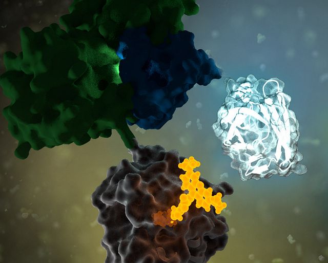
RNA doesn’t just carry genetic instructions—it also interacts with proteins to regulate nearly every aspect of gene expression, from splicing to translation. When those interactions go awry, the consequences can be devastating. In myotonic dystrophy type 1 (DM1), the most common adult-onset muscular dystrophy, a toxic RNA repeat expansion hijacks a critical protein called MBNL1, trapping it in nuclear clumps called foci. This leads to widespread splicing defects and progressive muscle wasting. But studying these toxic interactions inside living cells—and finding small molecules that can disrupt them—has been a significant challenge.
A recent study led by the Scripps Institute may have a solution. The study introduces a NanoBRET™ assay that can monitor the interaction between the expanded CUG RNA repeats and MBNL1 protein in real time, in live cells. Their findings demonstrate how this platform can be used not only to detect disease-driving RNA–protein complexes but also to identify small molecules that break them apart.
In biologics, cell therapy, and targeted protein degradation, the science is moving fast—and so are the tools. From GPCR-targeted therapies to real-time CAR-T manufacturing tools, new techniques are reshaping how scientists approach drug development, live-cell imaging, and protein degradation.
The “Bringing Light to Science”Discover Glo 2025 speaker series brought together researchers from across academia and industry to share real-world examples of how bioluminescent technologies are helping them advance their research. Now available on demand, these sessions offer fresh perspectives and actionable takeaways on the future of therapeutic development, cellular analysis and assay design.
We’ve distilled five key takeaways from the sessions—practical insights you can apply to your own work or use to stay current with where the field is heading.
This blog was written by guest contributor Tian Yang, Associate Product Manager, Promega, in collaboration with Kristin Huwiler, Manager, Small Molecule Drug Discovery, Promega.
During the development of chemical probes or small-molecule drugs, compound affinity (Kd) or potency (IC50) is used to characterize compound-target interactions, to guide structure-activity relationship analysis and lead optimization and to assess compound selectivity.
However, neither parameter provides information on how quickly a compound engages with and dissociates from the target. The dissociation constant Kd reflects the relative concentrations of unbound and bound state of the compound at thermodynamic equilibrium, and while IC50 is an empirical metric that measures the concentration at which a biochemical or cellular process is reduced to half of the maximum value, IC50 values are typically determined when the process is assumed to be at equilibrium or steady-state. For a closed system, like cells in a culture dish, these thermodynamic parameters are quite informative. In an open system like the human body, where compound-target interactions often do not reach equilibrium, the kinetic parameters, in addition to the thermodynamic parameters, are needed to better understand and characterize compound target engagement over time (1,2).
Understanding the expression, function and dynamics of
proteins in their native environment is a fundamental goal that’s common to
diverse aspects of molecular and cell biology. To study a protein, it must
first be labeled—either directly or indirectly—with a “tag” that allows
specific and sensitive detection.
Using a labeled antibody to the protein of interest is a
common method to study native proteins. However, antibody-based assays, such as
ELISAs and Western blots, are not suitable for use in live cells. These
techniques are also limited by throughput and sensitivity. Further, suitable
antibodies may not be available for the target protein of interest.
This blog was written by guest contributor Tian Yang, Associate Product Manager, Promega, in collaboration with Kristin Huwiler, Manager, Small Molecule Drug Discovery, Promega.
Determining the selectivity of a compound is critical during chemical probe or drug development. In the case of chemical probes, having a clearly defined mechanism of action and specific on-target activity are needed for a chemical probe to be useful in delineating the function of a biological target of interest in cells. Similarly, optimizing a drug candidate for on-target potency and reducing off-target interactions is important in the drug development process (1,2). A thorough understanding of the selectivity profile of a drug can facilitate drug repurposing, by enabling approved therapeutics to be applied to new indications (3). Interestingly, small molecule drugs do not necessarily require the same selectivity as a chemical probe, since some drugs may benefit from polypharmacology to achieve their desired clinical outcome.
Selectivity profiling panels based on biochemical methods have commonly been used to assess compound specificity for established target classes in drug discovery and chemical probe development. Biochemical assays are target-specific and often quantitative, enabling direct measurements of compound affinities for targets of interest and facilitate comparison of compound engagement to a panel of targets. As an example, several providers offer kinase selectivity profiling services using different assay formats and kinase panels comprised of 100 to 400 kinases (4). However, just as biochemical target engagement does not always translate to cellular activity, selectivity profiles based on biochemical platforms may not reflect compound selectivity in live cells (5).
This guest blog post is written by Tian Yang, Associate Product Manager at Promega.
In the realm of chemical probe development and drug discovery, understanding the interactions between drugs/compounds and their targets is crucial. Two frequently used metrics to characterize these interactions are IC50 and Kd, which guide researchers in evaluating the potential of compounds in effecting changes in target function. IC50 offers insights into a compound’s potency by quantifying its ability to inhibit a specific biological activity. Kd provides a measure of the affinity between a ligand and its receptor, reflecting how tightly a compound binds to its target (1). Together, these parameters are instrumental in the early stages of drug development, helping to identify promising candidates by assessing a compounds’s binding characteristics and its observed efficacy.
This blog was written by guest author Michael Curtin, Senior Product Manager, Small Molecule Drug Discovery.
ATP is the universal energy currency of cells, and thousands of proteins outside the kinase family “spend” it to move cargo, remodel nucleic acids, pump ions, or fold proteins. These ATP-hydrolyzing enzymes—collectively known as ATPases—span functional classes including motor proteins, transporters, chaperones, chromatin remodelers, ligases, and, crucially for genome stability, helicases.
From DNA replication to RNA processing, helicases are essential players. DNA/RNA helicases such as MCM, XPB/XPD, WRN, and members of the DDX family sit alongside AAA+ unfoldases, ABC transporters, and V-ATPases—all drawing on ATP to power their molecular work.
This guest blog post is written by Tian Yang, Associate Product Manager at Promega.
There are often challenges with translating results from a test tube into a living system, demanding more physiologically relevant assays. In drug discovery, demonstrating a compound’s ability to modulate its target protein in live cells is a critical step in the hit-to-lead workflow. A variety of cell-based assays can be used to assess a compound’s activity in live cells. Take kinase inhibitors as an example, these assays can range from substrate phosphorylation assays that more directly report on the activity of target kinases, to genetic reporter assays or cell viability assays that assess the downstream effects of target modulation.
In the case of Tivantinib, several pieces of data from its development were used to establish its role as an inhibitor of MET kinase. MET Kinase is a prominent target for anti-cancer therapeutics due to frequent MET dysregulation in a wide range of tumors. For example, over-activation of MET drives cancer proliferation and metastasis. In the initial report on Tivantinib, in addition to biochemical activity assays performed with purified MET, the activity of Tivantinib in cells was verified by several methods, including: 1) inhibition of phosphorylation of MET and downstream signaling pathways, 2) cytotoxicity in cancer cell lines expressing MET, and 3) antitumor activity in xenograft mouse models (1). Additionally, a co-crystal structure of the MET-Tivantinib complex was solved, seemingly confirming that Tivantinib is a bona fide MET inhibitor capable of engaging MET in live cells (2). Based on these observations and other pre-clinical data, Tivantinib appeared to be a promising drug candidate and was taken through phase 3 clinical trials targeting cancers with MET overexpression. However, Tivantinib ultimately was not approved as a new therapeutic, failing to show efficacy in these phase 3 clinical trials (3,4).
Within three years of the initial publication on Tivantinib, two separate articles challenged the mechanism of action in Tivantinib-induced cytotoxicity of tumor cells (5,6). Authors for both articles showed that Tivantinib can kill both MET-addicted and nonaddicted cells with similar potency. Both articles also concluded that perturbation of microtubule dynamics, instead of MET inhibition, is likely responsible for the cytotoxicity observed with Tivantinib. Considering the failed clinical trials and uncertainties regarding the mechanism of action, one may wonder if the original pre-clinical work adequately determined if Tivantinib effectively binds and inhibits MET in cells? If Tivantinib’s cellular engagement to MET was assessed directly rather than by MET phosphorylation analysis, would a different pre-clinical recommendation have been made?
In April 2024, Promega hosted the “Target Engagement in Chemical Biology Symposium” at the Kornberg Center, a research and development hub on Promega’s campus in Madison, Wisconsin. The goal of the symposium was to gather interdisciplinary researchers interested in the field of small molecule target engagement to foster collaboration through knowledge sharing and innovation. The symposium featured a 1.5-day agenda packed with 23 speakers, 4 workshops, poster sessions and social events. Over 130 attendees gathered to participate in the multifaceted event, with participants from different geographic regions and in different research sectors from academia to government to industry.
Attendees gather for the poster session in Kornberg Atrium. Photo by Anna Bennett (Promega Corporation)
The symposium highlighted the collective commitment to overcoming the challenges in drug discovery by developing more targeted and efficacious treatments, driven by a shared determination to create innovative solutions that address unmet medical needs. While we cannot share all the exciting research presented at the symposium, we are thrilled to highlight a few talks that exemplify the novel work and collaborative spirit of this research community.
Model organisms are essential tools in the pursuit of understanding biological processes, elucidating the mechanisms of diseases, and developing potential treatments and therapies. Use of these organisms in scientific research has paved the way for groundbreaking discoveries across various fields of biology. In particular, non-mammalian models can be valuable due to characteristics such as rapid life cycles, low cost, and amenability to use with advanced genetic tools, including bioluminescent reporters such as NanoLuc® Luciferase.
NanoLuc® is a small (19.1 kDa) luciferase enzyme originating from deep sea shrimp that is 100x brighter than firefly or Renilla luciferase. It utilizes a furimazine substrate to produce its bright glow-type luminescence. In the decade following its development, the NanoLuc® toolbox has expanded to include NanoBiT® complementation, NanoBRET™ energy transfer methods, and new reagents such as the Nano-Glo® Fluorofurimazine In Vivo Substrate (FFz) which was designed for in vivo detection of NanoLuc® Luciferase, NanoLuc® fusion proteins or reconstituted NanoBiT® Luciferase. In addition to the aqueous-soluble reagents increased substrate bioavailability in vivo, with fluorofurimazine, NanoLuc® and firefly luciferase can be used together in dual-luciferase molecular imaging studies.
Here we spotlight some recent research that demonstrates how the expanded NanoLuc® toolbox can be adapted to use in non-mammalian models, shedding new light on fundamental biological processes and advancing our understanding of complex mechanisms in these diverse organisms.
XWe use cookies and similar technologies to make our website work, run analytics, improve our website, and show you personalized content and advertising. Some of these cookies are essential for our website to work. For others, we won’t set them unless you accept them. To learn more about our approach to Privacy we invite you to Read More
By clicking “Accept All”, you consent to the use of ALL the cookies. However you may visit Cookie Settings to provide a controlled consent.
We use cookies and similar technologies to make our website work, run analytics, improve our website, and show you personalized content and advertising. Some of these cookies are essential for our website to work. For others, we won’t set them unless you accept them. To find out more about cookies and how to manage cookies, read our Cookie Policy.
If you are located in the EEA, the United Kingdom, or Switzerland, you can change your settings at any time by clicking Manage Cookie Consent in the footer of our website.
Necessary cookies are absolutely essential for the website to function properly. These cookies ensure basic functionalities and security features of the website, anonymously.
Cookie
Duration
Description
cookielawinfo-checbox-analytics
11 months
This cookie is set by GDPR Cookie Consent plugin. The cookie is used to store the user consent for the cookies in the category "Analytics".
cookielawinfo-checbox-functional
11 months
The cookie is set by GDPR cookie consent to record the user consent for the cookies in the category "Functional".
cookielawinfo-checbox-others
11 months
This cookie is set by GDPR Cookie Consent plugin. The cookie is used to store the user consent for the cookies in the category "Other.
cookielawinfo-checkbox-advertisement
1 year
The cookie is set by GDPR cookie consent to record the user consent for the cookies in the category "Advertisement".
cookielawinfo-checkbox-necessary
11 months
This cookie is set by GDPR Cookie Consent plugin. The cookies is used to store the user consent for the cookies in the category "Necessary".
cookielawinfo-checkbox-performance
11 months
This cookie is set by GDPR Cookie Consent plugin. The cookie is used to store the user consent for the cookies in the category "Performance".
gdpr_status
6 months 2 days
This cookie is set by the provider Media.net. This cookie is used to check the status whether the user has accepted the cookie consent box. It also helps in not showing the cookie consent box upon re-entry to the website.
lang
This cookie is used to store the language preferences of a user to serve up content in that stored language the next time user visit the website.
viewed_cookie_policy
11 months
The cookie is set by the GDPR Cookie Consent plugin and is used to store whether or not user has consented to the use of cookies. It does not store any personal data.
Analytical cookies are used to understand how visitors interact with the website. These cookies help provide information on metrics the number of visitors, bounce rate, traffic source, etc.
Cookie
Duration
Description
SC_ANALYTICS_GLOBAL_COOKIE
10 years
This cookie is associated with Sitecore content and personalization. This cookie is used to identify the repeat visit from a single user. Sitecore will send a persistent session cookie to the web client.
vuid
2 years
This domain of this cookie is owned by Vimeo. This cookie is used by vimeo to collect tracking information. It sets a unique ID to embed videos to the website.
WMF-Last-Access
1 month 18 hours 24 minutes
This cookie is used to calculate unique devices accessing the website.
_ga
2 years
This cookie is installed by Google Analytics. The cookie is used to calculate visitor, session, campaign data and keep track of site usage for the site's analytics report. The cookies store information anonymously and assign a randomly generated number to identify unique visitors.
_gid
1 day
This cookie is installed by Google Analytics. The cookie is used to store information of how visitors use a website and helps in creating an analytics report of how the website is doing. The data collected including the number visitors, the source where they have come from, and the pages visted in an anonymous form.
Advertisement cookies are used to provide visitors with relevant ads and marketing campaigns. These cookies track visitors across websites and collect information to provide customized ads.
Cookie
Duration
Description
IDE
1 year 24 days
Used by Google DoubleClick and stores information about how the user uses the website and any other advertisement before visiting the website. This is used to present users with ads that are relevant to them according to the user profile.
test_cookie
15 minutes
This cookie is set by doubleclick.net. The purpose of the cookie is to determine if the user's browser supports cookies.
VISITOR_INFO1_LIVE
5 months 27 days
This cookie is set by Youtube. Used to track the information of the embedded YouTube videos on a website.
Performance cookies are used to understand and analyze the key performance indexes of the website which helps in delivering a better user experience for the visitors.
Cookie
Duration
Description
YSC
session
This cookies is set by Youtube and is used to track the views of embedded videos.
_gat_UA-62336821-1
1 minute
This is a pattern type cookie set by Google Analytics, where the pattern element on the name contains the unique identity number of the account or website it relates to. It appears to be a variation of the _gat cookie which is used to limit the amount of data recorded by Google on high traffic volume websites.