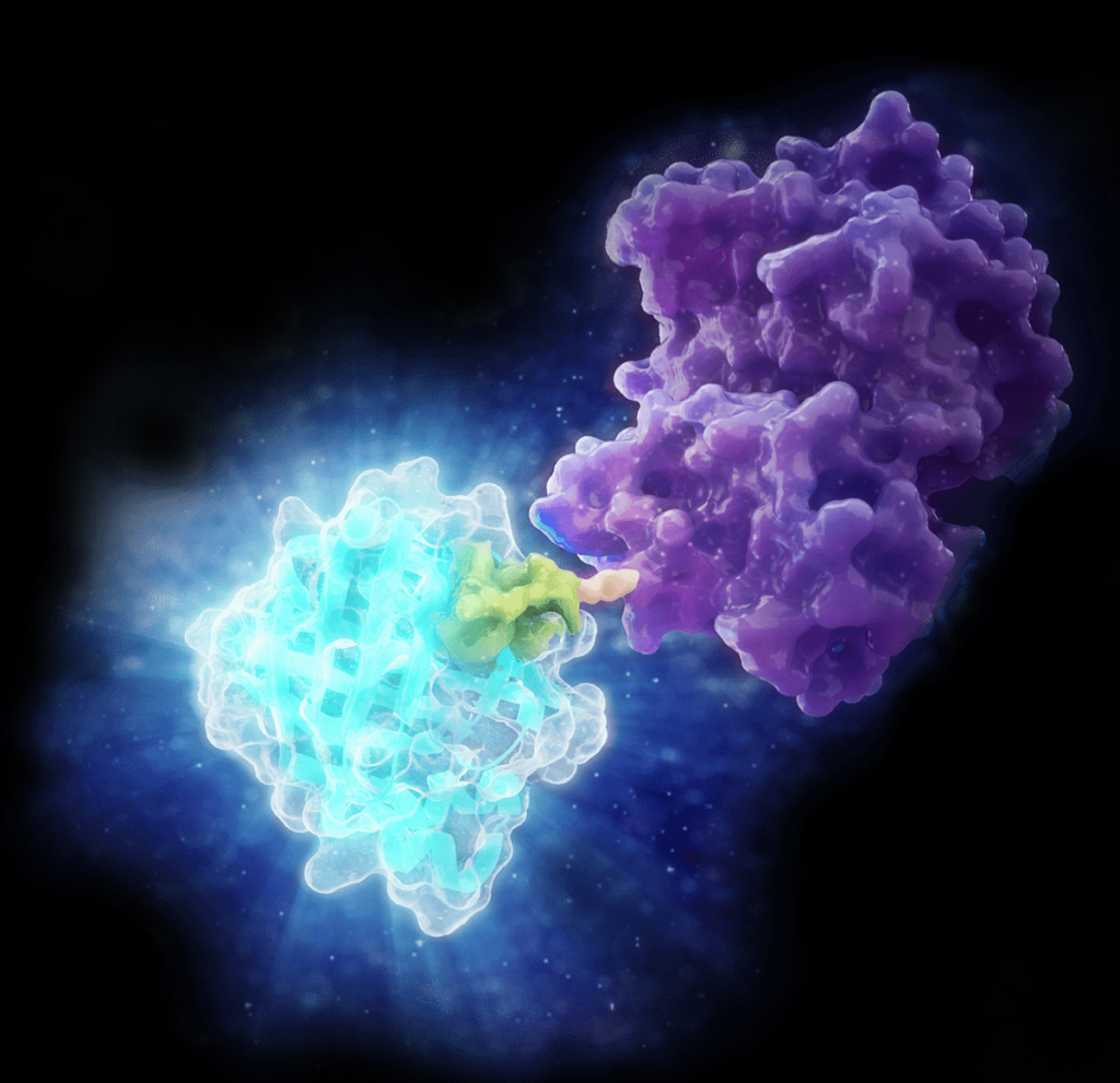
Most cancer-related deaths from solid tumors aren’t caused by the primary tumor itself, but are the result of metastasis. Metastasis is the process by which cancer cells break away from the primary tumor, travel through the bloodstream and establish new tumors in distant tissues. This is a complex, multi-step process and cancer researchers have spent decades trying to understand and disrupt the metastatic cascade. What is known: platelets and calcium play important roles in metastasis. What is unknown: How do these two factors interact?
Here, we explore a recent study published in Scientific Reports by researchers at George Washington University1. They investigate how calcium levels influence platelet-cancer cell interactions and what happens when both factors converge.
Cowpea (Vigna unguiculata), a humble tan and black legume, is one of the most important food crops in the world. Grown across sub-Saharan Africa, Asia, and parts of the Americas, Cowpea provides protein-rich nutrition for hundreds of millions of people, making it a cornerstone of smallholder agriculture. But cowpea production faces a persistent threat: the cowpea aphid-borne mosaic virus (CABMV), a common virus that can devastate yields across entire growing regions.
What makes CABMV particularly difficult to combat is how the virus infects its host. Instead of relying on viral translational machinery, the virus hijacks the plant’s systems to replicate. CABMV targets a protein called eIF4E, a translation initiation factor that the plant needs to read its own genetic instructions and produce proteins. The virus produces a protein, VPg, that binds directly to eIF4E and redirects the plant’s translational machinery to produce viral proteins instead. The plant can’t simply get rid of eIF4E. Without it, protein synthesis stalls. So how can cowpea defend itself against a virus that exploits one of its most essential proteins?
A new study published in Agronomy by researchers at the Federal University of Pernambuco, the Federal University of Minas Gerais, and Embrapa Recursos Genéticos e Biotecnologia takes a comprehensive look at this problem from the inside out1. The team characterized all three members of the eIF4E gene family in cowpea (eIF4E, eIF(iso)4E, and nCBP) across six cultivated varieties (cultivars) with known contrasting responses to CABMV infection. Two of those cultivars (Bajão and IT85F-2687) are resistant to the virus; the other four (Boca Negra, BR14 Mulato, Pingo de Ouro, and Santo Inácio) are susceptible to the virus.
Using a multi-omics approach that combined genomic, evolutionary and structural analyses, the researchers set out to answer a fundamental question: what makes some versions of eIF4E exploitable by the virus, and others not?
If you’ve read anything about the gut microbiome in the last decade, you’ve probably encountered a familiar setup: researchers collect stool samples, sequence the microbial DNA, and draw conclusions about gut health based on what microbes populate the gut. It’s a practical approach because stool is relatively easy to collect and doesn’t require invasive procedures. But how well does a stool sample represent the health of the entire intestinal tract?
A team of researchers at the Quadram Institute Bioscience and UK Health Security Agency set out to answer this question in primates1. They characterized the intestinal microbiome of cynomolgus macaques, a primate commonly used in biomedical research because of its genetic and physiological similarities to humans. Rather than relying on stool alone, the team collected samples from six distinct regions along the intestinal tract in 24 captive-bred animals ranging in age from 4 to 20 years.
Connecticut is a small yet ecologically interesting state. Over 85% of the human population lives in cities, yet more than 60% of the land is covered by forest, creating a diverse mix of habitats where wildlife and urban life overlap. In this landscape, bobcats have staged an impressive comeback over the past several decades, reclaiming their role as one of the region’s top predators. But as bobcat numbers rise, a quieter story is unfolding alongside them: the New England cottontail, the region’s only native rabbit, is vanishing.
Many consider enzymes the workhorses of biochemistry (move over, mitochondria)—catalyzing reactions, breaking down substrates, keeping the machinery of life humming along. But a growing number of researchers are re-envisioning what enzymes can do. Instead of facilitating chemistry, what if enzymes could steer and even guide tiny robots to a tumor?
That’s exactly what’s happening in the rapidly expanding field of enzyme-powered microscopic robots (a.k.a “microrobots”). Microrobots are tiny, engineered devices—often smaller than the width of a human hair—built to perform tasks inside the body that would be difficult or impossible at a larger scale, like delivering drugs to a specific tissue. A recent paper published in Nature Nanotechnology by a team of researchers at California Institute of Technology and the University of Southern California offers a particularly elegant example that we highlight below1.
Targeted protein degradation (TPD) is an emerging drug discovery strategy that offers an entirely different approach to tackle disease-relevant proteins, including classic “undruggable” targets. Instead of inhibiting protein function, small molecules like PROTACs and molecular glues co-opt the cell’s own ubiquitin-proteasome system to eliminate specific proteins altogether. But as this targeted approach gains traction, it also challenges existing methods for validating compound activity.
How do you confirm that degradation is happening in a biologically-relevant system? Can you validate protein degradation in real-time?
G-protein-coupled receptors (GPCRs) are among the most important drug targets in human biology, mediating signals across nearly every physiological system. But not all GPCRs are equally easy to study—especially those that interact with peptide ligands. These ligands tend to be flexible, fast-moving, and hard to trace in live cells by standard methods. Historically, radioligand binding assays have filled this gap, offering a way to measure peptide–receptor interactions with high sensitivity. However, these assays are typically performed using isolated membrane preparations or cells under non-physiological conditions, and they don’t allow for real-time or kinetic measurements.
The Wnt/β-catenin pathway, long studied in the context of developmental biology, has become increasingly recognized for its role in a wide range of human diseases. Its dysregulation has been implicated in cancer, fibrosis, immune modulation, and neurodegenerative conditions—making it a clinically actionable target across diverse therapeutic areas1. In this blog, we cover the fundamentals of Wnt/β-catenin signaling, highlight ongoing research efforts to understand its role in disease, and show how combining live-cell imaging with luminescent assays complements functional studies.
In our final blog post on double-stranded RNA (dsRNA), we turn our attention to the chemical building blocks of mRNA therapeutics—modified nucleotides. These seemingly minor changes to the RNA sequence play a crucial role in the success of mRNA-based vaccines and treatments. However, they also introduce complexities in accurately detecting and quantifying unwanted dsRNA byproducts— key steps in ensuring the therapeutic efficacy of your mRNA product.
What Are Modified Nucleotides?
Modified nucleotides are ribonucleotides containing chemically altered nucleosides — like specialty ingredients swapped into a classic recipe to improve taste and nutrition. Just as a chef might use a lactose-free milk or gluten-free flour to make a dish easier to digest without changing its core structure, scientists use chemically altered nucleosides during in vitro transcription (IVT) to improve how mRNA therapies perform. These modifications replace their natural counterparts (e.g., uridine or cytidine) in the final RNA product. Their incorporation improves the performance and safety of mRNA therapeutics in several ways:
As mRNA therapeutics continue to expand across clinical pipelines, one persistent challenge remains for developers: reducing double-stranded RNA (dsRNA) contaminants that can compromise safety and efficacy. These unintended byproducts of in vitro transcription (IVT) can trigger unwanted immune responses and reduce the potency of the final product. Developers must prioritize dsRNA detection and control as essential steps in the process. In our previous blog post we offered a high-level discussion of what is double-stranded RNA (dsRNA), its biological function, and importance of detection in a therapeutic context. Here, we’ll take a closer look at origins of dsRNA contamination, quality control measures, and improvement strategies.
Large-scale production of single-stranded RNA (ssRNA) for mRNA-based therapeutics is primarily done through in vitro transcription (IVT), an enzymatic process designed to generate high-yield, functional mRNA transcripts from a DNA template. This process uses purified RNA polymerase enzymes, such as T7, that recognize specific promoter sequences in the DNA template, generating the RNA transcripts of interest. However, IVT reactions also generate unwanted dsRNA byproduct. Below, we delve into some of the major quality control (QC) considerations and strategies to reduce dsRNA byproducts.
XWe use cookies and similar technologies to make our website work, run analytics, improve our website, and show you personalized content and advertising. Some of these cookies are essential for our website to work. For others, we won’t set them unless you accept them. To learn more about our approach to Privacy we invite you to Read More
By clicking “Accept All”, you consent to the use of ALL the cookies. However you may visit Cookie Settings to provide a controlled consent.
We use cookies and similar technologies to make our website work, run analytics, improve our website, and show you personalized content and advertising. Some of these cookies are essential for our website to work. For others, we won’t set them unless you accept them. To find out more about cookies and how to manage cookies, read our Cookie Policy.
If you are located in the EEA, the United Kingdom, or Switzerland, you can change your settings at any time by clicking Manage Cookie Consent in the footer of our website.
Necessary cookies are absolutely essential for the website to function properly. These cookies ensure basic functionalities and security features of the website, anonymously.
Cookie
Duration
Description
cookielawinfo-checbox-analytics
11 months
This cookie is set by GDPR Cookie Consent plugin. The cookie is used to store the user consent for the cookies in the category "Analytics".
cookielawinfo-checbox-functional
11 months
The cookie is set by GDPR cookie consent to record the user consent for the cookies in the category "Functional".
cookielawinfo-checbox-others
11 months
This cookie is set by GDPR Cookie Consent plugin. The cookie is used to store the user consent for the cookies in the category "Other.
cookielawinfo-checkbox-advertisement
1 year
The cookie is set by GDPR cookie consent to record the user consent for the cookies in the category "Advertisement".
cookielawinfo-checkbox-necessary
11 months
This cookie is set by GDPR Cookie Consent plugin. The cookies is used to store the user consent for the cookies in the category "Necessary".
cookielawinfo-checkbox-performance
11 months
This cookie is set by GDPR Cookie Consent plugin. The cookie is used to store the user consent for the cookies in the category "Performance".
gdpr_status
6 months 2 days
This cookie is set by the provider Media.net. This cookie is used to check the status whether the user has accepted the cookie consent box. It also helps in not showing the cookie consent box upon re-entry to the website.
lang
This cookie is used to store the language preferences of a user to serve up content in that stored language the next time user visit the website.
viewed_cookie_policy
11 months
The cookie is set by the GDPR Cookie Consent plugin and is used to store whether or not user has consented to the use of cookies. It does not store any personal data.
Analytical cookies are used to understand how visitors interact with the website. These cookies help provide information on metrics the number of visitors, bounce rate, traffic source, etc.
Cookie
Duration
Description
SC_ANALYTICS_GLOBAL_COOKIE
10 years
This cookie is associated with Sitecore content and personalization. This cookie is used to identify the repeat visit from a single user. Sitecore will send a persistent session cookie to the web client.
vuid
2 years
This domain of this cookie is owned by Vimeo. This cookie is used by vimeo to collect tracking information. It sets a unique ID to embed videos to the website.
WMF-Last-Access
1 month 18 hours 24 minutes
This cookie is used to calculate unique devices accessing the website.
_ga
2 years
This cookie is installed by Google Analytics. The cookie is used to calculate visitor, session, campaign data and keep track of site usage for the site's analytics report. The cookies store information anonymously and assign a randomly generated number to identify unique visitors.
_gid
1 day
This cookie is installed by Google Analytics. The cookie is used to store information of how visitors use a website and helps in creating an analytics report of how the website is doing. The data collected including the number visitors, the source where they have come from, and the pages visted in an anonymous form.
Advertisement cookies are used to provide visitors with relevant ads and marketing campaigns. These cookies track visitors across websites and collect information to provide customized ads.
Cookie
Duration
Description
IDE
1 year 24 days
Used by Google DoubleClick and stores information about how the user uses the website and any other advertisement before visiting the website. This is used to present users with ads that are relevant to them according to the user profile.
test_cookie
15 minutes
This cookie is set by doubleclick.net. The purpose of the cookie is to determine if the user's browser supports cookies.
VISITOR_INFO1_LIVE
5 months 27 days
This cookie is set by Youtube. Used to track the information of the embedded YouTube videos on a website.
Performance cookies are used to understand and analyze the key performance indexes of the website which helps in delivering a better user experience for the visitors.
Cookie
Duration
Description
YSC
session
This cookies is set by Youtube and is used to track the views of embedded videos.
_gat_UA-62336821-1
1 minute
This is a pattern type cookie set by Google Analytics, where the pattern element on the name contains the unique identity number of the account or website it relates to. It appears to be a variation of the _gat cookie which is used to limit the amount of data recorded by Google on high traffic volume websites.