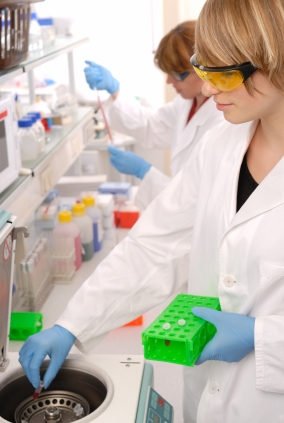
What are you thankful for in science?As the social media lead for Promega, I keep my eye on trends in new media. I have personal accounts that I keep mostly to see what other people are doing. I try hangouts, social networking and other things so that I have an idea of developing practices outside of the biotechnology industry. One activity that has been popular over the last couple of years during the month of November in the United States is the Facebook post of “30 days of thanksgiving”.
I wondered what “thanksgiving” looks like to the research scientist. So I asked:
What are the things you are thankful for in science?
The answers have been as varied as the people I talked to ranging from little things like water bath floats to really big things, like the renewal of your research funding or achieving tenure.
Here are some of the answers from my informal inquiries:
“Tube floaties for water baths.”
—E.V., genomics product manager
“I was always thankful for Geiger counters.”
—K. G., science writer
“Thermal cyclers and Taq Polymerase. As an undergrad I watched someone sit with a timer and move their tubes between water baths at 3 different temperatures, opening tubes and adding polymerase at the end of each cycle. Modern PCR is SOOO much easier.”
—M.M., research scientist
“I am thankful for competent cells. I remember preparing the CaCl2 and doing slow centrifugation. Also thankful for serum-compatible transfection, rapid ligations and online journal access (no longer have to traipse over to the university library to get papers photocopied- uuurrrgggghhh).”
—R.D., technical services scientist
“How about T-vectors for cloning? I was no molecular biologist, but could make a T-vector work.”
—K.K., science writer
“I am thankful for open-access journals and the ability to read the full article without an institutional subscription.”
—S.K., science writer
“I am ever so thankful for ONLINE ORDERING! So awesome. Throw in online technical manuals, on-line support tools, on-line calculators – all are awesome!!”
—A.P., director, scientific courses
“I am thankful for automated sequencing- manual sequencing was laborious and hazardous!!!”
—R.G., technical services scientist
Do any of these resonate with you? What are you thankful for as a scientist? Let us know in the comments.
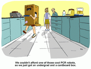
Cartoon by Ed Himelblau Copyright Ed Himelblau.When I was in the lab, there was more than a few times I held tubes in my hand (maybe even under my arm) to make them thaw faster, especially reaction buffers. However, I never considered whether this could be a strategy for actual incubation although humans run at about 37°C and many restriction enzyme reactions proceed most efficiently at 37°C. But research published in PLOS ONE by Crannell, Rohrman and Richards-Kortum took this idea and decided to experiment with the possibility of eliminating an instrument-based DNA amplification. Continue reading “DNA Amplification Using Body Heat, No Instrument Required”
The multiple Lombardi trophies won by Pittsburgh Steelers. Image used under Wikimedia Creative Commons, and attributed to daveynin.
It is fall and the season for American football. For this football fan, watching the game is a bit less enjoyable than it used to be, as more and more information is available about the serious and permanent brain injuries suffered by football players.
In the introduction to a recent paper in the journal Cell, “P7C3 Neuroprotective Chemicals Function by Activating the Rate-Limiting Enzyme in NAD Salvage”, not a word about American football is mentioned.
However, the paper begins, “No substantive therapeutics are available for the treatment of almost any form of disease entailing nerve death” (1). The authors list a range of neurodegenerative disorders such as Huntington’s, Alzheimers and Parkinson’s diseases, as well as ALS or Lou Gherig’s disease. They also note that there are currently no effective treatments for trauma to the brain or peripheral nervous system.
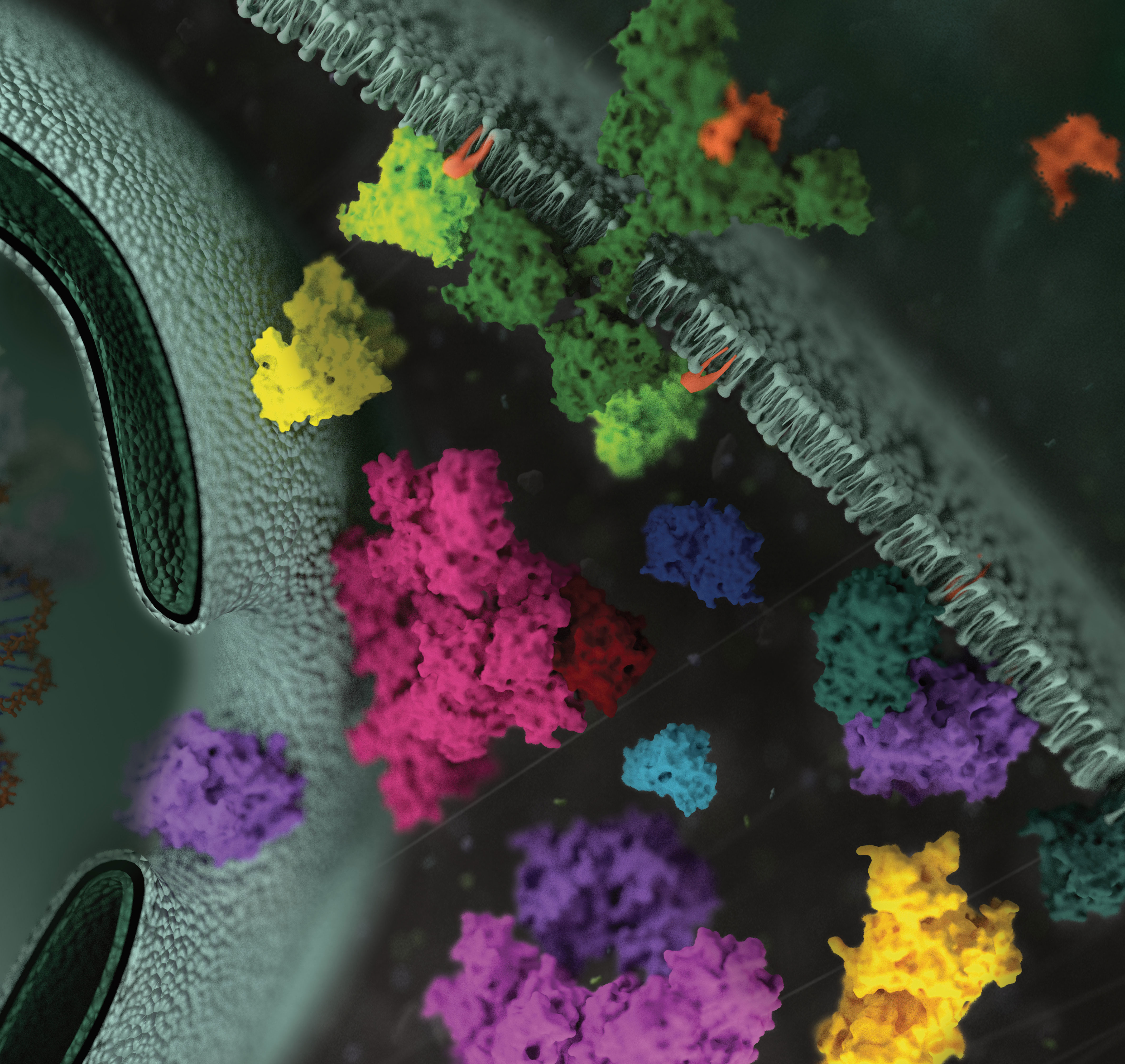
Protein phosphorylation is the most widespread type of post-translational modification. It affects every basic cellular process, including metabolism, growth, division, differentiation, motility, organelle trafficking, membrane transport, muscle contraction, immunity, learning and memory (1,2). Protein kinases catalyse the transfer of the phosphate from ATP to specific amino acids in proteins. In eukaryotes, these are usually Ser, Thr and Tyr residues. Due to the development of specific phosphopeptide enrichment techniques and highly sensitive MS instruments, phosphoproteomics has enabled researchers to gain a comprehensive view on the dynamics of protein phosphorylation and phosphorylation based signaling networks.
Due to its high cleavage specificity, trypsin is the commonly used proteolytic enzyme in MS-based proteomics, cleaving peptides carboxyterminal of the amino acids lysine and arginine. However, various factors such as the tertiary structure of a protein, adjacent basic amino acids or negatively charged residues close to cleavage sites as well as PTMs are known to impair proteolysis.
To gain closer insights into the impact of phosphorylation on tryptic digestion, a recent publication(3) systematically characterized the digestion efficiency of model peptide sequences that are known to be prone to incomplete digestion.
The results indicated that increasing trypsin concentrations up to a trypsin to peptide ratio of 1:10 led to a significant gain (1) in the overall number of phosphorylation sites (up to 9%) and in the intensities of individual phosphopeptides, thereby improving the sensitivity of phosphopeptide quantification.
The effect of organic solvents (ACN, acetonitrile and TFE trifuorethanol was also evaluated). Positive results were noted with TFE when determining the digestion of individual peptides. However TFE interfered with TiO2 phosphopeptide enrichment and therefore was not recommended for use with complex samples.
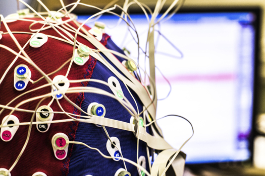
There is something very futuristic, and perhaps scary, about the idea of nonverbally transferring one person’s thoughts to another person, especially for the purpose of controlling or influencing a person’s actions and behaviors. Maybe that’s why telepathy and mind control are favorite topics of many science fiction movies. However, there are times when direct, nonverbal transfer of thoughts would be advantageous, for example when communicating complex concepts or feelings that are difficult to convey. Direct transfer also would circumvent the need to translate information from one language to another. For these reasons, scientists are currently developing technologies to allow such thought transfers. A recent PLOS ONE article describes a simple brain-to-brain interface in humans and shows how this interface can be used to capture a thought generated by one person and communicate that information directly to the brain of a second person and elicit a physical response (1).
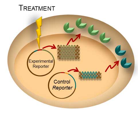
Transient transfection is often used to perform reporter assays. We have advocated using a dual-reporter system for decades to normalize the data obtained and gain a clearer understanding of your results. The experimental reporter should vary with treatment and the control reporter should vary little with treatment. The control reporter thus serves as a marker to help you understand the relative activity of your experimental reporter. The bioluminescent Dual-Luciferase® method allows for sequential detection of the second reporter in a single sample providing a simple two-step normalization method. Here are seven ways in which dual-reporter assays help you avoid misinterpreting results.
Simply comparing the ratio of the experimental to the control reporter can resolve differences in:
Number of Cells/Well: When manually pipeting cells into a 96-well plate, there is always a chance of having variable numbers of cells in each well. This variation is cell number will affect the experimental and control reporters equally, so the ratio of experimental:control reporter activity will eliminate false interpretation of the experimental data–whether it affects an entire row or column on the plate or individual wells.
Transfection Efficiency: The variations in transfection efficiency will equally affect both the experimental and control reporters so the ratio of activity in dual-reporter assays will normalize the data.
Cell Viability: Often, reporter assays look at the dose response curve of a particular compound with regard to gene expression. Ideally, if a compound causes a change in the experimental reporter the control reporter will demonstrate little effect. However, if the compound is toxic, both the experimental and control will be altered and the ratio will tell you whether the compound truly affects reporter activity or just kills the cells.
Lysis Efficiency: When lysing a plate of cells, you could encounter situations where rows or columns lyse differently, especially if you are using manual disruption or get interrupted mid-plate. The difference is lysis will affect the experimental and control equally so the ratio will remove the variation.
Temperature: Ideally, a plate should be equilibrated to ambient room temperature before proceeding to the reporter assay. Plates can cool at different rates or researchers anxious to record data may read the data early. Temperature variations will affect both reporters so the ratio will limit the affect on the data.
Measurement Time: Repetition of data is a hallmark of good science. You are often called upon to repeat experiments sometimes days or weeks apart. Let’s say you repeat your experiment one week after the initial experiment. The first time you measured the response, you waited 10 minutes after reagent addition to read, this week you waited 30 minutes. This will affect both reporters equally and therefore the ratio will allow you to more easily compare the data from this week and last week.
Bonus Benefit from Dual-Luciferase®, Dual-Glo® and the NanoGlo® Dual Luciferase Reporter Systems: NoLysate Splitting: Promega dual-reporter assays are designed for same-well multiplexing so there is no chance of variations creeping into your data due to unequal splitting of the cellular lysate to measure two separate reporter activities.
Since the introduction of the first bioluminescent dual-luciferase assay in 1995, this approach has been used in countless studies to advance our scientific understanding of cellular gene regulation.
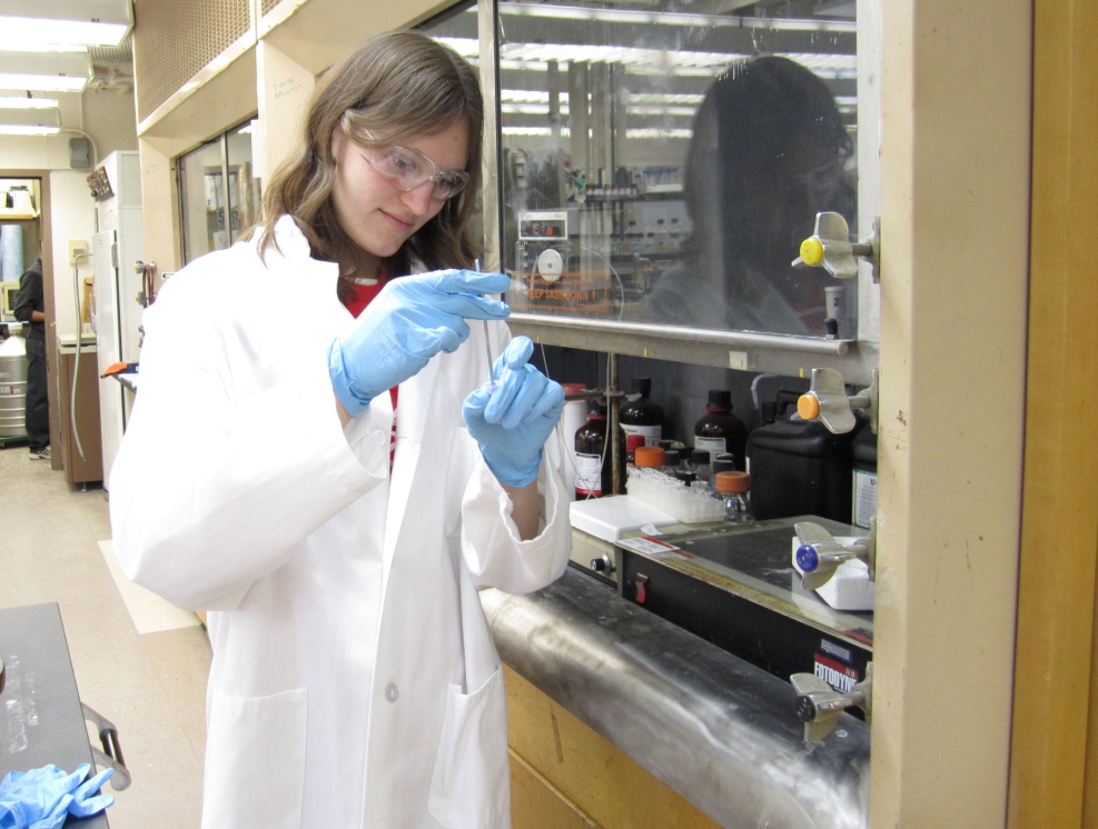
Photo credit: BTC Institute.Ellyn Lepinski is an intern at Promega who started her biotechnology career path five years ago as a high school junior taking a course from the BTC Institute (www.btci.org) as part of the Biotechnology Youth Apprenticeship Program.
Ellyn credits the program with helping her achieve her goals:
“Over the course of two years in which I was a Youth Apprentice, I obtained numerous skills, both inside and outside of the lab. I gained valuable scientific experience, including techniques like gel electrophoresis, nucleic acid purification, PCR, SDS-PAGE, Western blotting, cell culture and more.
On a personal level, I became very close with other students in the class and with our instructors, Barbara Bielec and Chad Zimprich. Everyone involved was always very approachable and willing to help with both laboratory tasks and in terms of giving advice for the future.
Through the program, I was placed in Dr. Que Lan’s entomology lab at UW-Madison, beginning in 2009. While there, I worked on a project involving sterol carrier protein-2, a protein involved in cholesterol uptake in mosquitoes.Notably, I am still working in Dr. Lan’s lab, however my research focus has shifted to bacterial fermentation. In between working in Dr. Lan’s lab, I also worked at the Forest Products Laboratory (USDA).
Additionally, this past June, I began an internship at Promega in the Scientific Applications department. Here I work to develop new applications for existing projects. This November marks five years of laboratory research for me, which would not have been possible without the Youth Apprenticeship Program and everyone involved. In addition to the specific labs that I have had the opportunity to work in, my experience in the Youth Apprenticeship Program has allowed me to emerge as a leader in my college lab courses. The program has clearly made a phenomenal impact on my life and is something I am very grateful for.”
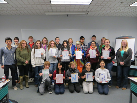
Photo credit: BTC Institute.Since 1993, the BTC Institute in partnership with the Dane County School Consortium has helped make such opportunities possible to nearly 300 students from public schools throughout Dane County. The program includes a paid apprenticeship in an industry or UW-Madison research lab and specialized instruction. In addition to being paid for their work, students receive high school credit for their participation in the worksite and the specialized biotechnology course held at the BTC Institute.
One aspect of the program that makes it so effective and unique is the amount of time that students spend working. Youth apprentices who start as juniors in the program must work 900 paid work hours to earn the Science, Technology, Engineering and Math (STEM) Skill Standards Certificate from the State of Wisconsin, youth apprentices who start work as seniors must earn 450 work hours. Students have had employment at a variety of companies and UW-Madison research labs, a few examples that have hired multiple apprentices include Genus PIC (ABS), MOFA Global, Promega and laboratories in the UW-Madison Departments of Bacteriology, Biochemistry, Entomology, Genetics, Horticulture, Plant Pathology and Surgery. Many of the students, like Ellyn, continue to be employed by their worksite long after they graduate from high school—proof of how effective this program is in helping to create the next generation STEM workforce.
Each year the BTC Institute hosts a Youth Apprenticeship Program preview night for all of the Dane County youth apprenticeship options: biotechnology, automotive technician, health services, and many more (www.dcsc.org). This year the preview nights will be held February 24 and 25 starting at 5:00pm. Students in grades 10 and 11 who are interested in learning more about the program are encouraged to attend one of the evening sessions with a parent.
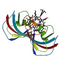
ImageSource=RCSB PDB; StructureID=1qpf; DOI=http://dx.doi.org/10.2210/pdb1qpf/pdb;
This article review was written by guest author, Amy Landreman, in the Cellular Analysis and Proteomics Group at Promega.
Charcot-Marie Tooth (CMT) disease is one of the most common inherited neurological disorders affecting approximately 2.8 million people worldwide. The most common form of CMT, CMT Type 1A, is caused by a 1.5Mb genomic duplication on Chr17 that results in trisomy of the critical myelin gene Peripheral Myelin Protein 22 (Pmp22). The extra copy of Pmp22 results in excessive PMP22 protein causing the neurophathy associated with CMT type 1A. Although there is no way to remove the extra copy of the gene, even subtle decreases in Pmp22 expression have shown promise against this inherited neuropathy in laboratory models.
In a recent paper, Inglese et al. 2014, describe an interesting new approach used to identify compounds that effectively decrease Pmp22 expression using a novel gene editing strategy and reporter-based screen. Their challenge was to create an assay that accurately represented endogenous Pmp22 expression including both transcriptional and post-transcriptional regulatory mechanisms, while maintaining the sensitivity required to detect subtle changes in expression in a loss of signal assay in a format compatible with microtiter 1536-well quantitative high-throughput screening (qHTS). Continue reading “Genome Editing and Reporter Technologies Enable Endogenous Pathway High-Throughput Assays”
As an HR professional, I attend different seminars and conferences to obtain credits for my HR certification. We had a SHRM (Society for Human Resources Management) state conference last week, and I learned all kinds of new strategies involving recruitment, succession planning, employee engagement and change management. One topic was present in every session I was in: social media. How to recruit through social media, engage employees in social media, and how to maintain your company and personal brand through social media.
Before you begin your subcloning, you need to know: The restriction enzyme (RE) sites available for subcloning in your parent vector multiple cloning region (or in the insert if you need to digest the insert); the RE sites available in the destination vector multiple cloning region (MCR); and if these same sites also occur in your insert. Once you know this information, you can use the chart below to decide which subcloning strategy to use.
XWe use cookies and similar technologies to make our website work, run analytics, improve our website, and show you personalized content and advertising. Some of these cookies are essential for our website to work. For others, we won’t set them unless you accept them. To learn more about our approach to Privacy we invite you to Read More
By clicking “Accept All”, you consent to the use of ALL the cookies. However you may visit Cookie Settings to provide a controlled consent.
We use cookies and similar technologies to make our website work, run analytics, improve our website, and show you personalized content and advertising. Some of these cookies are essential for our website to work. For others, we won’t set them unless you accept them. To find out more about cookies and how to manage cookies, read our Cookie Policy.
If you are located in the EEA, the United Kingdom, or Switzerland, you can change your settings at any time by clicking Manage Cookie Consent in the footer of our website.
Necessary cookies are absolutely essential for the website to function properly. These cookies ensure basic functionalities and security features of the website, anonymously.
Cookie
Duration
Description
cookielawinfo-checbox-analytics
11 months
This cookie is set by GDPR Cookie Consent plugin. The cookie is used to store the user consent for the cookies in the category "Analytics".
cookielawinfo-checbox-functional
11 months
The cookie is set by GDPR cookie consent to record the user consent for the cookies in the category "Functional".
cookielawinfo-checbox-others
11 months
This cookie is set by GDPR Cookie Consent plugin. The cookie is used to store the user consent for the cookies in the category "Other.
cookielawinfo-checkbox-advertisement
1 year
The cookie is set by GDPR cookie consent to record the user consent for the cookies in the category "Advertisement".
cookielawinfo-checkbox-necessary
11 months
This cookie is set by GDPR Cookie Consent plugin. The cookies is used to store the user consent for the cookies in the category "Necessary".
cookielawinfo-checkbox-performance
11 months
This cookie is set by GDPR Cookie Consent plugin. The cookie is used to store the user consent for the cookies in the category "Performance".
gdpr_status
6 months 2 days
This cookie is set by the provider Media.net. This cookie is used to check the status whether the user has accepted the cookie consent box. It also helps in not showing the cookie consent box upon re-entry to the website.
lang
This cookie is used to store the language preferences of a user to serve up content in that stored language the next time user visit the website.
viewed_cookie_policy
11 months
The cookie is set by the GDPR Cookie Consent plugin and is used to store whether or not user has consented to the use of cookies. It does not store any personal data.
Analytical cookies are used to understand how visitors interact with the website. These cookies help provide information on metrics the number of visitors, bounce rate, traffic source, etc.
Cookie
Duration
Description
SC_ANALYTICS_GLOBAL_COOKIE
10 years
This cookie is associated with Sitecore content and personalization. This cookie is used to identify the repeat visit from a single user. Sitecore will send a persistent session cookie to the web client.
vuid
2 years
This domain of this cookie is owned by Vimeo. This cookie is used by vimeo to collect tracking information. It sets a unique ID to embed videos to the website.
WMF-Last-Access
1 month 18 hours 24 minutes
This cookie is used to calculate unique devices accessing the website.
_ga
2 years
This cookie is installed by Google Analytics. The cookie is used to calculate visitor, session, campaign data and keep track of site usage for the site's analytics report. The cookies store information anonymously and assign a randomly generated number to identify unique visitors.
_gid
1 day
This cookie is installed by Google Analytics. The cookie is used to store information of how visitors use a website and helps in creating an analytics report of how the website is doing. The data collected including the number visitors, the source where they have come from, and the pages visted in an anonymous form.
Advertisement cookies are used to provide visitors with relevant ads and marketing campaigns. These cookies track visitors across websites and collect information to provide customized ads.
Cookie
Duration
Description
IDE
1 year 24 days
Used by Google DoubleClick and stores information about how the user uses the website and any other advertisement before visiting the website. This is used to present users with ads that are relevant to them according to the user profile.
test_cookie
15 minutes
This cookie is set by doubleclick.net. The purpose of the cookie is to determine if the user's browser supports cookies.
VISITOR_INFO1_LIVE
5 months 27 days
This cookie is set by Youtube. Used to track the information of the embedded YouTube videos on a website.
Performance cookies are used to understand and analyze the key performance indexes of the website which helps in delivering a better user experience for the visitors.
Cookie
Duration
Description
YSC
session
This cookies is set by Youtube and is used to track the views of embedded videos.
_gat_UA-62336821-1
1 minute
This is a pattern type cookie set by Google Analytics, where the pattern element on the name contains the unique identity number of the account or website it relates to. It appears to be a variation of the _gat cookie which is used to limit the amount of data recorded by Google on high traffic volume websites.

 As an HR professional, I attend different seminars and conferences to obtain credits for my HR certification. We had a SHRM (Society for Human Resources Management) state conference last week, and I learned all kinds of new strategies involving recruitment, succession planning, employee engagement and change management. One topic was present in every session I was in: social media. How to recruit through social media, engage employees in social media, and how to maintain your company and personal brand through social media.
![4498MA-[Converted]](https://www.promegaconnections.com/wp-content/uploads/2014/10/4498MA-Converted-e1413386878386.jpg)