Biotechnology Ice Breakers: A Few Conversation Starters

quiz pictureThe biotechnology industry is one of the most dynamic out there – in fact, it never stands still! For non-scientists this can be intimidating. For scientists, it can be challenging to explain what we do in ways that non-scientists can understand and appreciate.

Scientists have made great strides in improving our ability to use molecular processes to our advantage, from discovering the basics of how to isolate and manipulate DNA to gaining an understanding of how genes direct the creation of proteins in cells.  It’s clear that there is a lot we can contribute to the scientific literacy of the general public.

In this spirit, we’ve designed a short quiz for both non-scientists (you may learn something new) and scientists (you may find it useful for engaging in conversations with your non-scientist friends and family members).  Spoiler alert: answers are provided. Continue reading “Biotechnology Ice Breakers: A Few Conversation Starters”

Death in the Stars: A Virus Decimates Sea Star Populations Along the Pacific Coast

Starfish BlueA killer is lurking in the waters off the pacific coast. Silent and lethal, it leaves its decimated victims in tidal pools. They first began to appear in the early summer of 2013. Limp and curled, missing some or all of their limbs, the bodies were little more than globs of slimy tissue.  They were hardly recognizable as what they once were—Sea Stars. Continue reading “Death in the Stars: A Virus Decimates Sea Star Populations Along the Pacific Coast”

MicroRNAs as Circulating Biomarkers

12097693_lMicroRNAs (miRNAs) are short strands of RNA averaging between 19-24 nucleotides in length that were first discovered in C.elegans and subsequently shown to exist in species ranging from algae to humans (1). Speculated to be merely “junk” more than a decade ago, miRNAs have emerged as powerful regulators of a wide array of cellular processes because of their influence on gene expression at the posttrancriptional level. Dysregulation of these miRNAs is also associated with life-threatening conditions such as cancer and cardiovascular disease, which points to a potential use of miRNAs in diagnosis and treatment. Recently, it has been demonstrated that miRNAs are present in circulating blood plasma, protected from degradation by inclusion in lipid or lipoprotein complexes. This opens up the possibility to exploit miRNA as a useful diagnostic tool in clinical samples. Continue reading “MicroRNAs as Circulating Biomarkers”

2 Ways to Save Your Single Reporter Data

Reporter assays using a single reporter, be it from a stable cell line or transient transfection, can benefit from normalization. Obviously, we are not talking about adding a second control reporter but normalizing to the number of live or dead cells in the well.  Two cell health assays, CellTiter-Fluor™ Cell Viability Assay and CellTox™ Green Cytotoxicity Assay, are ideally suited for multiplexing with reporter assays. 

Continue reading “2 Ways to Save Your Single Reporter Data”

General Considerations for Transfection

Many studies, from reporter assays to protein localization to BRET and FRET, require successful transfection first. Yet, transfection can be tricky and difficult. There are many considerations when planning transfection of your cells including reagent selection, stable or transient experiment, type of molecule and endpoint assay used. Here we discuss these considerations to help you plan a successful transfection scheme for your experimental system. Continue reading “General Considerations for Transfection”

How Do You Defrost the Lab Freezer?

When I was in the lab, we usually started with an elaborate system of borrowed hairdryers and old chemistry ring stands. What is your preferred method of attacking a frost-full freezer?
eh_august2014_large

Differentiating but not Mature Adipocytes Provide a Defense Against S. aureus Infection

http://upload.wikimedia.org/wikipedia/commons/6/65/Blausen_0012_AdiposeTissue.png
Cross-section of skin and adipose tissue enlargement. Used courtesy of Wikimedia Commons, Blausen.

A basic tenet of immunology is that antibodies produced by B cells are very important and specific immunoprotective agents, released in response to infection.

However, antibodies do not supply immediate protection. The invading organism needs to get into the host, meet up with T cells and then B cells, in order for antibody production to occur. If the host has seen this particular pathogen previously, the antibody response occurs somewhat more quickly, but we’re still talking about days. If the invading organism is a bacterium, it can multiply and double in numbers in just hours. Thus an infection could potentially gain a foothold in a body prior to an antibody response.

Fortunately we have a more rapid, first line of defense to invading pathogens, a cellular response. In the case of a puncture or skin wound, epithelial cells, mast cells and leukocytes are activated quickly in response to pathogens. Neutrophils and monocytes also aid the cellular response.

Now a recently published report demonstrates that fat cells also play a part in the cellular response to invading bacteria. R. Gallo et al. published a study on Jan. 2 in Science, providing more in depth information on the role of adipocytes in the host response to the bacterium Staphylococcus aureus (S. aureus). Continue reading “Differentiating but not Mature Adipocytes Provide a Defense Against S. aureus Infection”

Improved Characterization and Quantification of Complex Cell Surface N-Glycans

MSextractcropped

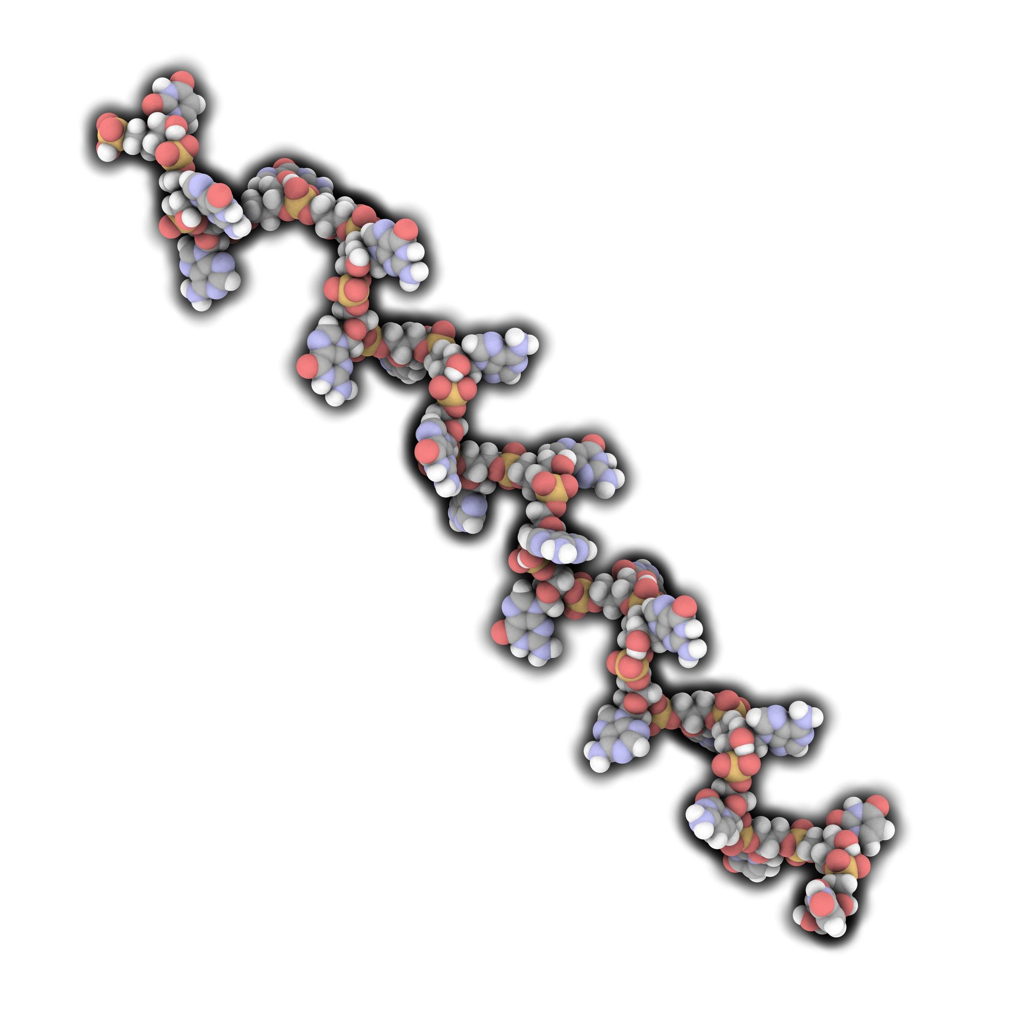
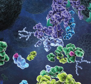
N-Glycosylation is a common protein post-translational modification occurring on asparagine residues of the consensus sequence asparagine-X-serine/threonine, where X may be any amino acid except proline. Protein N-glycosylation takes place in the endoplasmic reticulum (ER) as well as in the Golgi apparatus.

Approximately half of all proteins typically expressed in a cell undergo this modification, which entails the covalent addition of sugar moieties to specific amino acids. There are many potential functions of glycosylation. For instance, physical properties include: folding, trafficking, packing, stabilization and protease protection. N-glycans present at the cell surface are directly involved in cell−cell or cell−protein interactions that trigger various biological responses.

The standard method used to profile the N-glycosylation pattern of cells is glycoprotein isolation followed by denaturation and/or tryptic digestion of the glycoproteins and an enzymatic release of the N-glycans using PNGase F followed by analysis mass spec. This method has been reported to yield high levels of high-mannose N-glycans that stem from both membrane proteins as well as proteins from the ER.(1,2)

Are you looking for proteases to use in your research?
Explore our portfolio of proteases today.

For those researchers interested in characterizing only cell surface glycans (i.e.,  complex N-glycans)  a recent reference has developed a model system using HEK-292 cells that demonstrates a reproducible, sensitive, and fast method to profile surface N-glycosylation from living cells (3). The method involves standard centrifugation followed by enzymatic release of cell surface N-glycans. When compared to the standard methods the detection and quantification of complex-type N-glycans by increased their relative amount from 14 to 85%.

  1. North, S. J. et al. (2012) Glycomic analysis of human mast cells, eosinophils and basophils. Glycobiology. 2012, 22, 12–22.
  2. Reinke, S. O. et al. (2011) Analysis of cell surface N-glycosylation of the human embryonic
    kidney 293T cell line. J. Carbohydr. Chem.  30, 218–232.
  3. Hamouda, H. et al. (2014) Rapid Analysis of Cell Surface N‑Glycosylation from Living Cells Using Mass Spectrometry. J of Proteome Res. 13, 6144–51.

Monday SMILE

Monday lab meeting? Journal club? Long weekend in the lab?

Enjoy this classic from the Promega Cartoon Lab.

eh1

How MicroRNAs Have a Big Effect on Genetic Regulation

miR-133 microRNA (green) and myogenin mRNA (red) in differentiating C2C12 cells. Image by Ryan Jeffs, courtesy of Wikimedia Commons.

Some of us scientists who have been around for a while still think about RNA molecules falling into three categories: messenger RNA (mRNA), ribosomal RNA (rRNA) and transfer RNA (tRNA). However, I have revised my outdated RNA classification scheme as scientists discover exciting new classes of RNAs that do some fairly amazing things. For example, in the early 1980s, Thomas Cech discovered ribozymes, RNAs that have catalytic functions (1), and in the early 1990s, researchers began to take interest in short noncoding RNAs that act as a genetic regulators, the first of which was discovered in C. elegans (2). RNA is no longer simply a biological middleman between DNA and protein. These ephemeral nucleic acid molecules play a much bigger role of cellular physiology and gene regulation than we had previously ascribed to RNA.

Continue reading “How MicroRNAs Have a Big Effect on Genetic Regulation”