Lost in Translation? Tips for Preparing RNA for in vitro Translation Experiments

In vitro translation of proteins through cell-free expression systems using rabbit reticulocytes, E. coli S30, or wheat germ extracts can be invaluable in studying protein function.ย  If you only need a small amount (100s of nanograms), itโ€™s also faster and easier than synthesizing vast quantities in bacterial or mammalian cells (~ 90 minutes for cell-free vs. long growth times and extraction steps after an initial optimization for protein synthesized in larger scale). ย There are many systems out there, and knowing which to use can sometimes be difficult.ย  Many kits include components that combine transcription and translation in one-step, eliminating the need to provide your own RNA.ย  But when you want to make your own RNA templates to add to lysates, then there are additional concerns.
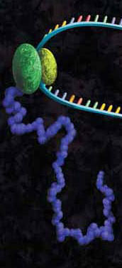
artists concept of in vitro translation
A protein chain being produced from a ribosome.

Many people donโ€™t want to work with RNA since the common lab lore suggests itโ€™s a finicky molecule, and for good reason.  Extracting it requires the utmost care in technique and elimination of nucleases.  Failing to do so results in degradation of the molecule, and so with it your experiments (see our recent blog by Terri Sundquist on tips for isolating RNA with ease).  Preparing RNA for cell-free expression is subject to the same concerns as extracted RNA, but with the proper care is not that much more of a challenge than using a DNA template.

The first step for using cell-free expression systems with RNA templates is to make the RNA.  Here are some tips that will ensure success.

Continue reading “Lost in Translation? Tips for Preparing RNA for in vitro Translation Experiments”
Get More Out of Your Lentiviral Production

fugene6_lvv_blogThis review is a guest blog by Amy Landreman, Product Specialist in Cellular Analysis at Promega Corporation.
Lentiviral vectors (LVV) have become a valuable research tool for delivering genetic content into a wide range of cell types. Commonly derived from the HIV-1 genome, LVV have the advantage of being able to infect both dividing and non-dividing cells. They can be particularly valuable for introducing genetic material into cell lines that are difficult to transfect using other methods and are also being used in gene therapy applications.

Unlike other gene delivery tools, transducing mammalian cells with LVV requires significant upfront effort since the LVV particles carrying the desired genetic content first need to be created. In general this involves co-transfecting a packaging cell line, such as HEK293T, with a set of three to four separate plasmids that encode the protein content required to generate the LVV particles: the transfer plasmid, which contains the transgene of interest, a packaging plasmid, and an envelope plasmid. After co-transfection, the packaging cell line is allowed to incubate for a couple of days during which time the LVV particles are produced and accumulating in the culture supernatant. The supernatant containing the recombinant LVV is then harvested and, following several concentration steps, the LVV particles are ready to be used for introducing the desired genetic content into the mammalian target cells. Continue reading “Get More Out of Your Lentiviral Production”

How to Isolate RNA like a Pro

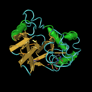
Ribbon diagram of RNAโ€™s biggest threat: a ribonuclease

Back in graduate school, I purified a lot of RNA, and after a while, I became fairly successful at it. My yields were good, and the RNA was intact. However, many of my early attempts at RNA isolation yielded degraded RNA that did not work well in many downstream applications. In my case, successfully isolating high-quality RNA required practice. During my trials and tribulations, I learned a lot of tricks and tips about how to obtain high-quality RNA. Here I share some of these tricks to help you speed through that โ€œpractice makes perfectโ€ phase so that you can isolate RNA like a pro.

Continue reading “How to Isolate RNA like a Pro”

Dealing with PCR Inhibitors

Inhibition

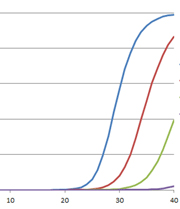
The polymerase chain reaction (PCR) has revolutionized modern biology as a quick and easy way to generate amazing amounts of genomic data. However, when PCR doesnโ€™t work, it can be frustrating. At these times, PCR and reverse transcription PCR (RT-PCR) inhibitors seem to be everywhere: They lie dormant in your starting material and can co-purify with the template of interest, and they can be introduced during sample handling or reaction setup. The effects of these inhibitors can range from partial inhibition and underestimation of the target nucleic acid amount to complete amplification failure. What is a scientist to do?

Continue reading “Dealing with PCR Inhibitors”

Practical Tips for HEK293 Cell Culture When Using cAMP-Glo™ Assay

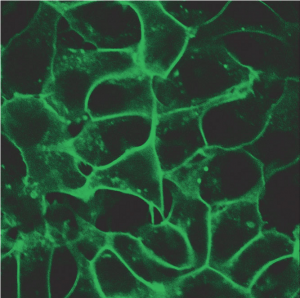
HEK293 cells stably expressing HaloTagยฎ-ECS (ExtraCellular Surface; comprised of a signal sequence and single transmembrane domain of ฮฒ1-integrin) fusion protein labeled with HaloTagยฎ Alexa Fluorยฎ 488 Ligand and then imaged.
HEK293 cells stably expressing HaloTagยฎ-ECS fusion protein labeled
with HaloTagยฎ Alexa Fluorยฎ 488 Ligand and then imaged.

G Protein Coupled Receptors represent one of the largest classes of cell surface receptors and one of the most important classes for drug targets. Fifty of the top 200 drugs target GPCRs. GPCRs respond to various stimuli like light, odors, hormones, neurotransmitters and others. They cover virtually all therapeutic areas. When a particular GPCR is implicated in a disease, researchers screen the GPCR and its signaling pathways, the hope being that promising therapeutic targets might be identified. Major G-protein families signal via secondary messengers like cAMP, which in turn activate a range of effector systems to change cell behavior and/or gene transcription. There are various approaches and methods to study GPCRs and measure the increase or decrease of intracellular cAMP. However, the fastest and the most sensitive among all methods is a plate based cAMP-Gloโ„ข Assay.

Continue reading “Practical Tips for HEK293 Cell Culture When Using cAMP-Glo™ Assay”

Piecing the Puzzle Together: Using Multiple Assays to Better Understand What Is Happening with Your Cells

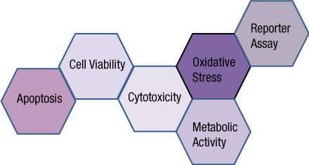
You often need several pieces of information to really understand what is happening within a cell or population of cells. If your cells are not proliferating, are they dying? Or, are you seeing cytostasis? If they are dying, what is the mechanism? Is it apoptosis or necrosis? If you are seeing apoptosis, what is the pathway: intrinsic or extrinsic?

If you are measuring expression of a reporter gene and you see a decrease in expression, is that decrease due to transfection inefficiencies, cytotoxicity, or true down regulation of your reporter gene?

To investigate these multiple parameters, you can run assays in parallel, but that requires more sample, and sample isn’t always abundant.

Multiplexing assays allows you to obtain information about multiple parameters or events (e.g., reporter gene expression and cell viability; caspase-3 activity and cell viability) from a single sample. Multiplexing saves sample, saves time and gives you a more complete picture of the biology that is happening with your experimental sample.

What information do you need about your cells to complete the picture?

Multiplexing assay reagents to measure biomarkers in the same sample has often been considered an application only accomplished with antibodies or dyes and sophisticated detection instrumentation. However, Promega has developed microwell plate based assays for cells in culture that allow multiplexed detection of biomarkers in the same sample well using standard multimode multiwell plate readers. Continue reading “Piecing the Puzzle Together: Using Multiple Assays to Better Understand What Is Happening with Your Cells”

Methods for Quantitating Your Nucleic Acid Sample

Nucleic acid quanitation webinar

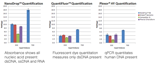
For most molecular biology applications, knowing the amount of nucleic acid present in your purified sample is important. However, one quantitation method might serve better than another, depending on your situation, or you may need to weigh the benefits of a second method to assess the information from the first. Our webinar โ€œTo NanoDropยฎ or Not to NanoDropยฎ: Choosing the Most Appropriate Method for Nucleic Acid Quantitationโ€ given by Doug Wieczorek, one of our Applications Scientists, discussed three methods for quantitating nucleic acid and outlined their strengths and weaknesses.

Continue reading “Methods for Quantitating Your Nucleic Acid Sample”

Top Ten Tips for Successful PCR

We decided to revisit a popular blog from our Promega Connections past for those of you in the amplification world. Enjoy:

magnesium-31

    • Modify reaction buffer composition to adjust pH and salt concentration.
    • Titrate the amount of DNA polymerase.
    • Add PCR enhancers such as BSA, betaine, DMSO, nonionic detergents, formamide or (NH4)2SO4.
    • Switch to hot-start PCR.
    • Optimize cycle number and cycling parameters, including denaturation and extension times.
    • Choose PCR primer sequences wisely.
    • Determine optimal DNA template quantity.
    • Clean up your DNA template to remove PCR inhibitors.
    • Determine the optimal annealing temperature of your PCR primer pair.

[Drum roll please]…and the ย mostย importantย thing you can do to improve your PCR results is:

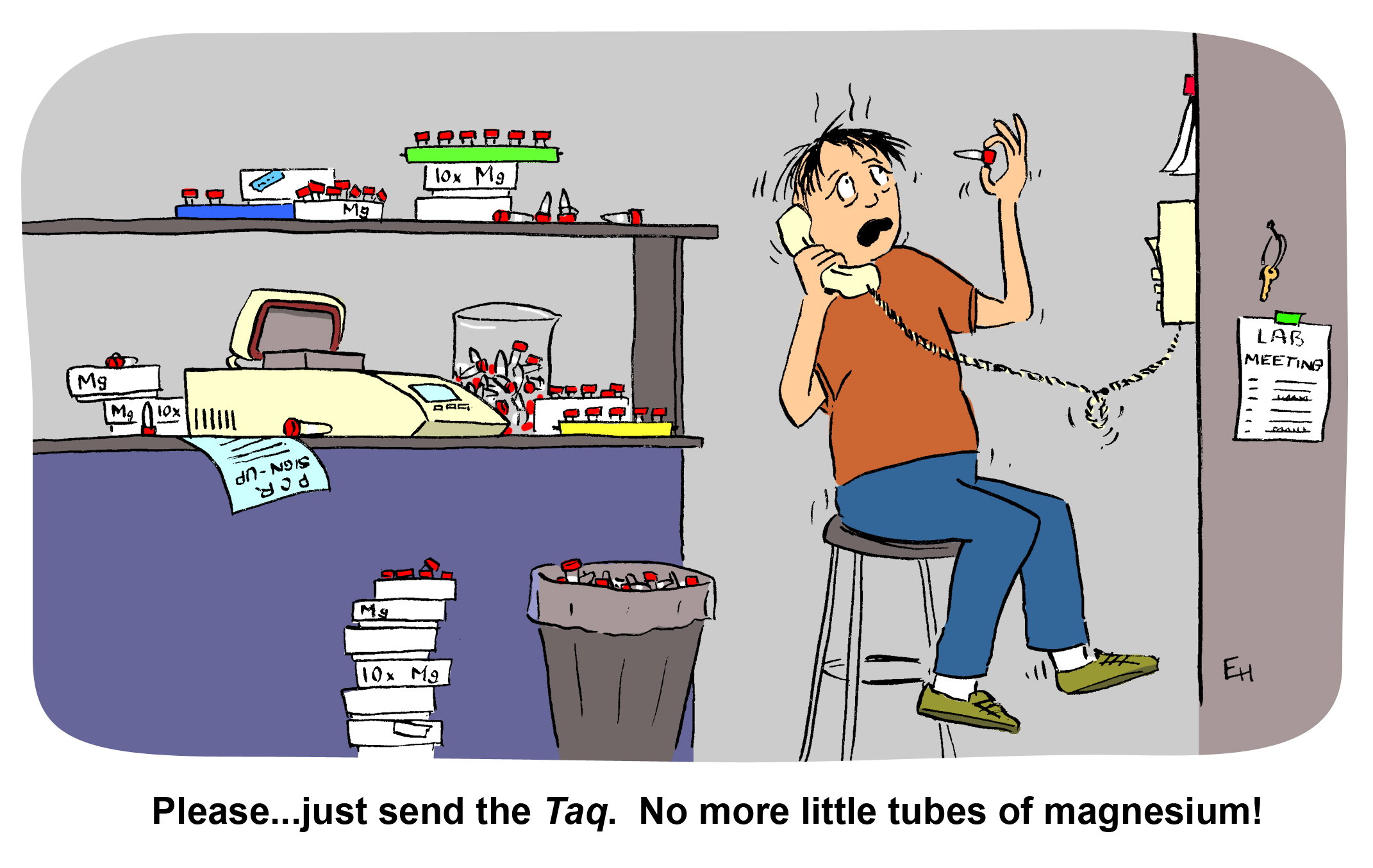
  • Titrate the magnesium concentration.

Successful Ligation and Cloning of Your Insert

Ligation and cloning
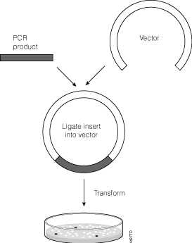
Cloning PCR product.

You have PCR amplified your insert of interest, made sure the PCR product is A tailed and are ready to clone into a T vector (e.g., pGEMยฎ-T Easy Vector). The next step is as simple as mixing a few microliters of your purified product with the cloning vector in the presence of DNA ligase, buffer and ATP, right? In fact, you may need to consider the molar ratio of T vector to insert.

Continue reading “Successful Ligation and Cloning of Your Insert”

Don’t Let Ribonucleases Ruin Your Week(end): Establish a Ribonuclease-free Environment

buffers_image1_274x218

My very first job in science was in a lab that worked exclusively with RNA, and it was only after I moved on to a different job that I learned just how much different the world of DNA research is from that of RNA. When working with DNA, for example, you rarely if ever have the sample you have labored over reduced to a fuzzy blur at the bottom of a gel because it has been degraded beyond rescue. With RNA, unfortunately, this happens all too frequently. In fact, a labmate of mine once put up a poll on the door to our lab asking if it was better to discover that your RNA sample was degraded on a Monday or a Friday.

The culprits in this scenario are Ribonucleases (RNases). They are everywhere. They are incredibly stable and difficult to inactivate. And, if you work with RNA, they are your enemy. Take heart though, they can be defeated if you follow some pretty simple steps.

Continue reading “Don’t Let Ribonucleases Ruin Your Week(end): Establish a Ribonuclease-free Environment”