Scientists have had a love-hate relationship with PCR amplification for decades. Real-time or quantitative PCR (qPCR) can be an amazingly powerful tool, but just like traditional PCR, it can be quite frustrating. There are several parameters that can influence the success of your PCR assay. We’ve highlighted ten things to consider when trying to improve your qPCR results.
Continue reading “Top 10 Tips to Improve Your qPCR or RT-qPCR Assays”Tips and Tools
A Normalization Method for Luciferase Reporter Assays of miRNA-Mediated Regulation
Today’s blog is from guest blogger Ken Doyle of Loquent, LLC. Here, Ken reviews a 2014 paper highlighting specific considerations for using reporter assays to study miRNA-mediated gene regulation.

The accelerated pace of research into noncoding RNAs has revealed multiple regulatory roles for microRNAs (miRNAs). These diminutive noncoding RNA speciesโtypically 20-24 nucleotides in lengthโare now known to mediate a broad range of biological functions in plants and animals. In humans, miRNAs have been implicated in various aspects of development, differentiation, and metabolism. They are known to regulate an assortment of genes involved in processes from neuronal development to stem cell division. Dysregulation of miRNA expression is associated with many disease states, including neurodegenerative disorders, cardiovascular disease, and cancer.
Typically, miRNAs act as post-transcriptional repressors of gene expression, either by targeted degradation of messenger RNA (mRNA) or by interfering with mRNA translation. Most miRNAs exert these effects by binding to specific sequences called microRNA response elements (MREs). These sequences are found most often within the 3ยด-untranslated regions (3ยด-UTRs) of animal genes, while they may occur within coding sequences in plant genes.
Studies of the regulatory roles played by miRNAs often involve cell-based assays that use a reporter gene system, such as luciferase or green fluorescent protein. In a standard assay, the reporter gene is cloned upstream of the 3ยด-UTR sequence being studied; this construct is then cotransfected with the miRNA into cells in culture. A study by Campos-Melo et al., published in September 2014, examined this experimental approach for miRNAs from spinal cord tissues, using firefly luciferase as the reporter gene and Renilla luciferase as a transfection control.
Continue reading “A Normalization Method for Luciferase Reporter Assays of miRNA-Mediated Regulation”Cloning Tips for Restriction Enzyme-Digested Vectors and Inserts

PCR Cloning: Answers to Some Frequently Asked Questions
Q: What is the easiest way to clone PCR Products?
A: The simplest way to clone PCR Products is to amplify the product using thermostable polymerases such as Taq, Tfl or Tth polymerase. These polymerases add a single deoxyadenosine to the 3ยด-end of the amplified products (3ยด-end overhang), and can be cloned directly into a linearized T-vector.
Q: What if my DNA polymerase has 3ยด to 5ยด exonuclease activity (i.e., proofreading activity) that removes the 3ยด-end overhang?
A: To clone PCR products that have been amplified with a polymerase that have proof reading activity into a T-vector, you will need to perform an A-tailing step using Taq DNA polymerase and dATP. Blunt ended restriction digest fragments can also be A-tailed using this method. The method below uses GoTaq Flexi DNA Polymerase (comes with a Mg-free reaction buffer), but anyย Taqย DNA polymerase can be used.
Set up the following reaction in a thin-walled PCR tube:
1โ4ยตl purified blunt-ended DNA fragment (from PCR or restriction enzyme digestion)
2ยตl of 5X GoTaq Reaction Buffer (Colorless or Green)
2ยตl of 1mM dATP (0.2mM final concentration)
1ยตl GoTaq Flexi DNA Polymerase (5u/ยตl)
0.6ยตl of 25mM MgCl2ย (1.5mM final concentration)
Nuclease-free water to a final volume of 10ยตl
Incubate at 70ยฐC for 15โ30 minutes in a water bath or thermal cycler.
Q: What is a T-vector, and why are they used for cloning PCR products?
A: T vectors are linearized plasmids that have been treated to add T overhangs to match the A overhangs of the PCR product. PCR fragments that contain an A overhang can be directly ligated to these T-tailed plasmid vectors with no need for further enzymatic treatment other than the action of T4 DNA ligase.
For a complete PCR Cloning protocol, Visit the Cloning Chapter of the Promega Protocols and Applications Guide.
General Considerations for Transfection
Many studies, from reporter assays to protein localization to BRET and FRET, require successful transfection first. Yet, transfection can be tricky and difficult. There are many considerations when planning transfection of your cells including reagent selection, stable or transient experiment, type of molecule and endpoint assay used. Here we discuss these considerations to help you plan a successful transfection scheme for your experimental system. Continue reading “General Considerations for Transfection”
6 + 1 Ways Dual-Reporter Assays Can Save Your Data
Updated 02/12/2021
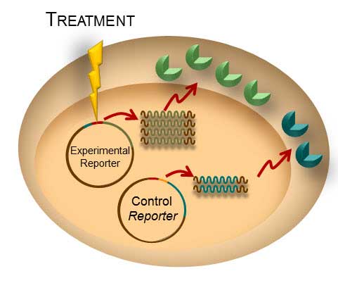
Transient transfection is often used to perform reporter assays. We have advocated using a dual-reporter system for decades to normalize the data obtained and gain a clearer understanding of your results. The experimental reporter should vary with treatment and the control reporter should vary little with treatment. The control reporter thus serves as a marker to help you understand the relative activity of your experimental reporter. The bioluminescent Dual-Luciferaseยฎ method allows for sequential detection of the second reporter in a single sample providing a simple two-step normalization method. Here are seven ways in which dual-reporter assays help you avoid misinterpreting results.
Simply comparing the ratio of the experimental to the control reporter can resolve differences in:
- Number of Cells/Well: When manually pipeting cells into a 96-well plate, there is always a chance of havingย variable numbers of cells in each well. This variation is cell number will affect the experimental and control reporters equally, so the ratio of experimental:control reporter activity will eliminate false interpretation of the experimental data–whether it affects an entire row or column on the plate or individual wells.
- Transfection Efficiency: The variations in transfection efficiency will equally affect both the experimental and control reporters so the ratio of activity in dual-reporter assays will normalize the data.
- Cell Viability: Often, reporter assays look at the dose response curve of a particular compound with regard to gene expression. Ideally, if a compound causes a change in the experimental reporter the control reporter will demonstrate little effect. However, if the compound is toxic, both the experimental and control will be altered and the ratio will tell you whether the compound truly affects reporter activity or just killsย the cells.
- Lysis Efficiency: When lysing a plate of cells, you could encounter situations where rows or columns lyse differently, especially if you are using manual disruption or get interrupted mid-plate. The difference is lysis will affect the experimental and control equally so the ratio will remove the variation.
- Temperature: Ideally, a plate should be equilibrated to ambient room temperature before proceeding to the reporter assay. Plates can cool at different rates or researchers anxious to record data may read the data early. Temperature variations will affect both reporters so the ratio will limit the affect on the data.
- Measurement Time: Repetition of data is a hallmark of good science. You are often called upon to repeat experiments sometimes days or weeks apart. Letโs say you repeat your experiment one week after the initial experiment. The first time you measured the response, you waited 10 minutes after reagent addition to read, this week you waited 30 minutes. This will affect both reporters equally and therefore the ratio will allow you to more easily compare the data from this week and last week.
Bonus Benefit from Dual-Luciferaseยฎ, Dual-Gloยฎ and the NanoGloยฎ Dual Luciferase Reporter Systems: No Lysate Splitting: Promega dual-reporter assays are designed for same-well multiplexing so there is no chance of variations creeping into your data due to unequal splitting of the cellular lysate to measure two separate reporter activities.
Since the introduction of the first bioluminescent dual-luciferase assay in 1995, this approach has been used in countless studies to advance our scientific understanding of cellular gene regulation.
Related Posts
Choosing Your Subcloning Strategy
Before you begin your subcloning, you need to know: The restriction enzyme (RE) sites available for subcloning in your parent vector multiple cloning region (or in the insert if you need to digest the insert); the RE sites available in the destination vector multiple cloning region (MCR); and if these same sites also occur in your insert. Once you know this information, you can use the chart below to decide which subcloning strategy to use.
To learn more about subcloning, visit our Subcloning Notebook.
To inject or not inject?

Luciferase assays are useful tools for studying a wide range of biological questions. They can be performed easily by adding a reagent that provides components necessary to generate a luminescent signal directly to cells or a cell lysate. However, once this reagent has been added, how long you wait to measure the signal becomes a key consideration in generating consistent data. Dependent on which luciferase assay you use, you may need a luminometer that can use injectors to deliver the assay reagents. The reason for this is simple, but can be confusing to new users.
Letโs start by discussing two types of luciferase assays: โflashโ vs. โglowโ. Continue reading “To inject or not inject?”
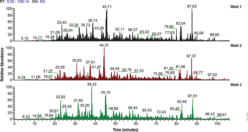
Monitoring Mass Spec Instrument Performance and Sample Preparation
Proteomics, the analysis of the entire protein content of a living system, has become a vital part of life science research, and mass spectrometry (MS) is the method for analyzing proteins. MS analysis of protein content allows researchers to identify proteins, sequence them and determine the nature of post translational modifications.

Mass spectrometry allows characterization of molecules by converting them to ions so that they can be manipulated in electrical and magnetic fields. Basically a small sample (analyte) is ionized, usually to cations by loss of an electron. After ionization, the charged particles (ions) are separated by mass and charge; the separated particles are measured and data displayed as a mass spectrum. The mass spectrum is typically presented as a bar graph where each peak represents a single charged particle having a specific mass-to-charge (m/z) ratio. The height of the peak represents the relative abundance of the particle. The number and relative abundance of the ions reveal how different parts of the molecule relate to each other.
For the study of large, organic macromolecules, matrix associated laser desorption/ionization (MALDI) or tandem mass spec/collision induced dissociation (MS/MS) techniques are often used to generate the charged particles from the analyte. MS analysis brings sensitivity and specificity to proteome analysis. The technique has excellent resolution and is able to distinguish one ion from another, even when their m/z ratios are similar. Macromolecules are present in extremely different concentrations in the cells, and MS analysis can detect biomolecules across five logs of concentration.
Continue reading “Monitoring Mass Spec Instrument Performance and Sample Preparation”Optimize Your Western Blot
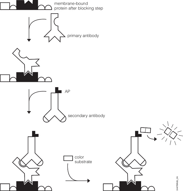
Youโve probably been there. Youโve got a new antibody or youโre testing out one youโve made yourself. After weeks or months of work, your antibody is going to help move your research project forward. As you excitedly head to the dark room to develop your film, your mood is crushed when you seeโฆbands, more bands, and smears. Alas, science has played one more cruel joke on you as you experience what so many of your fellow scientists have before. Despite such a dismal beginning, you often can still get good western blots by changing steps in your protocol.
Several steps in the western blot protocol can be optimized.
Continue reading “Optimize Your Western Blot”

![4498MA-[Converted]](https://www.promegaconnections.com/wp-content/uploads/2014/10/4498MA-Converted-e1413386878386.jpg)