Get More Out of Your Lentiviral Production

fugene6_lvv_blogThis review is a guest blog by Amy Landreman, Product Specialist in Cellular Analysis at Promega Corporation.

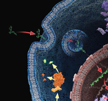
Lentiviral vectors (LVV) have become a valuable research tool for delivering genetic content into a wide range of cell types. Commonly derived from the HIV-1 genome, LVV have the advantage of being able to infect both dividing and non-dividing cells. They can be particularly valuable for introducing genetic material into cell lines that are difficult to transfect using other methods and are also being used in gene therapy applications.

Unlike other gene delivery tools, transducing mammalian cells with LVV requires significant upfront effort since the LVV particles carrying the desired genetic content first need to be created. In general this involves co-transfecting a packaging cell line, such as HEK293T, with a set of three to four separate plasmids that encode the protein content required to generate the LVV particles: the transfer plasmid, which contains the transgene of interest, a packaging plasmid, and an envelope plasmid. After co-transfection, the packaging cell line is allowed to incubate for a couple of days during which time the LVV particles are produced and accumulating in the culture supernatant. The supernatant containing the recombinant LVV is then harvested and, following several concentration steps, the LVV particles are ready to be used for introducing the desired genetic content into the mammalian target cells. Continue reading “Get More Out of Your Lentiviral Production”

Practical Tips for HEK293 Cell Culture When Using cAMP-Glo™ Assay

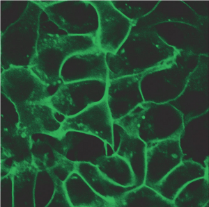
HEK293 cells stably expressing HaloTag®-ECS (ExtraCellular Surface; comprised of a signal sequence and single transmembrane domain of β1-integrin) fusion protein labeled with HaloTag® Alexa Fluor® 488 Ligand and then imaged.
HEK293 cells stably expressing HaloTag®-ECS fusion protein labeled
with HaloTag® Alexa Fluor® 488 Ligand and then imaged.

G Protein Coupled Receptors represent one of the largest classes of cell surface receptors and one of the most important classes for drug targets. Fifty of the top 200 drugs target GPCRs. GPCRs respond to various stimuli like light, odors, hormones, neurotransmitters and others. They cover virtually all therapeutic areas. When a particular GPCR is implicated in a disease, researchers screen the GPCR and its signaling pathways, the hope being that promising therapeutic targets might be identified. Major G-protein families signal via secondary messengers like cAMP, which in turn activate a range of effector systems to change cell behavior and/or gene transcription. There are various approaches and methods to study GPCRs and measure the increase or decrease of intracellular cAMP. However, the fastest and the most sensitive among all methods is a plate based cAMP-Glo™ Assay.

Continue reading “Practical Tips for HEK293 Cell Culture When Using cAMP-Glo™ Assay”

Preventing the Heartache of Cell Line Misidentification

Golden mask

It’s a scientist’s nightmare: Spending time and resources to investigate a biological phenomenon only to learn later that your cells are not what you think they are—their true identities hidden. As a result, all of the data that you’ve generated with those cells, published and unpublished, are cast into doubt. You thought that you knew your cells, that you could trust them, but your trust was misplaced. At some point, perhaps even before the traitorous cell line entered your laboratory, the cells were mislabeled, misidentified or contaminated with another cell line. It didn’t have to be this way. There are easy steps you can take to prevent the headache and heartache of cell line misidentification and contamination.

Continue reading “Preventing the Heartache of Cell Line Misidentification”

“Fingerprinting” Your Cell Lines

Working in the laboratoryResearchers working with immortalized cell lines would readily agree when I state that it is almost impossible to look at cells under the microscope and identify them by name. There are phenotypic traits, however they do change with change in media composition, passage number and in response to growth factors. I remember the pretty arborizations my neuroblastoma cell line SH-SY5Y exhibited in response to nerve growth factor treatment. Thus physical appearance is not a distinguishing feature. Currently, in many labs, researchers typically use more than one cell line, and more than likely, share the same lab space to passage cells and the same incubator to grow the cells. In such scenarios, it is not difficult to imagine that cell lines might get mislabeled or cross-contaminated. For example HeLa cells, one of the fastest growing cell lines have been shown to invade and overtake other cell lines.

Misidentification of cell lines has deep and severe implications. A review of cell lines used to study esophageal adenocarcinoma found that a large number of the cell lines were actually derived from lung or gastric cancers. Unfortunately, by the time this error was discovered, data from these cell line studies were already being used for clinical trials and other advanced studies and publications. Moreover, the cell lines were being to screen and design and test specific cancer drugs which ended up in flawed clinical trials. Continue reading ““Fingerprinting” Your Cell Lines”

Considerations for Successful Cell Based Assays II: Cell Culture Conditions

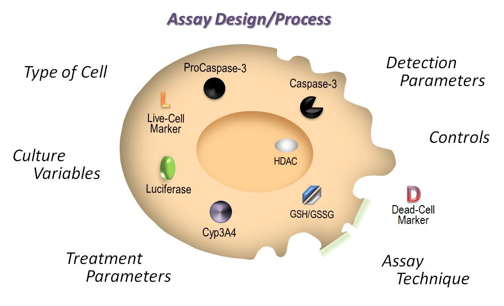
This is the second in a series of blog posts covering topics to consider when designing and performing cell-based assays. In the first installment, we discussed the importance of choosing the right cell type for your assay. Here we will discuss how cell culture conditions affect cell-based assays.

Continue reading “Considerations for Successful Cell Based Assays II: Cell Culture Conditions”