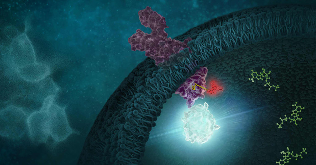
Cellular Selectivity Profiling: Unveiling Novel Interactions and More Accurate Compound Specificity

This blog was written by guest contributor Tian Yang, Associate Product Manager, Promega, in collaboration with Kristin Huwiler, Manager, Small Molecule Drug Discovery, Promega.

Determining the selectivity of a compound is critical during chemical probe or drug development. In the case of chemical probes, having a clearly defined mechanism of action and specific on-target activity are needed for a chemical probe to be useful in delineating the function of a biological target of interest in cells. Similarly, optimizing a drug candidate for on-target potency and reducing off-target interactions is important in the drug development process (1,2). A thorough understanding of the selectivity profile of a drug can facilitate drug repurposing, by enabling approved therapeutics to be applied to new indications (3). Interestingly, small molecule drugs do not necessarily require the same selectivity as a chemical probe, since some drugs may benefit from polypharmacology to achieve their desired clinical outcome.

Selectivity profiling panels based on biochemical methods have commonly been used to assess compound specificity for established target classes in drug discovery and chemical probe development. Biochemical assays are target-specific and often quantitative, enabling direct measurements of compound affinities for targets of interest and facilitate comparison of compound engagement to a panel of targets. As an example, several providers offer kinase selectivity profiling services using different assay formats and kinase panels comprised of 100 to 400 kinases (4). However, just as biochemical target engagement does not always translate to cellular activity, selectivity profiles based on biochemical platforms may not reflect compound selectivity in live cells (5).

Continue reading “Cellular Selectivity Profiling: Unveiling Novel Interactions and More Accurate Compound Specificity”
Exploring the Relationship Between IC50 and Kd in Pharmacology

This guest blog post is written by Tian Yang, Associate Product Manager at Promega.

In the realm of chemical probe development and drug discovery, understanding the interactions between drugs/compounds and their targets is crucial. Two frequently used metrics to characterize these interactions are IC50 and Kd, which guide researchers in evaluating the potential of compounds in effecting changes in target function. IC50 offers insights into a compound’s potency by quantifying its ability to inhibit a specific biological activity. Kd provides a measure of the affinity between a ligand and its receptor, reflecting how tightly a compound binds to its target (1). Together, these parameters are instrumental in the early stages of drug development, helping to identify promising candidates by assessing a compounds’s binding characteristics and its observed efficacy.

Continue reading “Exploring the Relationship Between IC50 and Kd in Pharmacology”

Drug Target Confirmed? Tivantinib’s Lesson on the Importance of Cellular Target Engagement

This guest blog post is written by Tian Yang, Associate Product Manager at Promega.

There are often challenges with translating results from a test tube into a living system, demanding more physiologically relevant assays. In drug discovery, demonstrating a compound’s ability to modulate its target protein in live cells is a critical step in the hit-to-lead workflow. A variety of cell-based assays can be used to assess a compound’s activity in live cells. Take kinase inhibitors as an example, these assays can range from substrate phosphorylation assays that more directly report on the activity of target kinases, to genetic reporter assays or cell viability assays that assess the downstream effects of target modulation.

In the case of Tivantinib, several pieces of data from its development were used to establish its role as an inhibitor of MET kinase. MET Kinase is a prominent target for anti-cancer therapeutics due to frequent MET dysregulation in a wide range of tumors. For example, over-activation of MET drives cancer proliferation and metastasis. In the initial report on Tivantinib, in addition to biochemical activity assays performed with purified MET, the activity of Tivantinib in cells was verified by several methods, including: 1) inhibition of phosphorylation of MET and downstream signaling pathways, 2) cytotoxicity in cancer cell lines expressing MET, and 3) antitumor activity in xenograft mouse models (1). Additionally, a co-crystal structure of the MET-Tivantinib complex was solved, seemingly confirming that Tivantinib is a bona fide MET inhibitor capable of engaging MET in live cells (2). Based on these observations and other pre-clinical data, Tivantinib appeared to be a promising drug candidate and was taken through phase 3 clinical trials targeting cancers with MET overexpression. However, Tivantinib ultimately was not approved as a new therapeutic, failing to show efficacy in these phase 3 clinical trials (3,4).  

Within three years of the initial publication on Tivantinib, two separate articles challenged the mechanism of action in Tivantinib-induced cytotoxicity of tumor cells (5,6). Authors for both articles showed that Tivantinib can kill both MET-addicted and nonaddicted cells with similar potency. Both articles also concluded that perturbation of microtubule dynamics, instead of MET inhibition, is likely responsible for the cytotoxicity observed with Tivantinib. Considering the failed clinical trials and uncertainties regarding the mechanism of action, one may wonder if the original pre-clinical work adequately determined if Tivantinib effectively binds and inhibits MET in cells? If Tivantinib’s cellular engagement to MET was assessed directly rather than by MET phosphorylation analysis, would a different pre-clinical recommendation have been made?

Continue reading “Drug Target Confirmed? Tivantinib’s Lesson on the Importance of Cellular Target Engagement”

Bioluminescence vs. Fluorescence: Choosing the Right Assay for Your Experiment 

From enzyme activity to gene expression, light-based assays have become foundational tools in life science research. Among these, fluorescence and bioluminescence are two of the most widely-used approaches for detecting and quantifying biological events. Both rely on the emission of light, but the mechanisms generating that light—and the practical implications for experimental design—are quite different. 

Choosing between a fluorescence or bioluminescence assay isn’t as simple as picking between two reagents off the shelf. Each has strengths and limitations depending on the application, instrumentation, and biological system. In this blog, we’ll walk through how each method works, where they shine (and where they don’t), and what to consider when deciding which approach is right for your experiment. 

Continue reading “Bioluminescence vs. Fluorescence: Choosing the Right Assay for Your Experiment “

From Tracers to Kinetic Selectivity: Highlights from the Target Engagement in Chemical Biology Symposium

In April 2024, Promega hosted the “Target Engagement in Chemical Biology Symposium” at the Kornberg Center, a research and development hub on Promega’s campus in Madison, Wisconsin. The goal of the symposium was to gather interdisciplinary researchers interested in the field of small molecule target engagement to foster collaboration through knowledge sharing and innovation. The symposium featured a 1.5-day agenda packed with 23 speakers, 4 workshops, poster sessions and social events. Over 130 attendees gathered to participate in the multifaceted event, with participants from different geographic regions and in different research sectors from academia to government to industry.  

People gather in a large atrium with scientific posters and table displays.
Attendees gather for the poster session in Kornberg Atrium. Photo by Anna Bennett (Promega Corporation)

The symposium highlighted the collective commitment to overcoming the challenges in drug discovery by developing more targeted and efficacious treatments, driven by a shared determination to create innovative solutions that address unmet medical needs. While we cannot share all the exciting research presented at the symposium, we are thrilled to highlight a few talks that exemplify the novel work and collaborative spirit of this research community.  

Continue reading “From Tracers to Kinetic Selectivity: Highlights from the Target Engagement in Chemical Biology Symposium”

Designing Science: A Behind-the-Scenes Look at Our Recent Journal Cover Art

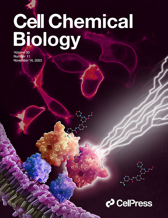
A 3D illustration showing RAF inhibitor LXH254 engages BRAF or CRAF protomers (orange), but spares ARAF (red). Unoccupied ARAF is competent to trigger downstream mitogenic signaling, which is demonstrated with lightning bolts. Red cells in the background are fluorescently labeled RAS proteins, expressed in live cells. The Cell Chemical Biology cover type superimposes the image.
Image adapted from original artwork by iSO-FORM LLC.

We made the cover! Of Cell Chemical Biology, that is.

This July, Cell Chemical Biology editors accepted a study from Promega scientists and invited the research team to submit cover art for the issue. The study in question details a BRET-based method to quantify drug-target occupancy within RAF-KRAS complexes in live cells. Promega scientists Matt Robers and Jim Vasta collaborated with one of our talented designers, Michael Stormberg, to craft an image that accurately represents the science in a dynamic and engaging way.

You can check out the paper and cover art in the November 16 issue of Cell Chemical Biology.

I spoke with Michael Stormberg to learn more about the creative process that went into creating this cover art and how he worked with the research team and other collaborators.

Continue reading “Designing Science: A Behind-the-Scenes Look at Our Recent Journal Cover Art”

RAF Inhibitors: Quantifying Drug-Target Occupancy at Active RAS-RAF Complexes in Live Cells

Mitogen-activated protein kinases (MAPKs) are a large family of proteins that regulate diverse cellular functions in eukaryotes, including gene expression, proliferation, differentiation and apoptosis (1). MAPK signaling pathways typically include three sequentially activated kinases, and these pathways are triggered in response to extracellular stimuli, such as cytokines, mitogens, growth factors and oxidative stress (1). Ultimately, the signal is transmitted to the nucleus, with the activation of a specific transcription factor that modulates the expression of one or more genes.

Among MAPK pathways, the RAS-RAF-MEK-ERK signaling pathway has been studied extensively. Mutations in RAS family proteins and resultant dysregulation of the signaling pathway are implicated in a variety of cancers. Therefore, this pathway is a popular target for anticancer drug development.

An overview of the RAS-RAF-MEK-ERK signaling pathway.
Continue reading “RAF Inhibitors: Quantifying Drug-Target Occupancy at Active RAS-RAF Complexes in Live Cells”

From Hit to Live-Cell Target Engagement Assay: DNA-Encoded Library Screens and NanoBRET Dye

Monitoring and quantifying drug-target binding in a live-cell setting is important to bridging the gap between in vitro assay results and the phenotypic outcome, and therefore represents a crucial step in target validation and drug development (1). The NanoBRET™ Target Engagement (TE) assay is a biophysical technique that enables quantitative assessment of small molecule-target protein binding in live cells. This live-cell target engagement assay uses the bioluminescence resonance energy transfer (BRET) from a NanoLuc® luciferase-tagged target protein and a cell-permeable fluorescent tracer that reversibly binds the target protein of interest. In the presence of unlabeled test compound that engages the target protein, the tracer is displaced, and a loss of BRET signal is observed. Due to the tight distance constraints for BRET, the signal measured is specific to the target fused to NanoLuc® luciferase.

Live-cell target engagement assay using NanoBRET to measure small molecule binding to a target transmembrane protein.

Promega offers over 400 ready-to-use assays for multiple target classes, including kinases, E3 ligases, RAS, and many others. For targets that do not have an existing NanoBRET™ TE assay, Promega offers NanoBRET™ dyes, NanoLuc® cloning vectors, and NanoBRET™ detection reagents to develop novel NanoBRET™ TE assays.

To learn more about the NanoBRET™ TE platform, see the NanoBRET™ Target Engagement Technology Page on our website.

One critical component in the development of novel NanoBRET™ TE assay is the creation of the cell-permeable fluorescent tracers (NanoBRET™ tracers) against the target protein of interest. The tracers are bifunctional, consisting of a NanoBRET™-compatible fluorophore and a target-binding moiety connected by a linker. While the NanoBRET™ 590 dyes have demonstrated consistently robust cell permeability and optimal spectral overlap with NanoLuc® for BRET, a ligand capable of binding to the target protein of interest needs to be identified to generate a NanoBRET™ tracer.

What Are DNA-Encoded Libraries?

DNA-Encoded Libraries, (DELs), have emerged as powerful tools for discovering small molecule ligands to target proteins of interest at an unprecedented scale. . owing to the ability of a DEL  to enable the synthesis of larger libraries of compounds and to target proteins without any prior structural knowledge of the proteins or their ligands (2). Because each member of a DEL contains a DNA barcode and a small molecule separated by a linker, DEL is primed for discovering leads within therapeutic modalities that rely on bifunctional chemistry, such as proteolysis targeting chimeras (PROTACs). Since NanoBRET™ tracers are also bifunctional, ligands identified from DEL selections could serve as ideal candidates for developing novel NanoBRET™ tracers that can enable NanoBRET™ TE assays for new targets.

Continue reading “From Hit to Live-Cell Target Engagement Assay: DNA-Encoded Library Screens and NanoBRET Dye”

Executing a NanoBRET™ Experiment: From Start to Data

This is a guest post from Katarzyna Dubiel, marketing intern in Cellular Analysis and Proteomics.

“The objective of my experiment was to test the NanoBRET™ assay as if I was a customer, independent of the research and development team which develops the assay.”

Designing and implementing a new assay can be a challenging process with many unexpected troubleshooting steps. We wanted to know what major snags a scientist new to the NanoBRET™ Assay would encounter. To determine this, we reached out to Laurence Delauriere, a senior applications scientist at Promega-France, who had never previously performed a NanoBRET™ assay. Laurence went step-by-step through the experimental process looking at the CRAF-BRAF interaction in multiple cell lines. In an interview, Laurence provided us with some tips and insights from her work implementing the new NanoBRET™ assay.

In a few words, can you explain NanoBRET?
“NanoBRET is used to monitor protein: protein interactions in live cells. It is a bioluminescence resonance energy transfer (BRET) based assay that uses NanoLuc® luciferase as the BRET energy donor and HaloTag® protein labeled with the HaloTag® NanoBRET™ 618 fluorescent ligand as the energy acceptor to measure the interaction of two binding partners.” Continue reading “Executing a NanoBRET™ Experiment: From Start to Data”

For Protein Complementation Assays, Design is Everything

Most, if not all, processes within a cell involve protein-protein interactions, and researchers are always looking for better tools to investigate and monitor these interactions. One such tool is the protein complementation assay (PCA). PCAs use  a reporter, like a luciferase or fluorescent protein, separated into two parts (A and B) that form an active reporter (AB) when brought together. Each part of the split reporter is attached to one of a pair of proteins (X and Y) forming X-A and Y-B. If X and Y interact, A and B are brought together to form the active enzyme (AB), creating a luminescent or fluorescent signal that can be measured. The readout from the PCA assay can help identify conditions or factors that drive the interaction together or apart.

A key consideration when splitting a reporter is to find a site that will allow the two parts to reform into an active enzyme, but not be so strongly attracted to each other that they self-associate and cause a signal, even in the absence of interaction between the primary proteins X and Y. This blog will briefly describe how NanoLuc® Luciferase was separated into large and small fragments (LgBiT and SmBiT) that were individually optimized to create the NanoBiT® Assay and show how the design assists in monitoring protein-protein interactions.

Continue reading “For Protein Complementation Assays, Design is Everything”