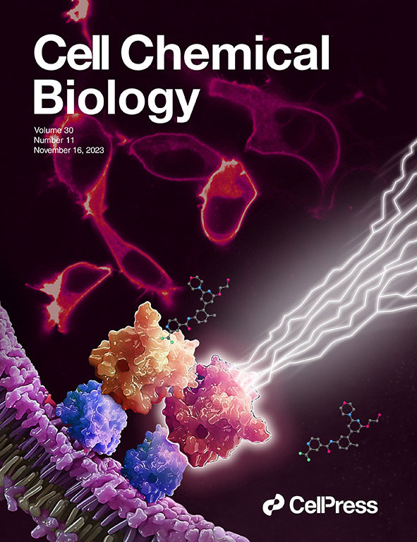
We made the cover! Of Cell Chemical Biology, that is.
This July, Cell Chemical Biology editors accepted a study from Promega scientists and invited the research team to submit cover art for the issue. The study in question details a BRET-based method to quantify drug-target occupancy within RAF-KRAS complexes in live cells. Promega scientists Matt Robers and Jim Vasta collaborated with one of our talented designers, Michael Stormberg, to craft an image that accurately represents the science in a dynamic and engaging way.
You can check out the paper and cover art in the November 16 issue of Cell Chemical Biology.
I spoke with Michael Stormberg to learn more about the creative process that went into creating this cover art and how he worked with the research team and other collaborators.
To learn more about the research study, check out our article summarizing the key take-aways: RAF Inhibitors: Quantifying Drug-Target Occupancy at Active RAS-RAF Complexes in Live Cells
Creating the Cover Art
After receiving the journal’s invitation for a cover art submission, Matt Robers connected with Michael Stormberg to begin the design process. They kicked things off with a brainstorming session that explored the visual style of past Cell Chemical Biology cover art, the details of the science they wanted to depict and what elements they could incorporate into the design.
The research presents a target occupancy assay for RAF heterodimers in complex with endogenous KRAS or KRAS mutants. Some of the key visual elements included:
- An image of fluorescently labelled KRAS proteins localized in the cell membrane of HEK293 cells—an image that was generated during the study and was provided by Ani Michaud, one of the study authors.
- A 3D rendering of membrane-bound protein complexes that had already been created for Promega by artists at iSO-FORM and could be repurposed for this project.
- A ball-and-stick chemical structure for LXH-254, a type II RAF inhibitor used in the study, which was provided by Cesear Corona, a Promega chemist.
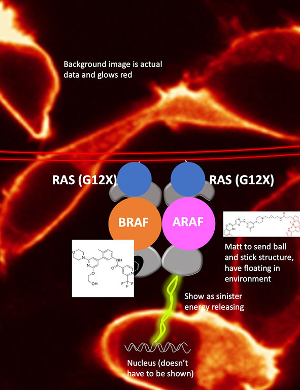
With their goals and these elements in hand, Michael created a first draft using Adobe Photoshop. He refined this draft based on feedback, toning down the energy beams and repositioning the RAF proteins.
Michael encountered a technical challenge in depicting the cell membrane. The 3D rendering shows proteins at the surface of a convex membrane. However, the KRAS-RAF complex is located on the internal, concave surface of a cell membrane—a point that was shown in the study using bioluminescent imaging.
A quick fix could “flip” the orientation of the membrane, but that would distort lighting. “It’s my goal to make sure it’s as accurate as we can make it,” Michael said. To achieve this accuracy, Michael used a Puppet Warp tool in Photoshop, which he had used in earlier projects. The tool allowed him to manipulate the curvature of the 3D rendering without distorting the lighting or other elements, yielding the final image.
In October, the team submitted the design to Cell Chemical Biology with the accompanying description:
“Target engagement of RAF dimer inhibitor LXH254 at RAF kinases, in complex with KRAS (blue). RAF inhibitor LXH254 engages BRAF or CRAF protomers (orange), but spares ARAF (red). Unoccupied ARAF is competent to trigger downstream mitogenic signaling (lightning bolts). Red cells in the background are fluorescently labeled RAS proteins, expressed in live cells.”
At the end of October, the team learned that the editors had selected their artwork for the cover of the November 16 issue.
“I’m so appreciative of the R&D team collaborative spirit and their ability to explain their work so well. They gave me everything I needed to put this piece together,” Michael said. “They’re just amazing to work with.”
What factors do you consider when communicating your research visually? What tools do you use?
Share in the comments below.
Latest posts by Jordan Nutting (see all)
- The Central Dogma of Promega: The Story and Science Behind Our Kit Packaging Design - May 7, 2024
- Silencing the Immunogenicity of AAV Vectors - April 4, 2024
- Discovering Cyclic Peptides with a “One-Pot” Synthesis and Screening Method - February 29, 2024
Title is grammatically incorrect! I think you mean “…Look At Our Recent…” (you are missing the “At”)