In the evolving field of forensic science, a study by Fantinato et al. has opened new avenues in using DNA extraction and analysis to recover important information from crime scenes. Their work, “The Invisible Witness: Air and Dust as DNA Evidence of Human Occupancy in Indoor Premises,” focuses on extracting DNA from air and dust. This novel approach could revolutionize how crime scenes are investigated, especially in scenarios where traditional evidence—like fingerprints or bodily fluids—is scarce, degraded or has been removed from surfaces.
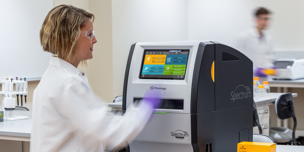
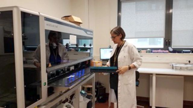
In genetic research, staying at the forefront of technology is crucial. The latest breakthrough in human identification comes in the form of 8-dye Short Tandem Repeat (STR) chemistry. This innovation promises unprecedented precision and accuracy in DNA analysis, revolutionizing the way we approach genetic studies. In this blog post, we’ll delve into the world of 8-color chemistry and explore how it seamlessly integrates with the game-changing Spectrum Compact CE System.
Understanding 8-Dye STR Chemistry
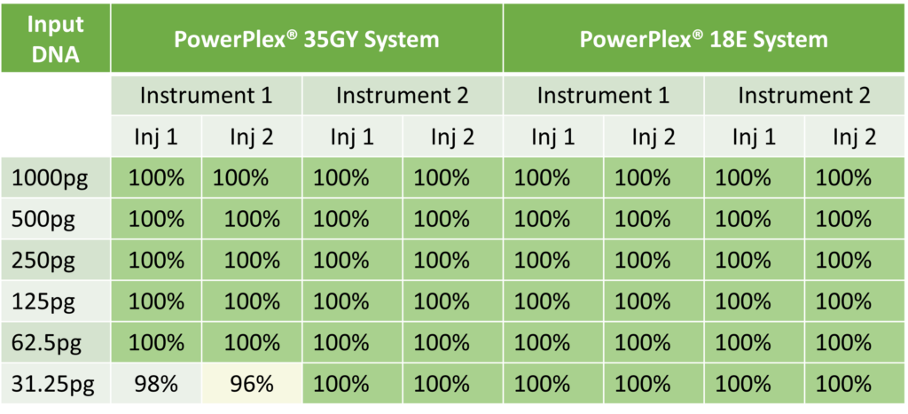
The introduction of 8-dye chemistry expands the capability of STR analysis, enabling researchers to analyze more DNA markers with smaller amplicons, providing more robust data from degraded or inhibited DNA samples. The performance of the 8-color dye chemistries from Promega on the Spectrum Compact CE System is sensitive, with both chemsitries (PowerPlex® 35 GY System and the upcoming PowerPlex® 18 E System) producing 100% profiles from their suggested inputs down to as little as 62.5 pg of DNA. The 18E system produced 100% profiles down to 31.25 pg of input DNA with minimal signal bleed through and low system noise.
Research into vaccines based on RNA began decades ago when scientists theorized that they could harness RNA to produce viral proteins within a cell, prompting a protective immune response. RNA vaccine research drew scientists’ attention during the development of SARS-CoV-2 vaccines during the COVID-19 pandemic, which opened the door for research targeting other diseases with RNA-based therapeutics.
The WSPCP works to provide seed potato growers with healthy planting stock
The mighty potato—the Midwest’s root vegetable of choice—is susceptible to a variety of diseases that, without proper safeguards, can spell doom for your favorite side dishes. Founded in 1913 and housed in the Department of Plant Pathology at the University of Wisconsin-Madison, the Wisconsin Seed Potato Certification Program (WSPCP) helps Wisconsin seed potato growers maintain healthy, profitable potato crops year-to-year through routine field inspections, a post-harvest grow-out and laboratory testing.
While WSPCP conducts visual inspections for various seed potato pathogens, their diagnostic laboratory testing is primarily focused on viruses such as Potato virus Y (PVY), which can cause yield reduction and tuber defects, along with select bacteria such as Dickeya and Pectobacterium species that cause symptoms like wilting, stem rot and tuber decay.
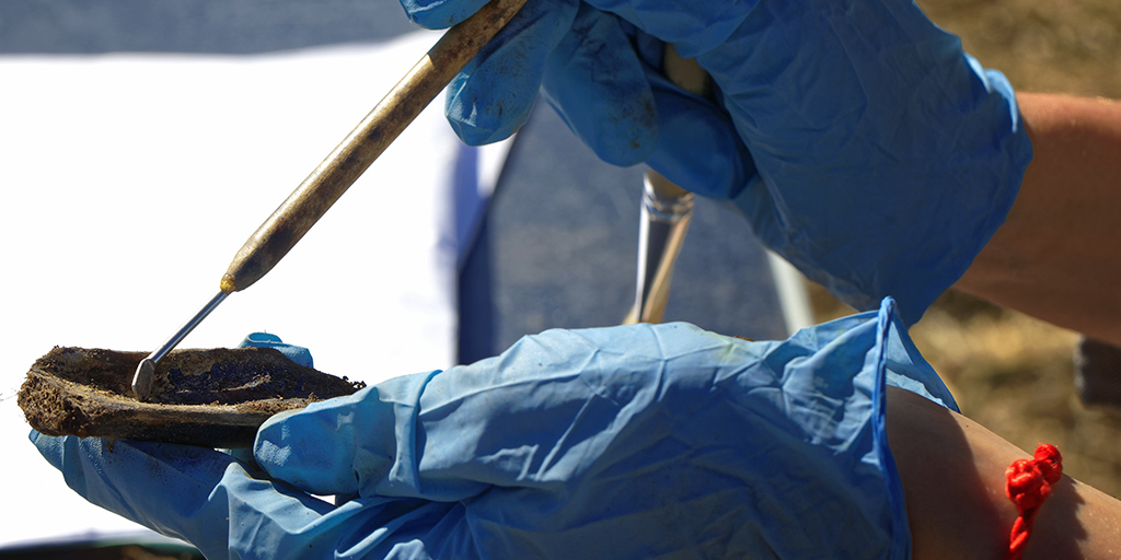
The elk tooth is small and ancient, with a crude hole bored through the top. It was likely worn as a pendant, but worn by whom? Was the owner male or female? Where did they come from? Did the pendant indicate their social status, mark a significant accomplishment, was it a gift, or was it worn as an expression of individuality?
Artifacts such as personal ornaments and tools play a pivotal role in helping us understand the migration, behavior and cultures of ancient peoples. To date, this information has stopped short of providing insight into things like the biological sex or genetic ancestry of the individuals who may have worn or used these items, and thus limited our ability to accurately characterize societal roles and behaviors. Recent advances in DNA techniques and technologies, and one little pendant, might be changing that.
The thought of an expensive instrument falling out of use and gathering dust on the shelf is enough to bring a tear to any lab manager’s eye. An instrument that once served a key purpose and now functions only as a “paperweight” is a tragic waste of valuable resources. Fortunately, it is sometimes possible to breathe new life into neglected tools and to retrofit or repurpose equipment to meet the new needs that will inevitably arise in a changing lab environment.
There’s a certain group of people (including this blog post author) who derive no small amount of amusement from seeing stock photos of DNA and pointing out flaws in the structure. It’s even more amusing when these photos are used in marketing by life science companies. The most common flaw: the DNA molecule is a left-handed double helix.
What does that even mean? DNA, like many organic chemicals in biology, is a chiral molecule. That is, it can exist in two structural forms that are mirror images of each other but are not superimposable (enantiomers). Just like your left and right hands are mirror images of each other, the two DNA structures are left-handed and right-handed double helices. The DNA double helix is chiral, because its building blocks (nucleotides) are chiral.
It can be challenging, at first glance, to tell whether an image of DNA is left-handed or right-handed. Various helpful hints are available; however, the one that I’ve found easiest to remember is described in a blog post by Professor Emeritus Larry Moran at the University of Toronto:
Imagine that the double helix is a spiral staircase, and you’re walking down the staircase. If you’re turning to the right as you descend, the DNA structure is right-handed; if turning to the left, it’s left-handed. In the image shown earlier, the DNA molecule on the right is a right-handed double helix, while its mirror image is left-handed.
Several different types of nucleic acids can be found circulating in human biofluids. Fragmented DNA and RNA are now routinely purified from plasma and other bodily fluids. These types of nucleic acids need to be purified from a cell-free fraction of the biofluids to ensure that the isolated nucleic acids are truly circulating and not from intact cells. In this blog post, we will learn a bit more about circulating nucleic acids (CNA) and how they can be used as biomarkers in research.
In oncology, tissue biopsies are commonly fixed in formalin and embedded in paraffin (FFPE). These FFPE samples can be used with immunohistochemical or molecular analysis for identifying biomarkers that guide the diagnosis and therapeutic management of patients. This fixation technique allows long-term storage of samples but impacts the integrity of nucleic acids. This makes extracting DNA and RNA from FFPE tissues in sufficient quantity and quality for molecular analysis techniques such as NGS analyses challenging for molecular oncology laboratories.
“At Rennes University Hospital, we receive many lung cancer samples with little material available, or samples of poor quality. The nucleic acid extraction step is therefore critical to get good yield. We have seen that it had a direct impact on the success of downstream analysis,” said Dr. Alexandra Lespagnol. Lespagnol is the Technical Manager of the Molecular Genetics of Cancer core lab at the University Hospital of Rennes in France.
In order to accommodate the increasing number of samples that needed to be analyzed, the Molecular Genetics of Cancer core lab of the University Hospital of Rennes initiated an automation project for extracting DNA from FFPE tissues. The lab also wanted to improve sample tracking and reproducibility of their results.
Tracking the spread of COVID-19 has been a tremendous challenge throughout the pandemic, but doing so is a key step toward containing the virus. Many communities have relied on patient testing and contact tracing, with limited success. In search of better methods, some countries have made inroads in a different form of disease surveillance: wastewater-based epidemiology (WBE). This approach involves testing wastewater for the presence of pathogens, primarily through DNA and RNA analysis, and has proved to be an accurate and highly effective way to keep tabs on the prevalence and progression of COVID-19 at the population level.
Switzerland is among those countries that have implemented WBE in their efforts to stay ahead of the pandemic. Since WBE first emerged in 2020 as a promising tool, several Swiss laboratories undertook wastewater testing, and protocols were established early.
“At the beginning, the methods to actually detect coronavirus in wastewater were rather laborious and complicated, and involved a lot of resources,” said Dr. Claudia Bagutti, microbiologist and molecular biologist in the State Laboratory of Basel-City, Switzerland.
Bagutti heads a small team performing applied biosafety research. In 2020, her lab was tasked with developing an assay for detecting COVID-19 in wastewater. However, the available methods were prohibitively complex and resource intensive.
In the meantime, researchers at Promega recognized that Promega products and methodologies could potentially be applied to WBE and set to work developing simpler and more efficient method for wastewater analysis. In the spring of 2021, Bagutti’s team decided to try adopting this method.
“Promega had a very nice method which was less laborious and much easier to handle, and that’s why we gave it a try,” said Bagutti.
In the ensuing study, Bagutti and her team analyzed effluent from the catchment area of one municipal wastewater plant in Switzerland. They examined the total wastewater output of around 270,000 people. Viral RNA was extracted using Promega’s Maxwell® RSC Environ Wastewater TNA Kit. The number of RNA copies present, representing the overall concentration of COVID-19 in each sample, was determined via quantitative reverse transcriptase (RT-qPCR) using the GoTaq® Enviro Wastewater SARS-CoV-2 Systems, also from Promega. The viral RNA was subsequently sequenced with next generation sequencing, and the results correlated quite well with the COVID-19 cases in the catchment area. Remarkably, this study detected the Omicron variant in a wastewater sample one day prior to the first reported case identified through patient testing.
“We observed a similar spread to most other western countries with respect to the time of the first discovery of these variants,” said Bagutti. “We were also able to demonstrate the presence [of Omicron] in the wastewater before it came up in a sample of a COVID-19 patient test, which of course shows the usefulness of wastewater monitoring for the prediction of new variants and infection dynamics.”
WBE is especially promising in that it provides population-level data independent of patient testing. Health departments can be alerted to the presence of COVID-19 earlier than would otherwise be possible with traditional testing and can take precautions to contain the spread. In creating a more user-friendly method for wastewater analysis, Promega has opened the door for more laboratories to conduct WBE, which could provide communities around the world with the information they need to preempt the progression of COVID-19.
“The Promega method is very straightforward to handle,” said Bagutti. “It only takes a small volume of wastewater, which makes it handy. It’s less time-consuming compared to the methods which were in the literature at the beginning of the pandemic, and it just works very well. We also did experience great support from Promega.”
At this point, much of the wastewater analysis performed in Switzerland is done with the Promega method, including in federal, state or private labs. The swift advance of WBE in Switzerland speaks to the colossal effort put forth both by Promega researchers in developing the necessary products and methodologies, as well by those labs that have made use of Promega’s products to monitor COVID-19 in wastewater.
“It’s really been a success story for us, from the beginning,” said Bagutti.
Learn more about Promega’s work with wastewater-based epidemiology.
XWe use cookies and similar technologies to make our website work, run analytics, improve our website, and show you personalized content and advertising. Some of these cookies are essential for our website to work. For others, we won’t set them unless you accept them. To learn more about our approach to Privacy we invite you to Read More
By clicking “Accept All”, you consent to the use of ALL the cookies. However you may visit Cookie Settings to provide a controlled consent.
We use cookies and similar technologies to make our website work, run analytics, improve our website, and show you personalized content and advertising. Some of these cookies are essential for our website to work. For others, we won’t set them unless you accept them. To find out more about cookies and how to manage cookies, read our Cookie Policy.
If you are located in the EEA, the United Kingdom, or Switzerland, you can change your settings at any time by clicking Manage Cookie Consent in the footer of our website.
Necessary cookies are absolutely essential for the website to function properly. These cookies ensure basic functionalities and security features of the website, anonymously.
Cookie
Duration
Description
cookielawinfo-checbox-analytics
11 months
This cookie is set by GDPR Cookie Consent plugin. The cookie is used to store the user consent for the cookies in the category "Analytics".
cookielawinfo-checbox-functional
11 months
The cookie is set by GDPR cookie consent to record the user consent for the cookies in the category "Functional".
cookielawinfo-checbox-others
11 months
This cookie is set by GDPR Cookie Consent plugin. The cookie is used to store the user consent for the cookies in the category "Other.
cookielawinfo-checkbox-advertisement
1 year
The cookie is set by GDPR cookie consent to record the user consent for the cookies in the category "Advertisement".
cookielawinfo-checkbox-necessary
11 months
This cookie is set by GDPR Cookie Consent plugin. The cookies is used to store the user consent for the cookies in the category "Necessary".
cookielawinfo-checkbox-performance
11 months
This cookie is set by GDPR Cookie Consent plugin. The cookie is used to store the user consent for the cookies in the category "Performance".
gdpr_status
6 months 2 days
This cookie is set by the provider Media.net. This cookie is used to check the status whether the user has accepted the cookie consent box. It also helps in not showing the cookie consent box upon re-entry to the website.
lang
This cookie is used to store the language preferences of a user to serve up content in that stored language the next time user visit the website.
viewed_cookie_policy
11 months
The cookie is set by the GDPR Cookie Consent plugin and is used to store whether or not user has consented to the use of cookies. It does not store any personal data.
Analytical cookies are used to understand how visitors interact with the website. These cookies help provide information on metrics the number of visitors, bounce rate, traffic source, etc.
Cookie
Duration
Description
SC_ANALYTICS_GLOBAL_COOKIE
10 years
This cookie is associated with Sitecore content and personalization. This cookie is used to identify the repeat visit from a single user. Sitecore will send a persistent session cookie to the web client.
vuid
2 years
This domain of this cookie is owned by Vimeo. This cookie is used by vimeo to collect tracking information. It sets a unique ID to embed videos to the website.
WMF-Last-Access
1 month 18 hours 24 minutes
This cookie is used to calculate unique devices accessing the website.
_ga
2 years
This cookie is installed by Google Analytics. The cookie is used to calculate visitor, session, campaign data and keep track of site usage for the site's analytics report. The cookies store information anonymously and assign a randomly generated number to identify unique visitors.
_gid
1 day
This cookie is installed by Google Analytics. The cookie is used to store information of how visitors use a website and helps in creating an analytics report of how the website is doing. The data collected including the number visitors, the source where they have come from, and the pages visted in an anonymous form.
Advertisement cookies are used to provide visitors with relevant ads and marketing campaigns. These cookies track visitors across websites and collect information to provide customized ads.
Cookie
Duration
Description
IDE
1 year 24 days
Used by Google DoubleClick and stores information about how the user uses the website and any other advertisement before visiting the website. This is used to present users with ads that are relevant to them according to the user profile.
test_cookie
15 minutes
This cookie is set by doubleclick.net. The purpose of the cookie is to determine if the user's browser supports cookies.
VISITOR_INFO1_LIVE
5 months 27 days
This cookie is set by Youtube. Used to track the information of the embedded YouTube videos on a website.
Performance cookies are used to understand and analyze the key performance indexes of the website which helps in delivering a better user experience for the visitors.
Cookie
Duration
Description
YSC
session
This cookies is set by Youtube and is used to track the views of embedded videos.
_gat_UA-62336821-1
1 minute
This is a pattern type cookie set by Google Analytics, where the pattern element on the name contains the unique identity number of the account or website it relates to. It appears to be a variation of the _gat cookie which is used to limit the amount of data recorded by Google on high traffic volume websites.