A Better GTPase Assay for Drug Development

Cover of October Issue of Assay and Drug Development Technologies featuring GTPase-Glo™ Assay.

The path to drug development is strewn with obstacles: Identifying targets; configuring assays to help identify targets or drugs; uncovering the right compound to affect the selected target without off-target effects and screening multiple compounds to eliminate or identify potential drugs. Without the right tools, compounds or target, identifying potential disease therapies becomes nearly impossible.

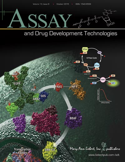
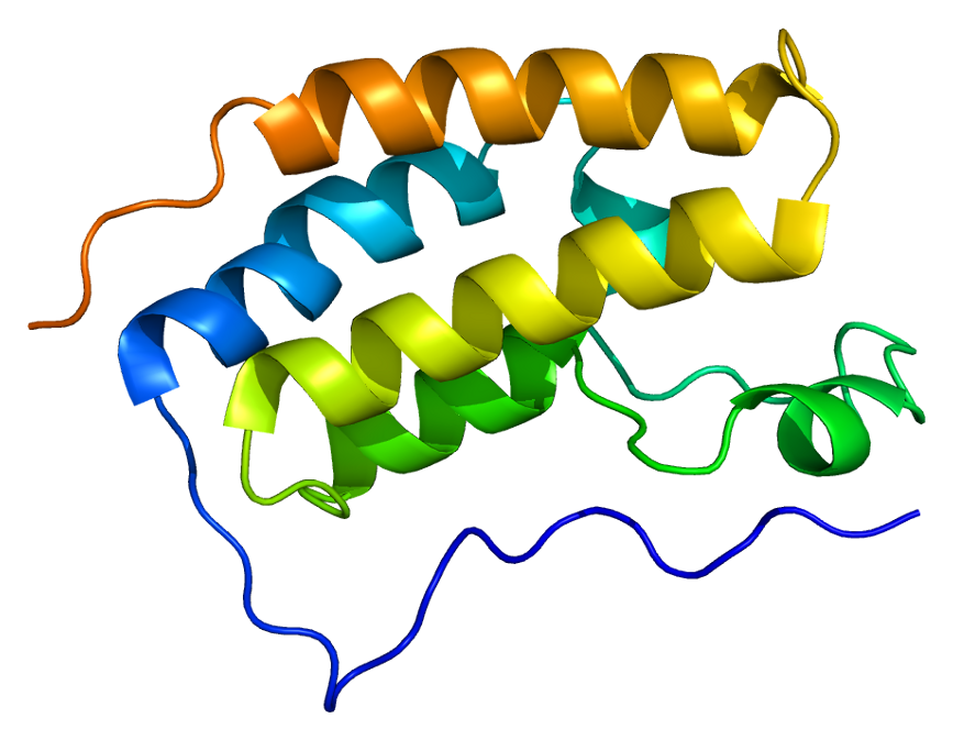
When it comes to a drug target for cancer, the Ras protein family is at the top of the list because the proteins are expressed ubiquitiously and found mutated in many types of cancer. Because Ras proteins are involved in transducing signals from the surface of cells, many of the resulting mutations produce an activated Ras, inducing uncontrolled expression of the genes that Ras controls. Ras proteins are small GTPases (20–25kDa) that comprise a larger superfamily of proteins divided into five subfamilies: Ras, Rho, Rab, Arf, and Ran. These proteins control diverse cellular activities, including cellular differentiation, proliferation, cell division, nuclear import and export, and vesicle transport. GTPases are guanosine-nucleotide-binding proteins with affinity for GDP or GTP and are able to hydrolyze GTP. When bound to GTP, GTPases are active (turned on) and interact with downstream proteins in the signaling cascade. When GTPases are bound to GDP, the proteins are inactivated (turned off) and no longer transduce signals.

Continue reading “A Better GTPase Assay for Drug Development”

How Fruit Flies (and maybe Pigeons?) Navigate; A New Report

A rock dove, similar in plumage to a pigeon.

Several years ago an intriguing story of successful navigation in complex situation, by pigeons, the birds most often compared to rats, caught my eye.

Our backyard once had a coop full of pigeons, so I’m not a total stranger to their navigation abilities (nor am I a pigeon expert). My favorites were the tumbling pigeons.

But it didn’t take much time researching that article from 2012, to learn that one of the more hotly debated how-do-they-do-it topics is animal navigation, in particular, the ability of pigeons to navigate back to home/point A when released at point B.

So when it appeared online today, in Nature Materials, the story “A Magnetic Protein Biocompass” caught my eye.

Continue reading “How Fruit Flies (and maybe Pigeons?) Navigate; A New Report”

Interrogating Protein Interactions: An Infographic for NanoBRET™ Assay Design

Yesterday my fellow blogger, Kari, posted a review of the ACS Chemical Biology paper describing a new BRET platform for analyzing protein-protein interactions. If you are interested in studying induction and inhibition of protein interactions in real time, take a look at the infographic below to learn how to develop a NanoBRET™ Assay to monitor your protein of interest.

21421668-IG-CR-NanoBRET-Infographic-Final

About the Development of an Improved BRET Assay: NanoBRET

"Protein BRD4 PDB 2oss" by Emw - Own work. Licensed under CC BY-SA 3.0 via Wikimedia Commons - https://commons.wikimedia.org/wiki/File:Protein_BRD4_PDB_2oss.png#/media/File:Protein_BRD4_PDB_2oss.png
“Protein BRD4 PDB 2oss” by Emw – Own work. Licensed under CC BY-SA 3.0 via Wikimedia Commons – https://commons.wikimedia.org/wiki/File:Protein_BRD4_PDB_2oss.png#/media/File:Protein_BRD4_PDB_2oss.png

One of the more exciting reporter molecules technologies available came online in the past year, with the launch of the Promega NanoBRET™ technology. While it’s easy for me, a science writer at Promega, to brag, seriously, this is a very cool protein interactions tool.

A few of the challenges facing protein-protein interactions researchers include:

  • The ability to quantitatively characterize protein-protein interactions
  • Ability to examine protein-protein interactions in situ, in the context of the living cell

A goal of the NanoBRET™ developers was to improve the sensitivity and dynamic range of traditional BRET technology, in order to address these challenges.

In May 2015 these researchers published an article outlining their efforts to create NanoBRET technology in ACS Chemical Biology, in an article entitled, “NanoBRET—A Novel BRET Platform for the Analysis of Protein-Protein Interactions”. Here is a brief look at their work.

Continue reading “About the Development of an Improved BRET Assay: NanoBRET”

Characterizing Immune-Modulating Antibodies Using Bioluminescence

Immune checkpoint pathways such as PD-1/PD-L1 and CTLA-4 are promising new immunotherapy targets for the treatment of cancer and autoimmunity. Immune checkpoint reporter-based bioassays provide a simple, consistent, and reliable cell-based assay to measure Ab function throughout the drug development pipeline.

The brief chalk talk below describes the assay principals of the reporter-based bioassay that monitors the functional blockade of PD-1/PD-L1 interactions.

Purify and Conjugate Antibodies in a Single Workflow

Isoform_Antibodies_LinkedIn
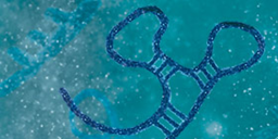
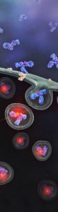
Antibodies labeled with small molecules such as fluorophore, biotin or drugs play a critical role in various areas of biological research,drug discovery and diagnostics. There are several limitations to current methods for labeling antibodies including the need for purified antibodies at high concentrations and multiple buffer exchange steps.

In a recent publication, a method (on-bead conjugation) is described that addresses these limitations by combining antibody purification and conjugation in a single workflow. This method uses high capacity-magnetic Protein A or Protein G beads to capture antibodies directly from cell media followed by conjugation with small molecules and elution of conjugated antibodies from the beads.

Using a variety of fluorophores the researchers show that the on-bead conjugation method is compatible with both thiol- and amine-based chemistry.

This method enables simple and rapid processing of multiple samples in parallel with high-efficiency antibody recovery. It is further shown that recovered antibodies are functional and compatible with downstream applications.

Literature Cited

Nidhi, N. et al. (2015) On-bead antibody-small molecule conjugation using high-capacity magnetic bead J. Immunol. Methods  http://dx.doi.org/10.1016/j.jim.2015.08.008

Rewriting the Histone Code: Searching for Treatments for Stage IV Thyroid Cancers

Chromatin fiber
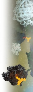
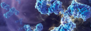
Often a diagnosis of thyroid cancer is associated with a good prognosis and fairly straightforward surgical treatments to remove the tumor followed by radioactive iodine ablation. Such treatment works well in tumors that have not metastasized and retain enough of their thyroid cell “identity” that they can still accumulate radioactive iodine.

However, aggressive thyroid cancers, which often metastasize and recur, do not respond to standard treatments because they are generally too dedifferentiated to accumulate iodine, so alternative treatments are needed.

One approach is to look for compounds that will reverse dedifferentiation, making tumor cells more likely to take up and concentrate radioactive iodine regardless of their location in the body. One possible target to effect dedifferentiation is epigenetic modification of histone proteins.

Histone proteins are more than the structural components of the nucleosome that organizes the chromatin inside cells. Histone proteins are subject to a host of protein modifications on their N-terminal tails such as acetylation, phosphorylation, methylation, ubiquitination and ADP-ribosylation. These various modifications are seen as creating a “histone code” that is read by other proteins and protein complexes (1). This code regulates patterns of gene expression and activity for a cell—in part resulting in a differentiated phenotype. Previous studies have suggested that some histone deacetylase (HDAC) inhibitors (e.g., valproic acid) can reverse some of the dedifferentiation associated with aggressive cancers (2).

Jang, et al. in a recent paper (3) published in Cancer Gene Therapy synthesized a group of HDAC inhibitor analogs (AB1–AB13) and tested them for their ability to inhibit growth of three aggressive human thyroid cancer cell lines and induce partial re-differentiation to the thyroid cell phenotype. Continue reading “Rewriting the Histone Code: Searching for Treatments for Stage IV Thyroid Cancers”

A Normalization Method for Luciferase Reporter Assays of miRNA-Mediated Regulation

Today’s blog is from guest blogger Ken Doyle of Loquent, LLC. Here, Ken reviews a 2014 paper highlighting specific considerations for using reporter assays to study miRNA-mediated gene regulation.

mirna

The accelerated pace of research into noncoding RNAs has revealed multiple regulatory roles for microRNAs (miRNAs). These diminutive noncoding RNA species—typically 20-24 nucleotides in length—are now known to mediate a broad range of biological functions in plants and animals. In humans, miRNAs have been implicated in various aspects of development, differentiation, and metabolism. They are known to regulate an assortment of genes involved in processes from neuronal development to stem cell division. Dysregulation of miRNA expression is associated with many disease states, including neurodegenerative disorders, cardiovascular disease, and cancer.

Typically, miRNAs act as post-transcriptional repressors of gene expression, either by targeted degradation of messenger RNA (mRNA) or by interfering with mRNA translation. Most miRNAs exert these effects by binding to specific sequences called microRNA response elements (MREs). These sequences are found most often within the 3´-untranslated regions (3´-UTRs) of animal genes, while they may occur within coding sequences in plant genes.

Studies of the regulatory roles played by miRNAs often involve cell-based assays that use a reporter gene system, such as luciferase or green fluorescent protein. In a standard assay, the reporter gene is cloned upstream of the 3´-UTR sequence being studied; this construct is then cotransfected with the miRNA into cells in culture. A study by Campos-Melo et al., published in September 2014, examined this experimental approach for miRNAs from spinal cord tissues, using firefly luciferase as the reporter gene and Renilla luciferase as a transfection control.

Continue reading “A Normalization Method for Luciferase Reporter Assays of miRNA-Mediated Regulation”

If We Could But Peek Inside the Cell …Quantifying, Characterizing and Visualizing Protein:Protein  Interactions

14231183 WB MS Protein Interactions Hero Image 600x214

Robert Hooke first coined the term “cell” after observing  plant cell walls through a light microscope—little empty chambers, fixed in time and space. However,  cells are anything but fixed.

Cells are dynamic: continually responding to a shifting context of time, environment, and signals from within and without. Interactions between the macromolecules within cells, including proteins, are ever changing—with complexes forming, breaking up, and reforming in new ways. These interactions provide a temporal and special framework for the work of the cell, controlling gene expression, protein production, growth, cell division and cell death.

Visualizing and measuring protein:protein interactions at the level of the cell without perturbing them is the goal of every cell biologist.

A recent article by Thomas Machleidt et al. published in ACS Chemical Biology, describes a new technology that brings us closer to being able to realize that goal.

Continue reading “If We Could But Peek Inside the Cell …Quantifying, Characterizing and Visualizing Protein:Protein  Interactions”

pH Reactive Dyes for Screening Antibody Internalization

19244302_phabaminebanner_900x246

Antibody drug conjugates (ADCs) are a new class of therapeutic drugs that uses antibodies to deliver highly toxic drug molecules specifically to the cancer cells. A key requirement for ADCs is the ability of antibody to bind to the cancer cells followed by internalization and subsequent release of drug inside the cells leading to cell apoptosis.

Traditionally, selection of lead antibody candidates for ADCs was done in a sequential workflow where antibodies were first selected based on their affinity followed by characterization involving antibody internalization and drug conjugation. However, there is evidence that high affinity doesn’t always correlate with good internalization and hence there is a need to screen antibodies for internalization properties in addition to their affinities.

Promega has developed a method that allows antibody to be screened for their internalization properties in a simple, plate-based format. The method uses pH sensor dyes (pHAb dyes), which are not fluorescent at neutral pH but become highly fluorescent at acidic pH. When antibody conjugated with pHAb dye binds to its antigen on the cancer cell membrane they are not fluorescent but upon internalization and trafficking into endosomal and lysosomal vesicles the pH drops and dye becomes fluorescent.

Fluorescence signal, for pHAb dyes conjugated using either amine or thiol chemistry, is minimal at pH>7 and increase significantly as the pH drops to pH 5.0, which is a typical pH in cell endosomal compartment. Moreover, pH response of free pHAb dye is similar to that of conjugated dye indicating that conjugation chemistry doesn’t influence the pH response of the dye.

Due to the high signal-to-background ratios of the dyes, plate-based internalization assays can be performed, enabling screening of large libraries of antibodies for their internalization properties, hopefully leading to improved identification of lead candidates for ADC applications.