Use of ProAlanase Digestion Increases Number of Identified Methylglyoxal (MGO)-Modified Proteins in Whole-Cell Lysates

space filling structural model methylglyoxal (MGO)
Methylglyoxal is responsible for post translational protein modifications, that result in advanced glycation endproducts (AGEs), which are associated with aging and disease.

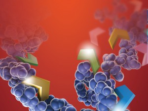
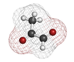
Post-translational modifications (PTM) of proteins are essential for the function of many proteins, but aberrant modification of protein residues also can interfere with protein function. PTMs occur in two ways. Proteins may be modified through the activity of enzymes such as kinases, phosphorylases, glycosylases and others that add or remove specific chemical moieties to amino acid residues. PTMs can also result from non-enzymatic reaction between electrophilic compounds and nucleophilic arginine and lysine residues within a protein. Metabolites and metabolic by products produced during glycolysis, especially glyoxal and methylglyoxal (MGO), are examples of such electrophilic compounds. These compounds can react with arginine and lysine to produce advanced glycation end products (AGEs), which are biomarkers associated with aging and degenerative diseases such as Alzheimer disease, diabetes and others. MGO is also elevated in tumors that have switched from oxidative phosphorylation to glycolysis as their main energy production pathway.

Only limited information is available about site-specific MGO PTMs in mammal cells, and most studies have focused on measuring the amount of MGO modifications in a treatment scenario compared to a control. Donnellan and colleagues recently published work to identify specific MGO protein modifications.  They used a “bottom-up” proteomic analysis of WIL2-NS B lymphoblastoid whole-cell lysates to identify specific MGO-modified proteins. In particular, the group was looking for modifications in proteins that might explain how MGO activity contributes to aneuploidy.

For the study, 100µg of cellular protein extract was reduced with dithiothreitol and then alkylated with chloroacetamide. The sample was diluted to reduce urea concentration. Trypsin Gold was added and samples were digested for 8 hours at 37°C. Digestion was terminated by adding formic acid. For ProAlanase digestion, 20µg of protein was reduced, alkylated and diluted to reduce urea concentration before adding digesting with ProAlanase for 4 hours at 37°C.

The authors identified 519 MGO-modified proteins.  Most of the modifications were identified in the trypsin digestion reactions; however, ProAlanase digestion increased the number of MGO modifications identified by approximately 25% (with less than 4% of the modification sites being detected in both the ProAlanase and trypsin digestion reactions. The authors suggest that ProAlanase increased sequence coverage to reveal sites not detected in the trypsin digestions. Therefore, they conclude that ProAlanase can be used along with trypsin digestion to increase the identification of MGO modifications.

ProAlanase can be used along with trypsin digestion to increase the identification of MGO modifications.

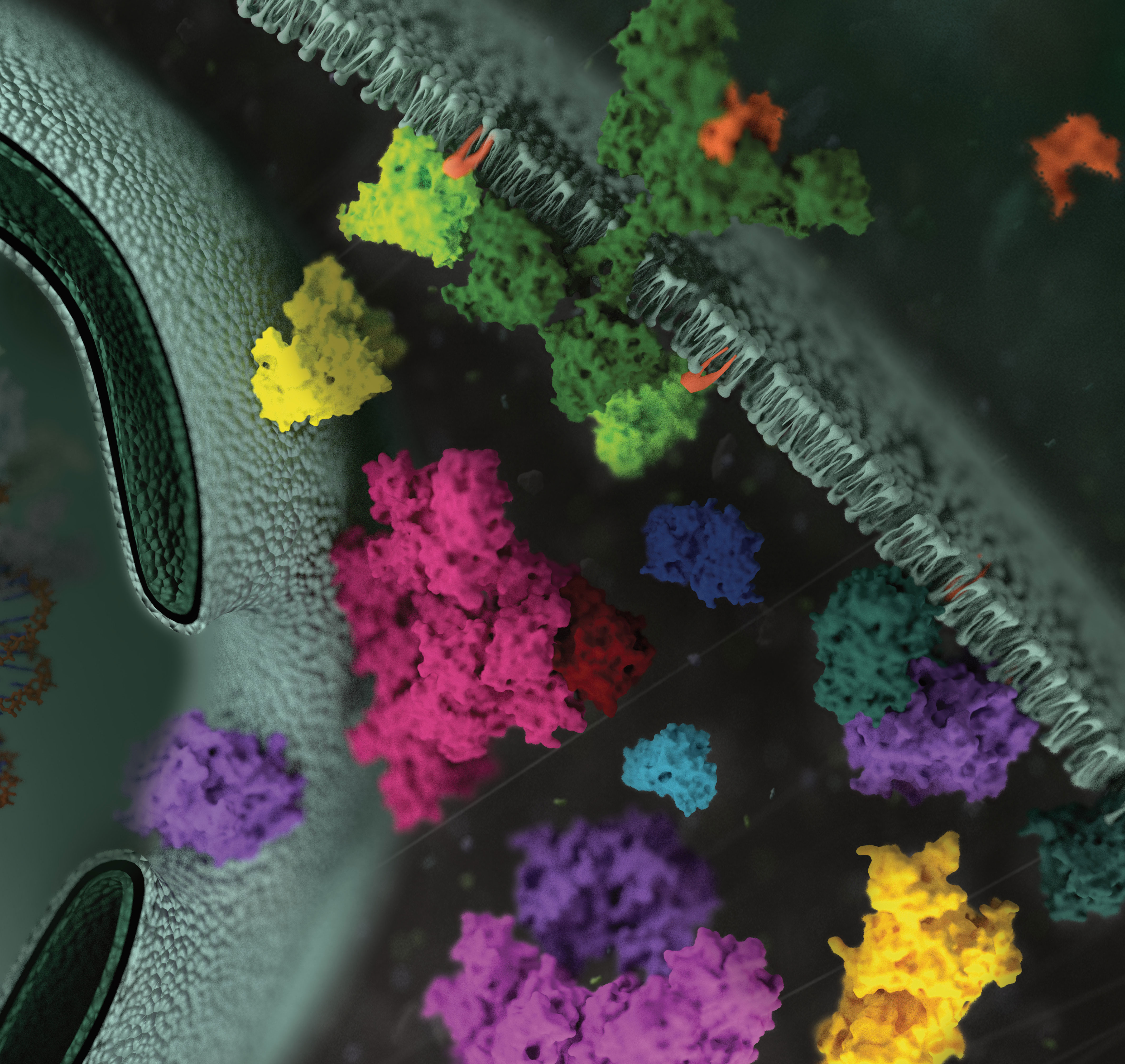
MGO-modified proteins from the WIL2-NS whole cell lysates included proteins involved in glycolysis, translation initiation, protein folding, mRNA splicing, cell-to-cell adhesion, heat response, nucleosome assembly, protein SUMOylation and the G2/M cell cycle transition. More work to further characterize the sites of these modifications and their potential effects on the function of the modified proteins is ongoing.


Read more about ProAlanase, a new site-specific endoprotease from Promega.


Literature Cited

Donnelian, L et al. (2022) Proteomic Analysis of Methylglyoxal Modifications Reveals Susceptibility of Glycolytic Enzymes to Dicarbonyl Stress Int. J. Mol. Sci. 23(7), 3689 doi.org/10.3390/ijms23073689

Detecting Disulfide Bond Shuffling in Biologics Using Trypsin Platinum

Biologic therapeutics such as monoclonal antibodies and biosimilars are complex proteins that are susceptible to post-translational modifications (PTMs). These chemical modifications can affect the performance and activity of the biologic, potentially resulting in decreased potency and increased immunogenicity. Such modifications include glycosylation, deamidation, oxidation and disulfide bond shuffling. These PTMs can be signs of protein degradation, manufacturing issues or improper storage. Several of these modifications are well characterized, and methods exist for detecting them during biologic manufacture. However, disulfide shuffling is not particularly well characterized for biologics, and no methods exist to easily detect and quantify disulfide bond shuffling in biologics.

Disulfide bond shuffling occurs when the S-S linkage is not between a Cys and its normal partner
Disulfide bonds are important for protein conformation and function

Normally the cysteines in a protein will pair with a predictable or “normal” partner residue either within a polypeptide chain or between two polypeptide chains when they form disulfide bonds. These normal disulfide bonds are important for final protein conformation and stability. Indeed, disulfide bonds are considered an important quality indicator for biologics.

In a recently published study, Coghlan and colleagues designed a semi-automated method for characterizing disulfide bond shuffling on two IgG1 biologics: rituximab (originator drug Rituxan® and biosimilar Acellbia®) and bevacizumab (originator Avastin® and biosimilar Avegra®).

Continue reading “Detecting Disulfide Bond Shuffling in Biologics Using Trypsin Platinum”

How Can You Improve Protein Digests for Mass Spectrometry Analysis?

Can predigestion with trypisin (ribbon structure shown) improve protein digests for mass spectrometry analysis?
Can pre-digestion with trypsin improve mass spec analysis?

The trypsin protease cleaves proteins on the carboxyterminus of Arginine (Arg) and Lysine (Lys). This cleavage reaction leaves a positive charge on the C-terminus of the resulting peptide, which enhances mass spectrometry analysis (1,2). Because of this advantage, trypsin has become the most commonly used protease for mass spectrometry analysis. Other proteases which cleave differently from trypsin, yielding complementary data are also used in mass spec analysis: these include Asp-N and Glu-C , which cleave acidic residues, and chymotrypsin which cleaves at aromatic residues. The broad spectrum protease, proteinase K is also used for some proteomic analyses. In a recent study, Dau and colleagues investigated whether sequential digestion with trypsin followed by the complementary proteases could improve protein digests for mass spectrometry analysis.

Continue reading “How Can You Improve Protein Digests for Mass Spectrometry Analysis?”

Mass Spec for Glycosylation Analysis of SARS-CoV-2 Proteins Implicated in Host-Cell Entry

The spike protein of the SARS-CoV-2 virus is a very commonly researched target in COVID-19 vaccine and therapeutic studies because it is an integral part of host cell entry through interactions between the S1 subunit of the spike protein with the ACE2 protein on the target cell surface. Viral proteins important in host cell entry are typically highly glycosylated. Looking at the sequence of the SARS-CoV-2 virus, researchers predict that the spike protein is highly glycosylated. In a recent study, researchers conducted a glycosylation analysis of SARS-CoV-2 proteins using mass spec analysis to determine the N-glycosylation profile of the subunits that make up the spike protein.

3d model of coronavirus covid-19 showing the spike protein. A recent study performed a glycosylation analysis of SARS-CoV-2 protein.

Glycans assist in protein folding and help the virus avoid immune recognition by the host. Glycosylation can also have an impact on the antigenicity of the virus, as well as potential effects on vaccine safety and efficacy. Mass spectrometry is widely used for viral characterization studies of influenza viruses. Specifically, mass spec has been used to study influenza protein glycosylation, antigen quantification, and determination of vaccine potency.

Continue reading “Mass Spec for Glycosylation Analysis of SARS-CoV-2 Proteins Implicated in Host-Cell Entry”

More muscle from eggs? Proteo-lipid complex may help prevent age-associated loss of muscle-mass

In older people, low muscle mass is strongly associated with reduced functional capacity and an increased risk of disability. Myostatin is a negative regulator of muscle growth and has become an important target for pharmaceutical companies designing therapeutics to address age-associated muscle loss.

Anti-myostatin drugs increase muscle size and strength in preclinical studies. Fortetropin is a proteo-lipid complex made from fertilized egg yolk and shows anti-myostatin activity. When Fortetropin is provided as a supplement, lowered circulating myostatin levels are observed both in rodents and in young men. Fortetropin in combination with resistance exercise also lowers myostatin and increased lean body mass.

Continue reading “More muscle from eggs? Proteo-lipid complex may help prevent age-associated loss of muscle-mass”

Proteomics from a Different Point of View: Introducing ProAlanase, the Newest Mass-Spec Grade Protease from Promega

Sometimes, when using trypsin to study a protein sequence or protein modifications, sequence coverage just isn’t quite as complete as you’d like. Looking for a protease with novel cleavage specificity or a protease that functions well in a low pH environment? Promega has a protease for that.

ProAlanase is a new site-specific endoprotease that preferentially cleaves proteins on the C-terminal side of proline and alanine amino acids. The unique cleavage specificity of ProAlanase (also known as An-PEP or EndoPro; 1–3) can help to uncover parts of the proteome not previously accessible with proteases typically used in proteomic studies.

Continue reading “Proteomics from a Different Point of View: Introducing ProAlanase, the Newest Mass-Spec Grade Protease from Promega”

Why Wait? Sample Prep/Protein Digestion in as Little as 30 Minutes!

While many proteases are used in bottom-up mass spectrometric (MS) analysis, trypsin (4,5) is the de facto protease of choice for most applications. There are several reasons for this: Trypsin is highly efficient, active, and specific. Tryptic peptides produced after proteolysis are ideally suited, in terms of both size (350–1,600 Daltons) and charge (+2 to +4), for MS analysis. One significant drawback to trypsin digestion is the long sample preparation times, which typically range from 4 hours to overnight for most protocols. Achieving efficient digestion usually requires that protein substrates first be unfolded either with surfactants or denaturants such as urea or guanidine. These chemical additives can have negative effects, including protein modification, inhibition of trypsin or incompatibility with downstream LC-MS/MS. Accordingly, additional steps are typically required to remove these compounds prior to analysis.

Continue reading “Why Wait? Sample Prep/Protein Digestion in as Little as 30 Minutes!”

Widening the Proteolysis Bottleneck: A New Protein Sample Preparation Tool

The poster featured in this blog provides background information and data on development of Rapid Digestion-Trypsin.
The poster featured in this blog provides background information and data on the development of Rapid Digestion-Trypsin.

Improvements in Protein Bioprocessing

As more and more protein-based therapeutics enter research pipelines, more efficient protocols are needed. In particular, we need better protocols for the characterization of protein structure and function, as well as means of quantitation. One main step in this pipeline, proteolysis of these proteins into peptides, presents a bottleneck and can require optimization of multiple steps including reduction, alkylation and digestion time.

We have developed a new trypsin reagent, Rapid Digestion–Trypsin, that streamlines the protein sample preparation process. This new development significantly reduces the time to achieve proteolysis to about 1 hour, a remarkable improvement over existing overnight sample preparation times.

How Does it Work?

With this new trypsin product, proteolysis is performed at 70°C, incorporating both denaturation and rapid digestion. The protocol can be used with multiple protein types, including pure proteins and complex mixtures, and is compatible with digestion under native, reduced or nonreduced conditions.

Continue reading “Widening the Proteolysis Bottleneck: A New Protein Sample Preparation Tool”

Optimization of Alternative Proteases for Bottom-Up Proteomics

Alternate Proteases Cover

Bottom-up proteomics focuses on the analysis of protein mixtures after enzymatic digestion of the proteins into peptides. The resulting complex mixture of peptides is analyzed by reverse-phase liquid chromatography (RP-LC) coupled to tandem mass spectrometry (MS/MS). Identification of peptides and subsequently proteins is completed by matching peptide fragment ion spectra to theoretical spectra generated from protein databases.

Trypsin has become the gold standard for protein digestion to peptides for shotgun proteomics. Trypsin is a serine protease. It cleaves proteins into peptides with an average size of 700-1500 daltons, which is in the ideal range for MS (1). It is highly specific, cutting at the carboxyl side of arginine and lysine residues. The C-terminal arginine and lysine peptides are charged, making them detectable by MS. Trypsin is highly active and tolerant of many additives.

Even with these technical features, the use of trypsin in bottom-up proteomics may impose certain limits in the ability to grasp the full proteome, Tightly-folded proteins can resist trypsin digestion. Post-translational modifications (PTMs) present a different challenge for trypsin because glycans often limit trypsin access to cleavage sites, and acetylation makes lysine and arginine residues resistant to trypsin digestion.

To overcome these problems, the proteomics community has begun to explore alternative proteases to complement trypsin. However, protocols, as well as expected results generated when using these alternative proteases have not been systematically documented.

In a recent reference (2), optimized protocols for six alternative proteases that have already shown promise in their applicability in proteomics, namely chymotrypsin, Lys-C, Lys-N, Asp-N, Glu-C and Arg-C have been created.

Data describe the appropriate MS data analysis methods and the anticipated results in the case of the analysis of a single protein (BSA) and a more complex cellular lysate (Escherichia coli). The digestion protocol presented here is convenient and robust and can be completed in approximately in 2 days.


Try a sample of high-efficiency Trypsin Platinum today!

Visit our website for more on Trypsin Platinum, Mass Spectrometry Grade, with enhanced proteolytic efficiency and superior autoproteolytic resistance.


Reference

  1. Laskay, U. et al. (2013) Proteome Digestion Specificity Analysis for the Rational Design of Extended Bottom-up and middle-down proteomics experiments. J of Proteome Res. 12, 5558–69.
  2. Giansanti, P. et. al. (2016) Six alternative protease for mass spectrometry based proteomics beyond trypsin. Nat. Protocols 11, 993–6

Related Posts

Optimizing Tryptic Digestions for Phosphoproteomics Analysis

11296971-DC-CR-KinaseProtein phosphorylation is the most widespread type of post-translational modification. It affects every basic cellular process, including metabolism, growth, division, differentiation, motility, organelle trafficking, membrane transport, muscle contraction, immunity, learning and memory (1,2). Protein kinases catalyse the transfer of the phosphate from ATP to specific amino acids in proteins. In eukaryotes, these are usually Ser, Thr and Tyr residues. Due to the development of specific phosphopeptide enrichment techniques and highly sensitive MS instruments, phosphoproteomics has enabled researchers to gain a comprehensive view on the dynamics of protein phosphorylation and phosphorylation based signaling networks.

Due to its high cleavage specificity, trypsin is the commonly used proteolytic enzyme in MS-based proteomics, cleaving peptides carboxyterminal of the amino acids lysine and arginine. However, various factors such as the tertiary structure of a protein, adjacent basic amino acids or negatively charged residues close to cleavage sites as well as PTMs are known to impair proteolysis.

To gain closer insights into the impact of phosphorylation on tryptic digestion, a recent publication(3) systematically characterized the digestion efficiency of model peptide sequences that are known to be prone to incomplete digestion.

The results indicated that increasing trypsin concentrations up to a trypsin to peptide ratio of 1:10 led to a significant gain (1) in the overall number of phosphorylation sites (up to 9%) and in the intensities of individual phosphopeptides, thereby improving the sensitivity of phosphopeptide quantification.

The effect of organic solvents (ACN, acetonitrile and TFE trifuorethanol was also evaluated). Positive results were noted with TFE when determining the digestion of individual peptides. However TFE interfered with TiO2 phosphopeptide enrichment and therefore was not recommended for use with complex samples.

  1. Engholm-Keller, K and Larsen, M.R. (2013) Technologies and challenges in large scale phosphoroproteomics. Proteomics 13, 910–31.
  2. Beausoleil, S. A. et al. (2010) Tissue-specific atlas of mouse protein phosphorylation and expression. Cell 143, 1174–89.
  3. Dickhut, C. et al. (2014) Impact of Digestion Conditions on phosphoproteomics. J. Proteome Res. 13, 2761–70.