Illuminating the Brain with a New Bioluminescence Imaging Substrate

Bioluminescence imaging is a powerful tool for non-invasive studies of the effect of treatments on cells and tissues. The luminescent signal is strong, and can be used in vivo, enabling repeated observations over time, allowing longitudinal study of cellular changes for hours or days. Bioluminescence imaging can be used in live animals over varying periods of time, without interfering with normal cellular processes.

Fluorescence imaging is also used in cellular studies. Although it can provide a stronger signal than luminescence, fluorescence requires light for excitation, and thus its in vivo use is limited at a tissue or cell depth greater than 1mm.

NanoLuc® Luciferase. Small, bright and now useful in brain bioluminescence imaging.

In addition, autofluorescence can be an issue with fluorescence imaging, as cellular components and surrounding proteins and cells can fluoresce when exposed to light. Autofluorescence can result in high background signals, making it difficult to distinguish true fluorescence from background.

Continue reading “Illuminating the Brain with a New Bioluminescence Imaging Substrate”

Seeing is Believing: How NanoLuc® Luciferase Illuminates Virus Infections

Artists interpretation of in vivo imaging of viral infections in mice using NanoLuc luciferase.

Wearing blue surgical gowns and white respirator hoods, research scientist Pradeep Uchil and post-doctoral fellow Irfan Ullah carry an anesthetized mouse to the lab’s imaging unit. Two days ago, the mouse was infected with a SARS-CoV-2 virus engineered to produce a bioluminescent protein. After an injection of a bioluminescence substrate, a blue glow starts to emanate from within the mouse’s nasal cavity and chest, visible to the imaging unit’s camera and Uchil’s eyes.

“We were never able to see this kind of signal with retrovirus infections.” Uchil is a research scientist at the Yale School of Medicine whose work focuses on the in vivo imaging of retroviral infections. Normally, the mouse would have to be sacrificed and “opened up” for viral bioluminescent signals from internal tissues to be imaged directly.

Continue reading “Seeing is Believing: How NanoLuc® Luciferase Illuminates Virus Infections”

NanoLuc® Luciferase: Brighter Days Ahead for In Vivo Imaging

nanoluc in vivo imaging

The development of NanoLuc® luciferase technology has provided researchers with new and better tools to study endogenous biology: how proteins behave in their native environments within cells and tissues. This small (~19kDa) luciferase enzyme, derived from the deep-sea shrimp Oplophorus gracilirostris, offers several advantages over firefly or Renilla luciferase. For an overview of NanoLuc® luciferase applications, see: NanoLuc® Luciferase Powers More than Reporter Assays.

The small size of NanoLuc® luciferase, as well the lack of a requirement for ATP to generate a bioluminescent signal, make it particularly attractive as a reporter for in vivo bioluminescent imaging, both in cells and live animals. Expression of a small reporter molecule as a fusion protein is less likely to interfere with the biological function of the target protein. NanoLuc® Binary Technology (NanoBiT®) takes this approach a step further by creating a complementation reporter system where one subunit is just 11 amino acids in length. This video explains how the high-affinity version of NanoBiT® complementation (HiBiT) works:

Continue reading “NanoLuc® Luciferase: Brighter Days Ahead for In Vivo Imaging”

HaloTag® Research Application: Detection of Cancer Biomarkers

10242TAAntibodies labelled with radioisotopes or the sequential administrationof an antibody and a radioactive secondary agent facilitate the in vivo detection and/or characterisation of cancers by positron emission tomography (PET) or by single-photon emission computed tomography (SPECT) imaging.

There are drawbacks to both methods, including prolonged exposure to radiation and  ensuring that both the antibody and the radiolabelled secondary agent are suitably designed so that they bind rapidly upon contact at the tumor.

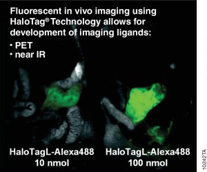
A recent publication (1) investigated a alternative method utilizing the HaloTag® dehalogenase enzyme HaloTag® is a dehalogenase enzyme (33 kDa) that contains an engineered cavity designed to accommodate the reactive chloroalkane group of a HaloTag® ligand (HTL). Upon entering the enzyme cavity, the terminal chlorine atom rapidly undergoes nucleophilic displacement, and a covalent adduct is formed, effectively anchoring the HaloTag® ligand in a precise location.

Three new HaloTag® ligands were synthesized and each labelled with the SPECT radionuclide indium-111  111In-HTL-1  and the dual-modality HaloTag® ligands,111In-HTL-2 and111;In-HTL-3 containing TMR which allows complementary imaging data).

For the validation of the pretargeting strategy based on these HaloTag® ligands, the target human epidermal growth factor receptor 2 (HER2)was selected. Trastuzumab (Herceptin®) was selected as the primary targeting agent and was modified with HaloTag® protein via the trans-cyclooctene/tetrazine ligation.

All three 111In-labelled HaloTa®g ligands exhibited significantly higher binding to the HER2 expressing when compared to negative controls.

Literature Cited

Knight, J. C et al.(2015) Development of an enzymatic pretargeting strategy for dual-modality imagingChem. Commun. 51, 4055–8.

Dual-Luciferase Imaging in vivo

22159-2268-CR-WC-NanolucWebNavImage280x140We are used to seeing multicolored fluorescence images labeling either specific events or structures within cells. When compared to imaging with fluorescent methods, bioluminescence imaging methods provide the advantages of low background and subsequent higher signal to noise ratio—enhancing sensitivity. A key prerequisite for dual-imaging experiments is the ability to distinguish the signal from each event separately and clearly. However, compared to the large number of available fluorescent compounds (many spectrally distinct fluorescent proteins and dyes), there are not many different luciferases to choose from. This lack of variety has limited the capabilities of bioluminescence for imaging multiple molecular events in the same sample. Therefore, there is a need for new luciferases with substrates and emission spectra that are different from the beetle luciferases currently in widespread use.

A paper published in Molecular Imaging in October 2013 describes use of firefly and the new NanoLuc® Luciferase to image cell signaling events in cultured cells and in a mouse model system. The paper, authored by Stacer et al. of the University of Michigan, details a proof-of-concept experiment using firefly and NanoLuc luciferases to image two distinct events in the TGF-beta1 signaling pathway. Continue reading “Dual-Luciferase Imaging in vivo”

Tracking the Progression of Plague Using Bioluminescence

Sequencing Yersinia pestis, the bacterium that caused the Black Plague in Europe during 1348–50, is an amazing accomplishment. Y. pestis infection still occurs sporatically and causes fatalities despite the Age of Antibiotics. Even with animal models, there are questions remaining about the progression of infection. Nham et al. used in vivo imaging to examine the course of infection in a mouse animal model using a bioluminescent clone of Y. pestis. Continue reading “Tracking the Progression of Plague Using Bioluminescence”