Bioluminescent in vivo imaging tools

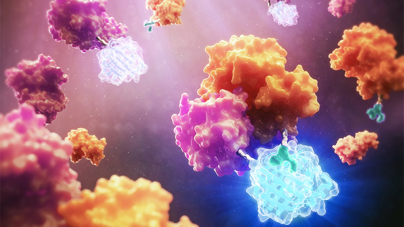
With advancements made over the past few decades, the future of in vivo bioluminescence imaging (BLI) continues to gain momentum. In vivo BLI provides a non-invasive way to image endogenous biological processes in whole animals. This provides an easier method to assess relevant systems and functions. Unlike fluorescent imaging, BLI relies on a combination of enzymes and substrates to produce light, greatly reducing background signal (Refaat et al., 2022). Traditional fluorescent tags are also quite large and may interfere with normal biological function. In vivo BLI research has been around for quite some time, primarily utilizing Firefly luciferase (Luc2/luciferin). A recent advancement was the creation of the small and bright NanoLuc® luciferase (NLuc). Promega offers an wide portfolio of NLuc products that provide ways to study genes, protein dynamics, and protein:protein interactions. To fully grasp the power of these tools, I interviewed several key investigators to determine their perspectives on the future of in vivo BLI. I was specifically interested in their thoughts on NLuc multiplexing potential with Firefly (FLuc), and future research areas. These two investigators are Dr. Thomas Kirkland, Sr. Scientific Investigator at Promega, and Dr. Laura Mezzanotte, Associate Professor at Erasmus MC.
Multiplexing NanoLuc® for in vivo Bioluminescence Imaging
NLuc has many alluring traits for its use within in vivo models. Its small size (19kDa) makes it an efficient tag less likely to impact biological function. NLuc is ATP independent, opening up areas traditionally difficult to assess, such as exosomes. Promega recently developed fluorofurimazine (FFz) as a substrate optimally suited for in vivo detection of NLuc. The characteristics of NLuc uniquely place it within the world of in vivo research.
An exciting approach for in vivo work is multiplexing it with other reporters, such as Firefly. Firefly (FLuc), and its substrate luciferin have dominated the world of in vivo BLI for decades (Kim et al., 2015). The advancements that multiplexing Nluc/FFz provide however, distinguish specific uses for each of these reporter systems. Tom elaborated on this point by stating, “NLuc is a good fit for multiplexing due to its complete orthogonality to FLuc, the dominant enzyme used in BLI. The substrates are completely bidirectionally orthogonal – each substrate will only emit light when processed by its cognate luciferase with no cross reactivity”.
Outside of the lack of substrate cross-reactivity, the distinct differences between FLuc and NLuc provide several advantages to multiplex. Tom highlighted this by saying “The best uses of NLuc/FLuc multiplexes are when each luciferase is used in a way that takes advantage of its strengths. FLuc is excellent at indicating tumor size since it is active intracellularly and its substrate luciferin can be injected in very high amounts. NLuc is active intra and extracellularly and is very bright, so it can be used to, for instance, label T-Cells in an immunooncology experiment or label therapeutic antibodies”.
Promega offers substrates for in vivo detection of both NanoLuc® and Firefly.
Key Considerations For In Vivo BLI
Laura brought up some caveats when planning on making the most from your reporters. “You really have to check the dynamic curve of light emission, it’s different depending on the tissue you want to image”. Various factors greatly impact the success of different reporters within whole animals. Deep tissues prove challenging for blue light emission to reach the surface. Fusion reporters combining NLuc with red/orange reporters, such as Antares (CyoGFP) have been developed to address these challenges(Su et al., 2020).
When conducting in vivo studies with several reporters, Laura highly encouraged longitudinal studies and stated, “it’s always better to do kinetic imaging, and then at the end of the longitudinal study you have more comprehensive data”. Longitudinal in vivo studies of NLuc are greatly improved due to the decreased toxicity of the new fluorofurimazine (FFz) substrate. Tom stated that overall “NLuc is superior to other marine luciferases like Renilla because it is much brighter and it effectively uses substrates that have better pharmacokinetics than coelenterazine, such as fluorofurimazine”.
Future Thoughts
Both Tom and Laura expressed excitement in the future use of NLuc for in vivo bioluminescence imaging. During our interview, Laura stated she remembered how excited she was to see NLuc used for analyzing viruses. She followed this up by laughing stating she had recently seen publications about that exact topic. When asked about what other fields she was looking forward to seeing NLuc used, Laura noted she was excited about “antibody labeling, particle tracking, and […] endogenous tagging”. Laura has transitioned into using organoids for her work with NLuc, but noted she sees a lot of potential exploring the brains of animals.
Tom works very closely with NLuc through his internal development of FFz at Promega. When asked about research areas he was excited about, he responded saying “I would love to see things move beyond cell tracking. BLI can be used to detect interactions and enzymatic activity through luciferase-based sensors (an area particularly suited for NLuc), BRET, and prosubstrates. These can all be detected with NLuc based systems, and one of the challenges is to normalize the signal. This is relatively easily done with FLuc as an orthogonal, ubiquitous reporter.”
Insights from leaders in the field, such as Laura and Tom, highlight the excitement and momentum behind in vivo BLI. With recent advances, such as the launch of FFz, multiplexing several reporters provides novel avenues to understand complex biological processes.
Citations
Kim, J. E., Kalimuthu, S., & Ahn, B. C. (2015). In Vivo Cell Tracking with Bioluminescence Imaging. In Nuclear Medicine and Molecular Imaging (Vol. 49, Issue 1, pp. 3–10). Springer Verlag. https://doi.org/10.1007/s13139-014-0309-x
Refaat, A., Yap, M. L., Pietersz, G., Walsh, A. P. G., Zeller, J., del Rosal, B., Wang, X., & Peter, K. (2022). In vivo fluorescence imaging: success in preclinical imaging paves the way for clinical applications. In Journal of Nanobiotechnology (Vol. 20, Issue 1). BioMed Central Ltd. https://doi.org/10.1186/s12951-022-01648-7
Su, Y., Walker, J. R., Park, Y., Smith, T. P., Liu, L. X., Hall, M. P., Labanieh, L., Hurst, R., Wang, D. C., Encell, L. P., Kim, N., Zhang, F., Kay, M. A., Casey, K. M., Majzner, R. G., Cochran, J. R., Mackall, C. L., Kirkland, T. A., & Lin, M. Z. (2020). Novel NanoLuc substrates enable bright two-population bioluminescence imaging in animals. Nature Methods, 17(8), 852–860. https://doi.org/10.1038/s41592-020-0889-6

