From Chromosomes to the Brain: How HaloTag Is Expanding What We Can See

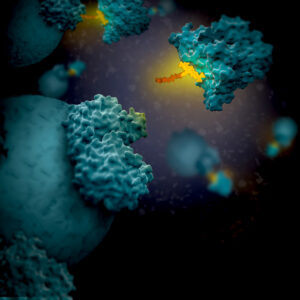
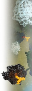
Artist's 3D concept of HaloTag with linker attaching to a target protein

Sometimes in the life sciences, itโ€™s really tempting to look at a process like mitosis and think, โ€œWow, we really understand that,โ€ and stop asking questions. In other cases, significant barriers like reliable expression of a non-endogenous reporter in an organ such as the brain is a challenge that seems impossible to overcome. However, those are precisely the moments when science and scientists need to push the boundaries of what we can see. In this post, I discuss two studies where researchers are literally seeing what could not be seen before by developing new methods for looking at their model systems.

In both papers discussed here, the HaloTagยฎ protein and ligands were key to developing new methods allowing scientists to visualize biological phenomena that had been previously inaccessible. HaloTagยฎ protein allows different functionalities to be linked onto a single genetic construct, rapidly and covalently under physiological conditions for experiments ranging from live-cell imaging to capture of protein interaction complexes (1). In the first study, HaloTag-CENP-A + JF635 ligand was the specific tool that let them directly see, in real time, that chromosome-attached microtubules pivot as the spindle elongates. In the second study, HaloTag was used to create a protocol for PET imaging of reporter gene systems in the brain.

Continue reading “From Chromosomes to the Brain: How HaloTag Is Expanding What We Can See”

Under Pressure: The Nuclear Choreography upon Replicative Stress

Todayโ€™s blog is guest-written by Jana Krietsch (University of Zurich), edited by Nour Mozaffari (Promega)

Each time a cell divides, it must make an accurate copy of its entire genomeโ€”in human cells, this means roughly 6.2 billion individual DNA building blocks to duplicate. This enormous molecular task takes place inside the nucleus, a crowded, highly organized, yet remarkably dynamic environment. As the DNA-copying machinery moves along the genome, it may encounter roadblocks such as damaged DNA, sequences that are difficult to copy, tightly packed chromatin, or other molecular processes using the same DNA at the same time. These obstacles can slow or stall replication, a phenomenon known as DNA replication stress.

A Hidden Vulnerability of Cancer Cells

Cells are well equipped to deal with replication stress. Depending on the type and severity of the problem, they activate specialized signaling and repair mechanisms that protect replicating DNA. Failure of these responses can jeopardize genome integrity and result in permanent genetic changes that contribute to disease development.

In cancer, oncogenic changes and rapid proliferation places tumor cells under persistent replication stress. This promotes genome instability, a cancer hallmark, and drives tumor evolution. At the same time, it makes cancer cells hyper-dependent on replication stress response mechanisms to survive, creating a hidden vulnerability. Several treatments exploit this Achillesโ€™ heel by increasing replication stress levels beyond what cancer cells can tolerate.

From Sequential Model to Dynamic Choreography

Many stress response pathways have been successfully reconstituted in the test tube. They are typically described as linear sequence of events triggered when an active replication site, termed replication fork, encounters a roadblock: the fork encounters an obstacle, its structure changes, signaling proteins are activated, repair factors are recruited, and DNA synthesis eventually resumes.

Continue reading “Under Pressure: The Nuclear Choreography upon Replicative Stress”

Polyserine Targeting: A New Strategy Against Neurodegeneration

Neurodegenerative diseases like Alzheimerโ€™s are marked by the accumulation of misfolded proteins that wreak havoc on neurons. One of the most notorious culprits is tau, a structural protein that, in its diseased form, clumps together into aggregates that spread throughout the brain. These aggregates interfere with normal cellular processes, leading to memory loss, behavioral changes, and other devastating symptoms. Preventing tau aggregation is therefore a key strategy for slowing the progression of these symptoms.

What if we could recruit molecular โ€œhelpersโ€ to stop tau from accumulating?

Continue reading “Polyserine Targeting: A New Strategy Against Neurodegeneration”

Mapping the Mind: In Vivo Imaging of Synaptic Plasticity with HaloTagยฎ Ligands

The brain is constantly rewiring itself, fine-tuning connections that shape how we think, learn, and remember. But capturing those fleeting molecular changes as they happen โ€” at the level of individual synapses and across entire brain regions โ€” has long been a challenge in neuroscience. Now, thanks to recent advances in HaloTagยฎ dye technology, researchers can visualize protein dynamics in living brains with stunning clarity and specificity.

Continue reading “Mapping the Mind: In Vivo Imaging of Synaptic Plasticity with HaloTagยฎ Ligands”

Cell Tracking Using HaloTag: Why are Scientists Chasing Cells?

Cells, commonly considered the smallest unit of life, provide structure and function for all living things (3).

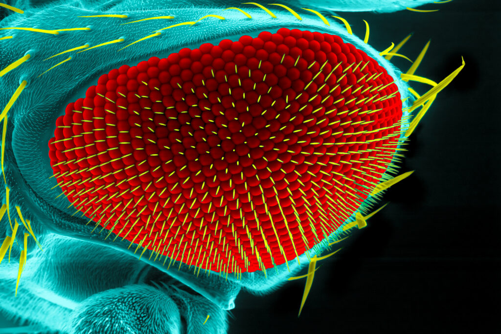
Eye of a fruit fly, Drosophila melanogaster, scanning electron microscopy. Scientists used HaloTag for cell tracking during eye development.
Eye of a fruit fly, Drosophila melanogaster, scanning electron microscopy

Because cells contain the fundamental molecules of life, in some situations such as yeast, a single cell can be considered the complete organism. In other situations, for more complex multicellular organisms, a multitude of cells can mature and acquire different, specialized functions (3).

Cells developing specificity are undergoing differentiation, a process where a cellโ€™s genes are either turned โ€œonโ€ or โ€œoffโ€ resultant in a more specific cell type. As these differentiated cells start to exhibit their identity, they organize themselves into the tissues, organs, and organ systems integral to the functioning of a multicellular, developing organism. This process in which order and form is created within a developing organism is referred to as morphogenesis (5).

Continue reading “Cell Tracking Using HaloTag: Why are Scientists Chasing Cells?”

RNA-Protein Interactions: A New Frontier for Drug Discovery

Almost 90% of the human genome is transcribed into RNA, but only 3% is ultimately translated into a protein. Some non-translated RNA is thought to be useless, while some play a significant yet often mysterious role in cancer and other diseases. Despite its abundance and biological significance, RNA is rarely the target of therapeutics.

โ€œWe say itโ€™s undruggable, but I would say that โ€˜not-yet-druggedโ€™ is a better way to put it,โ€ says Amanda Garner, Associate Professor of Medicinal Chemistry at the University of Michigan. โ€œWe know that RNA biology is important, but we donโ€™t yet know how to target it.โ€

Amandaโ€™s lab develops systems to study RNA biology. She employs a variety of approaches to analyze the functions of different RNAs and study their interactions with proteins. Her lab recently published a paper describing a novel method for studying RNA-protein interactions (RPI) in live cells. Amanda says that with the right tools, RPI could become a critical target for drug discovery.

โ€œItโ€™s amazing that current drugs ever work, because theyโ€™re all based on really old approaches,โ€ Amanda says. โ€œThis isnโ€™t going to be like developing a small molecule kinase inhibitor. Itโ€™s a whole new world.โ€

Continue reading “RNA-Protein Interactions: A New Frontier for Drug Discovery”

Choosing a Tag for Your Protein

You have identified and cloned your protein of interest, but you want to explore its function. A protein fusion tag might help with your investigation. However, choosing a tag for your protein depends on what experiments you are planning. Do you want to purify the protein? Would you like to identify interacting proteins by performing pull-down assays? Are you interested in examining the endogenous biology of the protein? Here we cover the advantages and disadvantages of some protein tags to help you select the one that best suits your needs.

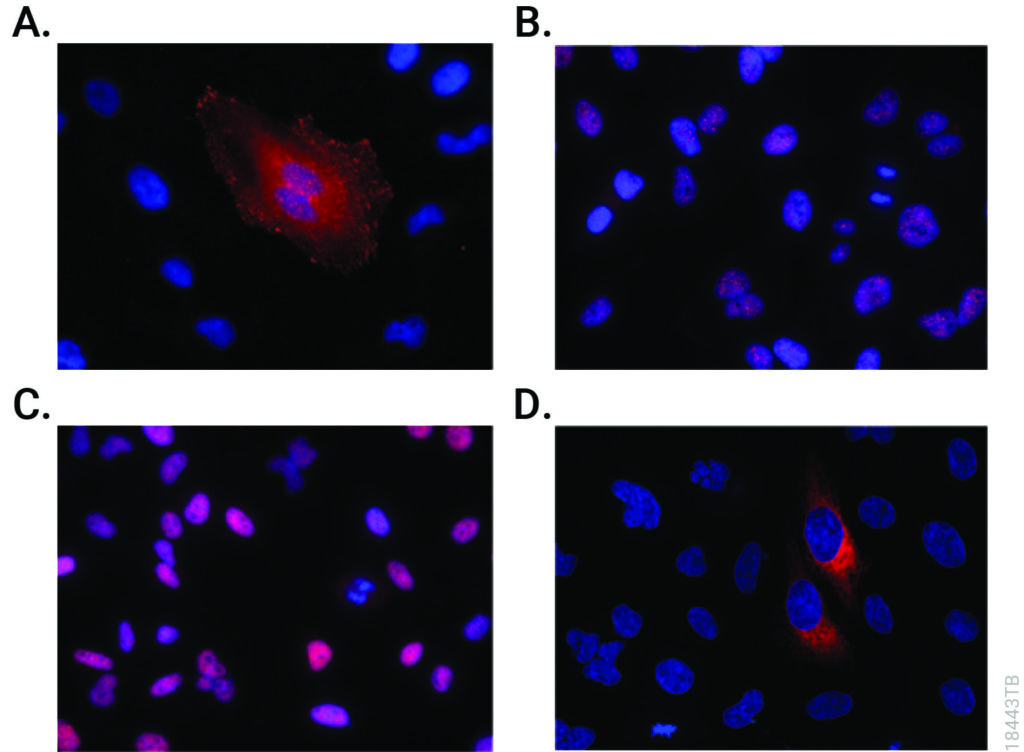
Immunofluorescent detection of HiBiT-tagged proteins in CRISPR-edited cell pools and clones using the Anti-HiBiT Monoclonal Antibody.
CRISPR-Cas9 editing knocked-in HiBiT at the endogenous locus of proteins with varying subcellular localization. Fixed CRISPR-modified clones or pools of cells were imaged by immunofluorescent staining using the Anti-HiBiT Monoclonal Antibody (red) and Hoechst dye (blue). Panel A. VCL-HiBiT pool. Panel B. SMARCA4-HiBiT clone. Panel C. HDAC2-HiBiT clone. Panel D. HSP90B1-HiBiT pool.

Affinity Tags

The most commonly used protein tags fall under the category of affinity tags. This means that the tag binds to another molecule or metal ion, making it easy to purify or pull down your protein of interest. In all cases, the tag will be fused to your protein of interest at either the amino (N) or carboxy (C) terminus by cloning into an expression vector. This protein fusion can then be expressed in cells or cell-free systems, depending on the promoter the vector contains.

Continue reading “Choosing a Tag for Your Protein”

Executing a NanoBRET™ Experiment: From Start to Data

This is a guest post from Katarzyna Dubiel, marketing intern in Cellular Analysis and Proteomics.

โ€œThe objective of my experiment was to test the NanoBRET™ assay as if I was a customer, independent of the research and development team which develops the assay.โ€

Designing and implementing a new assay can be a challenging process with many unexpected troubleshooting steps. We wanted to know what major snags a scientist new to the NanoBRET™ Assay would encounter. To determine this, we reached out to Laurence Delauriere, a senior applications scientist at Promega-France, who had never previously performed a NanoBRET™ assay. Laurence went step-by-step through the experimental process looking at the CRAF-BRAF interaction in multiple cell lines. In an interview, Laurence provided us with some tips and insights from her work implementing the new NanoBRET™ assay.

In a few words, can you explain NanoBRET?
โ€œNanoBRET is used to monitor protein: protein interactions in live cells. It is a bioluminescence resonance energy transfer (BRET) based assay that uses NanoLucยฎ luciferase as the BRET energy donor and HaloTagยฎ protein labeled with the HaloTagยฎ NanoBRET™ 618 fluorescent ligand as the energy acceptor to measure the interaction of two binding partners.โ€ Continue reading “Executing a NanoBRET™ Experiment: From Start to Data”

Purify and Conjugate Antibodies in a Single Workflow

Isoform_Antibodies_LinkedInAntibodies labeled with small molecules such as fluorophore, biotin or drugs play a critical role in various areas of biological research,drug discovery and diagnostics. There are several limitations to current methods for labeling antibodies including the need for purified antibodies at high concentrations and multiple buffer exchange steps.

In a recent publication, a method (on-bead conjugation) is described that addresses these limitations by combining antibody purification and conjugation in a single workflow. This method uses high capacity-magnetic Protein A or Protein G beads to capture antibodies directly from cell media followed by conjugation with small molecules and elution of conjugated antibodies from the beads.

Usingย a variety of fluorophores the researchers show that the on-bead conjugation method is compatible with both thiol- and amine-based chemistry.

This method enables simple and rapid processing of multiple samples in parallel with high-efficiency antibody recovery. It is further shown that recovered antibodies are functional and compatible with downstream applications.

Literature Cited

Nidhi, N. et al. (2015) On-bead antibody-small molecule conjugation using high-capacity magnetic bead J. Immunol. Methodsย  http://dx.doi.org/10.1016/j.jim.2015.08.008

If We Could But Peek Inside the Cell โ€ฆQuantifying, Characterizing and Visualizing Protein:Protein ย Interactions

14231183 WB MS Protein Interactions Hero Image 600x214

Robert Hooke first coined the term โ€œcellโ€ after observing  plant cell walls through a light microscopeโ€”little empty chambers, fixed in time and space. However,  cells are anything but fixed.

Cells are dynamic: continually responding to a shifting context of time, environment, and signals from within and without. Interactions between the macromolecules within cells, including proteins, are ever changingโ€”with complexes forming, breaking up, and reforming in new ways. These interactions provide a temporal and special framework for the work of the cell, controlling gene expression, protein production, growth, cell division and cell death.

Visualizing and measuring protein:protein interactions at the level of the cell without perturbing them is the goal of every cell biologist.

A recent article by Thomas Machleidt et al. published in ACS Chemical Biology, describes a new technology that brings us closer to being able to realize that goal.

Continue reading “If We Could But Peek Inside the Cell โ€ฆQuantifying, Characterizing and Visualizing Protein:Protein ย Interactions”