HaloTag® Research Application: Detection of Cancer Biomarkers

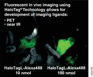
10242TAAntibodies labelled with radioisotopes or the sequential administrationof an antibody and a radioactive secondary agent facilitate the in vivo detection and/or characterisation of cancers by positron emission tomography (PET) or by single-photon emission computed tomography (SPECT) imaging.

There are drawbacks to both methods, including prolonged exposure to radiation and  ensuring that both the antibody and the radiolabelled secondary agent are suitably designed so that they bind rapidly upon contact at the tumor.

A recent publication (1) investigated a alternative method utilizing the HaloTag® dehalogenase enzyme HaloTag® is a dehalogenase enzyme (33 kDa) that contains an engineered cavity designed to accommodate the reactive chloroalkane group of a HaloTag® ligand (HTL). Upon entering the enzyme cavity, the terminal chlorine atom rapidly undergoes nucleophilic displacement, and a covalent adduct is formed, effectively anchoring the HaloTag® ligand in a precise location.

Three new HaloTag® ligands were synthesized and each labelled with the SPECT radionuclide indium-111  111In-HTL-1  and the dual-modality HaloTag® ligands,111In-HTL-2 and111;In-HTL-3 containing TMR which allows complementary imaging data).

For the validation of the pretargeting strategy based on these HaloTag® ligands, the target human epidermal growth factor receptor 2 (HER2)was selected. Trastuzumab (Herceptin®) was selected as the primary targeting agent and was modified with HaloTag® protein via the trans-cyclooctene/tetrazine ligation.

All three 111In-labelled HaloTa®g ligands exhibited significantly higher binding to the HER2 expressing when compared to negative controls.

Literature Cited

Knight, J. C et al.(2015) Development of an enzymatic pretargeting strategy for dual-modality imagingChem. Commun. 51, 4055–8.

Shedding Light on Protein:Protein Interactions with NanoBRET™ Technique

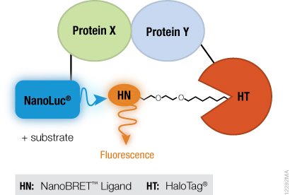
NanoBRET™ TechnologyIf you are trying to investigate protein:protein interactions inside cells, you know how important physiologically relevant results are. If you overload your cells with fusion constructs, your protein interactions may not actually reflect what is going on in the cell, and if your BRET energy donor and acceptor do not have sufficiently separated spectra, you can pick up a fair amount of noise in your experiment. Using the new superbright NanoLuc® Luciferase, and the HaloTag® Technology, we have developed a sensitive BRET system to help you take a better look specific protein interactions that interest you. Promega research scientist, Danette Daniels, describes the system in the Chalk Talk below:

Site-specific copy number variations in cancer: A story begins to unfold

Designed by Nick Klein for ISO-form, courtesy of Promega.

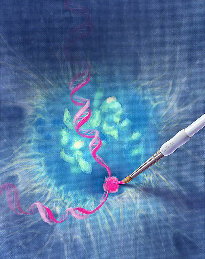
Tumor cells are characterized by many features: including uncontrolled proliferation, to loss of contact inhibition, acquired chromosomal instability and gene copy number changes among them. Some of those copy number changes are site-specific, but very little is known about the mechanisms or proteins involved in creating site-specific copy number changes. In a recently published Cell paper, Black and colleagues, propose a mechanism for site-specific copy number variations involving histone methylation proteins and replication complexes.

Previous work from Klang et al. had shown that local amplification of chromosomal regions occurs during S phase and that chromatin structure plays a critical role in this amplification (2), and other work by Black and colleagues (3) implicated KDM4A in changing timing of replication by altering chromatin accessibility in specific regions. Other research also had shown that KDM4A protein levels influence replication initiation and that KDM4A has a role in some DNA damage response pathways (4,5).  Looking at the body of work, Black et al. hypothesized that KDM4A, with its roles in replication, might possibly provide link into the mechanism of site-specific copy number variation in cancer. Continue reading “Site-specific copy number variations in cancer: A story begins to unfold”

Another Step Closer to Understanding Epigenetic Gene Regulation

Chromatin fiber

Back when I was a graduate student (more than a few years ago), I remember hearing another student joke that if a member of his thesis committee asked him to explain an unexpected or unusual result, he was going to “blame” epigenetics. At that time, the study of epigenetic gene regulation was in its infancy, and scientists had much to learn about this mysterious regulatory process. Fast forward to today, and you’ll find that scientists know a lot more about basic epigenetic mechanisms, although there is still plenty to learn as scientists discover that the topic is much more complicated than initially thought, as is often the case in science. A recent EMBO Journal article is contributing to our knowledge by shedding light on the role of the TET family of DNA-modifying enzymes in epigenetics (1).

Continue reading “Another Step Closer to Understanding Epigenetic Gene Regulation”

Screening for Protein Activity Using Cell-Free Expression

The analysis of functional protein typically requires lengthy laborious cell based protein expression that can be complicated by the lack of stability or solubility of the purified protein. Cell free protein expression eliminates the requirement for cell culture thus providing quick access to the protein of interest (1).

The HaloTag® Technology provides efficient, covalent and oriented protein immobilization of the fusion protein to solid surfaces (2).

Leippe et al. demonstrated the feasibility of using cell free expression and the HaloTag technology to express and capture a fusion protein for the rapid screening of protein kinase activity (3). The catalytic subunit of human cAMP dependent protein kinase was expressed in a variety of cell free expression formats as a HaloTag fusion protein. The immobilized cPKA fusion protein was assayed directly on magnetic beads in the active form and was shown to be inhibited by known PKA inhibitory compounds.

Therefore this unique combination of protein expression and capture technologies can greatly facilitate the process of activity screening and characterization of potential inhibitors

References

  1. Zhao, K.Q. et al. (2007) Functional protein expression from a DNA based wheat germ cell-free system. J. Struc. Funct. Genomics. 8, 199-208.
  2. Los, G.V. and Wood, K. (2007) The HaloTag: A novel technology for cell imaging and protein analysis. Meth. Mol. Biol. 356, 195-208
  3. Leippe DM, Zhao KQ, Hsiao K, & Slater MR (2010). Cell-free expression of protein kinase a for rapid activity assays. Analytical chemistry insights, 5, 25-36 PMID: 20520741