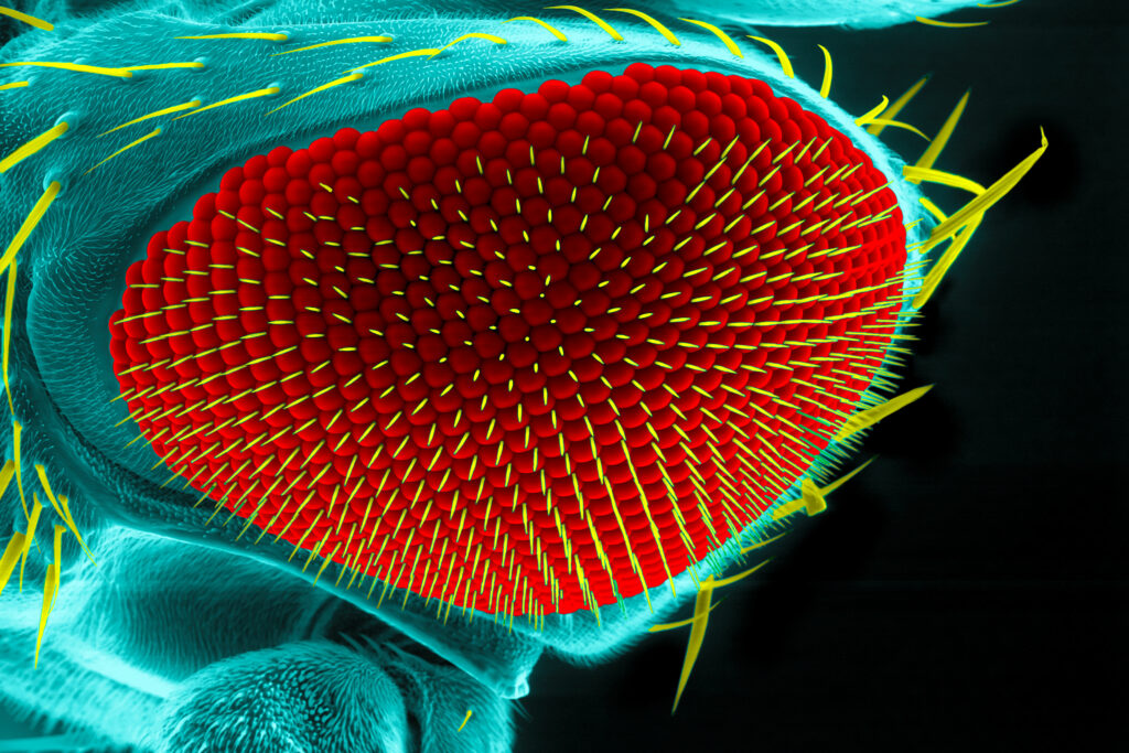
Cells, commonly considered the smallest unit of life, provide structure and function for all living things (3).

Because cells contain the fundamental molecules of life, in some situations such as yeast, a single cell can be considered the complete organism. In other situations, for more complex multicellular organisms, a multitude of cells can mature and acquire different, specialized functions (3).
Cells developing specificity are undergoing differentiation, a process where a cell’s genes are either turned “on” or “off” resultant in a more specific cell type. As these differentiated cells start to exhibit their identity, they organize themselves into the tissues, organs, and organ systems integral to the functioning of a multicellular, developing organism. This process in which order and form is created within a developing organism is referred to as morphogenesis (5).
Morphogenesis:
Morphogenesis remains a hot topic in developmental biology today, and is surrounded in a multitude of questions including (6):
- How do differentiated cells organize themselves into tissues and organs?
- How is growth coordinated over time, when and where do events happen to control this?
- How does your body know you are the right size, or your organs are the right size and shape?
- How do migrating cells orient themselves?
To find the answer to these questions scientists must first find small-scale ways to track cell movement in real time, with future intention of large-scale automated cell tracking.
Recently, scientists have been looking into different ways to track cells, including using sparse and random labelling of cells using HaloTag® technology in developing Drosophila tissues to track the behavior of a subset of cells over time.
Why HaloTag Labeling?
Currently there is some success with light-sheet microscopy and the image analysis of tissue formation in mouse embryos. This has enabled generation of dynamic fate maps and the ability to quantitively examine cell division and behavior during neural tube formation in the mouse embryo. However light microscopy does have its own limitations, especially in anatomical regions where lack of transparency and difficult optical properties make imaging challenging. (4).
A solution to this challenge?
One solution to this problem, the use of fluorescent reporters, in this case HaloTag technology, to track single or subsets of cells as described in a study by Couturier, L. et al (1).
Scientists interested in cell tracking during Drosophila development used HaloTag proteins, which have the capacity to covalently bind a cell-permeable synthetic ligand to generate fluorescent labels (1). In this study Drosophila eye imaginal disc explants were utilized, and conditional expression of HaloTag7 protein, nlsHalo, was initiated through FLP/FRT site specific recombination and the addition of heat shock.
The FLP/FRT system consisted of a FLP-out (flippase recombinase), an intramolecular excision spacer of DNA, surrounded by two FRT (FLP recombination target) sites (2). Heat-induced recombination of the FLP/FRT system allowed for a downstream gene, nlsHalo, to be brought together with a promoter, ubi, through the FLP-mediated excision of a transcriptional stop surrounded by the two FRT’s (1).
The study found that a brief heat shock was enough to cause 10–30% of the cells to recombine and express nlsHalo, with fluorescence after addition of the ligand observed after ~45–60 minutes and recombination being detected up to 7–8 hours later (1). This quick and long fluorescence was anticipated, as these ligands have a high signal-to-noise ratio and can easily be fused to a protein of interest, enabling them to produce a quicker, stronger, and more stable signal than red or infra-red fluorescent proteins. Under these conditions, HaloTag labeling enabled a total of 6-8 hours of imaging capable of tracking cell fate in Drosophila eye imaginal discs (1).
A final characteristic that makes HaloTag labeling a particularly useful tool, is its capability to adjust the frequency of recombination for labelling cells by tuning the heat-shock parameters. This would be advantageous in hard-to-label tissues, especially where cells move around rapidly (1).
Overall, this research provides a concept for future cell tracking and real time quantitative analysis of cell movement.
A Final Caveat to Mention
Though HaloTag is a great cell tracker, there is a current lack of ability to work in vivo, as the ligands needed to be provided directly to cells and tissues, resulting in an ex vivo experiment (1).
Works Cited
- Couturier, L et al. (2022) HaloTag-based reporters for sparse labeling and cell tracking. Fly. 16(1), 360-366.
- Davis, M. W. et al. (2008). Gene activation using FLP recombinase in C. elegans. PLoS Genetics. 4(3)
- Ellinger, I., & Ellinger, A. (2014). Smallest unit of life: Cell biology. Comparative Medicine. (pp. 19-33)
- McDole, K. et al. (2018). In Toto imaging and reconstruction of post-implantation mouse development at the single-cell level. Cell. 175(3), 859-876.e33.
- Sunderland, M. (2008). Morphogenesis. The Embryo Project Encyclopedia. Retrieved April 12, 2023.
- Saunders, T. E., & Ingham, P. W. (2019). Open Questions: how to get developmental biology into shape? BMC Biology. 17(1), 17.
Latest posts by Shannon Sindermann (see all)
- What do Exosomes have to do with Cancer Research? - May 30, 2024
- SLIM Chances: Upside-down, but not Out on the Lunar Surface - February 27, 2024
- Uniting Diverse Minds, Vibrant Ideas, and Collaborative Spirit at the 2023 Biologics Symposium - December 13, 2023
