Illuminating the Function of a Dark Kinase (DCLK1) with a Selective Chemical Probe

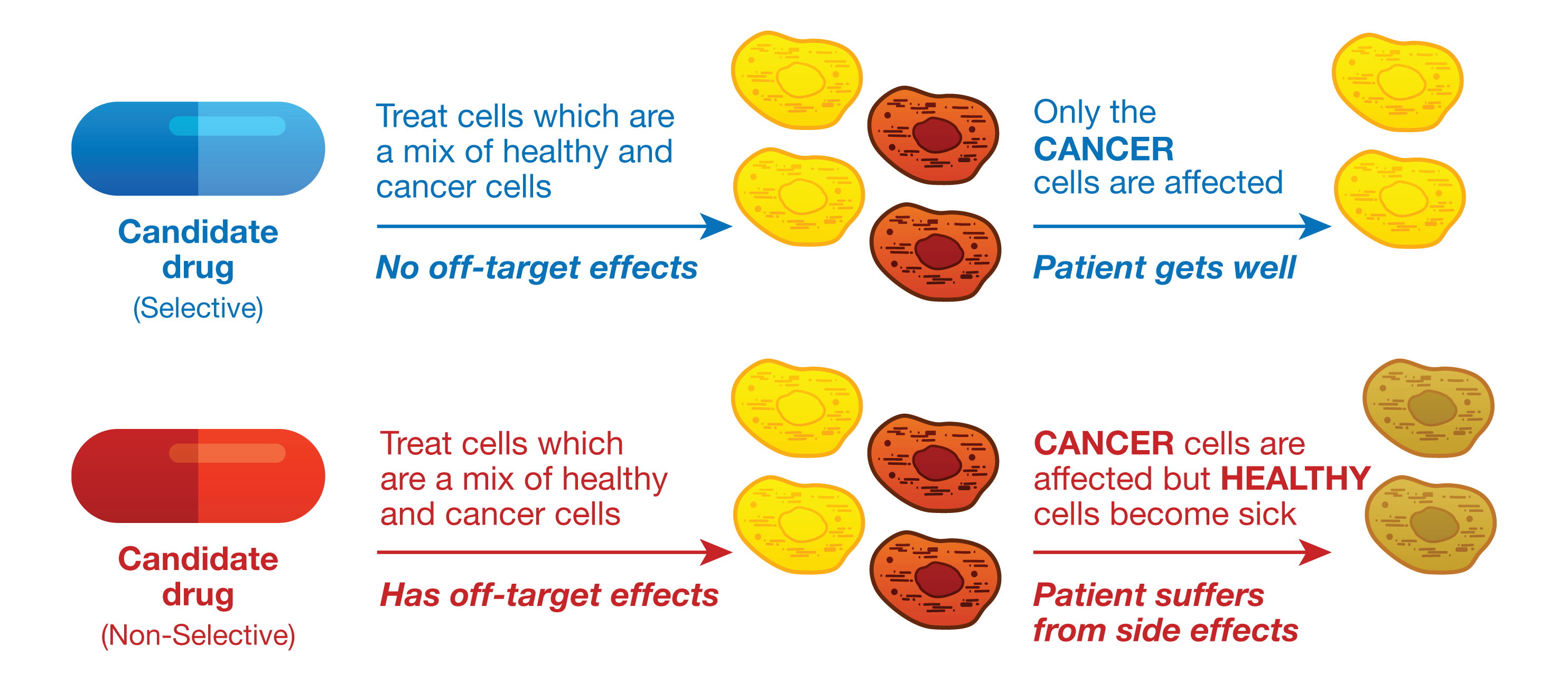
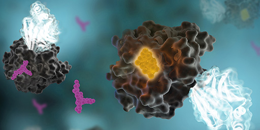
The understudied kinome represents a major challenge as well as an exciting opportunity in drug discovery. A team of researchers lead by Nathanael Gray at the Dana Farber Cancer Institute was able to partially elucidate the function of an understudied kinase, Doublecortin-like kinase 1 (DCLK1), in pancreatic ductal adenocarcinoma cells (PDAC). The characterization of DCLK1 in PDAC was realized by developing a highly specific chemical probe (1). Promega NanoBRET™ Target Engagement (TE) technology enabled intracellular characterization of this chemical probe.

The Dark Kinome

NanoBRET target engagement

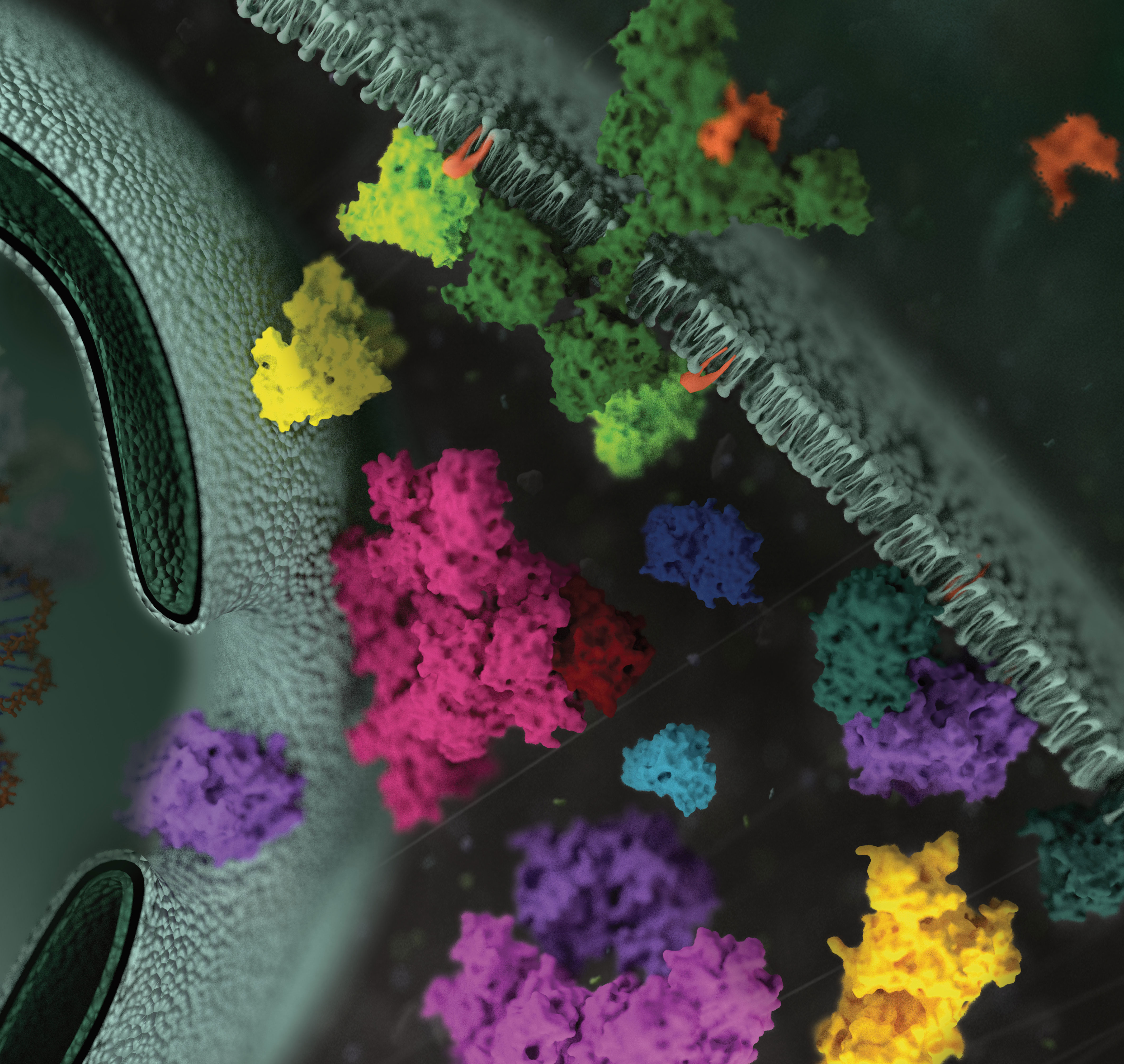
Comprised of over 500 proteins, the human kinome is among the broadest class of enzymes in humans and is rife with targets for small molecule therapeutics. Indeed, to date, over 50 small molecule kinase inhibitors have achieved FDA approval for use in treating cancer and inflammatory diseases, with nearly 200 kinase inhibitors in various stages of clinical evaluation (2). Moreover, broad genomic screening efforts have implicated the involvement of a large fraction of kinases in human pathologies (3). Despite such advancements, our knowledge of the kinome is limited to only a fraction of its family members (3,4). For example, currently less than 20% of human kinases are being targeted with drugs in clinical trials. Moreover, only a subset of kinases historically has garnered substantial citations in academic research journals (4). As a result, a large proportion of the human kinome lacks functional annotation; as such, these understudied or “dark” kinases remain elusive to therapeutic intervention (4).

Continue reading “Illuminating the Function of a Dark Kinase (DCLK1) with a Selective Chemical Probe”

Giant Rodent, Lowered Cancer Rates: What Genetic Analysis Reveals about the Capybara and Cancer

Do you find the thought of a giant rodent off-putting? Do your thoughts go to huge rats running amuck in dark allies, threatening unsuspecting passers by?

I personally hold rodents in low esteem. Rats, mice…who needs them? With the exception of cavies. I spent countless hours as a child playing with guinea pigs. We had as many as 16 of these little rodents at one time (the males are very capable of chewing or climbing out of cardboard boxes to reach a female in the next box). The baby guinea pigs were very cute and the adults had quite pronounced personalities, and a lot of attitude.

It was this history with guinea pigs that made me interested in learning more about the largest rodent in the world, the South American capybara (Hydrochoerus hydrochaeris). These family-oriented herbivores are found in savannas and forested areas, living in groups of as many as 100 members. They are excellent swimmers and can remain underwater for as long as 5 minutes. In fact, capybara mate only in the water. (Perhaps it’s not surprising then that the South American alligator, the caiman, is one of the capybara’s greatest predators.)

Photos of an adult male capybara.
A male capybara, with a scent gland (called a morillo) on his head. Photo by: Charles J Sharp – Own work, from Sharp Photography, sharpphotography, CC BY-SA 4.0.

With their squared-off nose and lack of tail, capybaras actually resemble guinea pigs. However, these oversized cavies weigh as much as 40 pounds. and can reach 24” at the shoulder, the size of an average standard poodle. Guinea pigs, on the other hand, weigh in at 2–3 pounds, and are 3–4” tall.

Their proportions make capybaras 60 times more massive than their closest relatives, rock cavies (Kerodon sp.) and 2,000 times more massive than the common mouse (Mus musculus). This tremendous size difference is why Herrera-Álvarez et al. took a closer look at the capybara, studying its propensity to develop cancer and other tradeoffs that would seem to coincide with its exceptional size.

Continue reading “Giant Rodent, Lowered Cancer Rates: What Genetic Analysis Reveals about the Capybara and Cancer”

Mutation Analysis Using HaloTag Fusion Proteins

In a recent reference, Kinoshita and colleagues characterized the phosphorylation dynamics of MEK1 in human cells by using the phosphate affinity electrophoresis technique, Phos-tag sodium dodecyl sulfate–polyacrylamide gel electrophoresis (Phos-tag SDS-PAGE; 1). They found that multiple variants of MEK1 with diferent phosphorylation states are constitutively present in typical human cells.

To investigate the relationships between kinase activity and drug efficacy researchers from the same laboratory group conducted phosphorylation profling of various MEK1 mutants by using Phos-tag SDS- PAGE (2).

They introduced mutations in of the MEK-1 coding gene that are associated with spontaneous melanoma, lung cancer, gastric cancer, colon cancer and ovarian cancer were introduced into Flexi HaloTag clone pFN21AE0668, which is suitable for expression of N-terminal HaloTag-fused MEK1 in mammalian cells. Continue reading “Mutation Analysis Using HaloTag Fusion Proteins”

Glycobiology Research and Training Opportunities are Plentiful

glycans on cell surface
Artist’s rendering of asymmetrically-branched carbohydrates on cell surface proteins.

Glycobiology is the study of glycans, the carbohydrate molecules that cover the surface of most human cells. Glycans attach to cell surface proteins and lipids, in a process called glycosylation. These cell surface structures are responsible for processes as varied at protein folding, cell signaling and cell-cell recognition, including sperm-egg recognition and immune cell interactions. Glycans play important roles in the red blood cell antigens that distinguish blood types O, A and B.

Opportunities in Glycomics Research
As more is learned about the role of glycans in cell communication, they are becoming important disease research targets, particularly the role of glycans in cancer and inflammatory diseases (2).

Some of the open questions surrounding glycans and glycosylation include glycan structural diversity. While some carbohydrates exist as straight or symmetrically branched chains, those populating the human glycome are asymmetrically branched, making them difficult to create and study in the laboratory (3). Continue reading “Glycobiology Research and Training Opportunities are Plentiful”

Making Drug Discovery More Efficient: Predicting Drug Side Effects in Early Screening Efforts

26911030-Laymans-KSPS-figure-WEB-R4Drug research and development is a complex and expensive process that begins with initial screening steps of candidate chemical compounds, and compounds that appear to have the desired potency against a specific cellular target or pathway are further evaluated. Candidate compounds that fail late in development or during clinical trials because of off-target effects are costly, and can be dangerous. Therefore drug developers not only need to ensure that a candidate compound is effective as a therapy, but also they need to predict any potential undesirable side effects due to off-target activities as early as possible in the drug discovery and development process. Continue reading “Making Drug Discovery More Efficient: Predicting Drug Side Effects in Early Screening Efforts”

A NanoBRET™ Biosensor for GPCR:G protein Interaction with the Kinetics and Temporal Resolution of Patch Clamping

Electrophysiologists are talented scientists/artists who see into the events of the cell with amazing detail.
Electrophysiology experiments provide a view into the cell with amazing detail. The paper reviewed here describes a molecular reporter biosensor (NanoBRET) that can offer the same kind of temporal and spatial resolution traditionally reserved for extremely labor-intensive experiments like patch clamp analysis.

I confess that I struggled through biophysics, and my Bertil Hille textbook Ion Channels of Excitable Membranes lies neglected somewhere in a box in my basement (I have not tossed it into the recycle bin—I can’t bear too, I spent too much time bonding with that book in graduate school).

My struggles in that graduate class and my attendance at the seminars of my grad school colleagues who were conducting electrophysiological studies left me with a sincere awe and appreciation of both the genius and the artistry required to produce nice electrophysiology data. The people who are good at these experiments are artists—they have the golden touch when it comes to generating that megaohm seal between a piece of cell membrane and a finely pulled glass pipette. And, they are brilliant scientists, they really understand the physics, the chemistry and the biology of the cells they study from a perspective that very few scientists ever develop.

Electrophysiology data, which often demonstrate the gating of a single channel protein in response to a single stimulus in real time–ions crossing a membrane through a single protein–are amazing for their ability, unlike virtually any other experimental data for the story they can tell about what is going on in a cell in real time under physiological conditions.

So when I read the paper recently published by Mashuo et al. in Science SignalingDistinct profiles of functional discrimination among G proteins determine the action of G protein-coupled receptors”, this sentence really caught my attention:

When constructs were ectopically expressed in HEK 293T/17 cells, we obtained very similar kinetics for the GPCR-driven responses between NanoBRET™ biosensors and the patch clamp recordings.

They continue:

Indeed, the activation rates that we observed were very similar to those of GPCR-stimulated GIRKs [G protein-coupled, inwardly rectifying K+ channel] in native cells, suggesting that the conditions of this assay closely match the in vivo setting. This finding further demonstrates the ability of the system to resolve the fast, physiological relevant kinetics of GPCR signaling.

A reporter biosensor that can resolve events similarly to patch clamping?!  Amazing. Continue reading “A NanoBRET™ Biosensor for GPCR:G protein Interaction with the Kinetics and Temporal Resolution of Patch Clamping”

Customized Kinase Selectivity Profiling Just Got Easier

11296971-DC-CR-KinaseOff-target activities of target compounds can become costly if they aren’t discovered until late in the drug research and discovery process. Therefore, knowing the inhibitory profile of your test compounds across a broad collection of kinases as quickly as possible is highly desirable.

However, screening against many kinases at once requires a universal platform that is still sensitive enough to detect inhibitor activity and assess selectivity and potency on kinases of different classes. The luminescent ADP-Glo™ Kinase Assay is a universal platform that measures kinase activity by quantifying the amount of ADP produced during a kinase reaction.

We have used the ADP-Glo™ Chemistry to develop highly sensitive assays for more than 170 kinases across the human kinome and further enhanced the assays for ease-of-use by developing the Kinase Selectivity Profiling Systems. These systems provide an easy-to-use, reliable platform for kinase inhibitor profiling in house.

And even better, we now provide an online Kinase Profiling System Designer so that you can design a custom Kinase Selectivity Profiling System to fit your exact experimental needs. Simply drag and drop the combination of kinases you need to create an 8-kinase strip and submit your order. Continue reading “Customized Kinase Selectivity Profiling Just Got Easier”

Targeting MYC: The Need to Study Protein:Protein Interactions in Cells

Crystal Structure of MYC MAX Heterodimer bound to DNA ImageSource=RCSB PDB; StructureID=1nkp; DOI=http://dx.doi.org/10.2210/pdb1nkp/pdb;

In 1982, picked up because of its homology to chicken virus genes that could transform cells, MYC became one of the first human genes identified that could drive cellular transformation (1,2). Since that time countless laboratories have prodded and poked the human MYC gene, the MYC protein, their homologs in other animal models, and their transforming viral counterparts.

MYC is a transcription factor and forms heterodimers with a required protein partner, MAX, before binding to the E box sequences of DNA regulatory regions (3). MYC regulates gene expression of many targets through interactions with a host of proteins, often referred to as the MYC Interactome (2).  In fact, MYC is estimated to bind 10–15% of the genome, and it regulates the expression of genes that  are transcribed by by each of the three RNA polymerases (2).

MYC plays a central role in regulating cell growth, proliferation, apoptosis, differentiation and transformation, acting as a central integrator of cellular signals. MYC is tightly regulated at multiple levels from gene expression to protein stability. Dysregulation (usually upregulation) of the amount and stability of Myc protein is observed in many human cancers. Even in cancers in which MYC is not directly involved in transforming cells, its normal expression is often required to support the extracellular matrix and/or vascularization necessary for tumor growth and formation (4).

Because MYC is such a central player cancer pathology, it is an attractive target for cancer therapeutics  (2) . Continue reading “Targeting MYC: The Need to Study Protein:Protein Interactions in Cells”

Uncovering Protein Autoinhibition Using NanoBRET™ Technology

13305818-protein ligandIntroducing new assays or technologies is meant to make it easier for you to perform research and craft experiments to test hypotheses. However, scientists are creative people, and new technologies or assays may just be the catalyst for a crucial experiment or new data you are seeking. In the case of a recent Proceedings of the National Academy of Sciences USA article, Wang et al. used the principle of our NanoBRET™ assay to understand how ERK1/2 phosphorylation of Rabin8, a guanine nucleotide exchange factor, influenced its configuration and subsequent activation of Rab8, a protein that regulates exocytosis. Continue reading “Uncovering Protein Autoinhibition Using NanoBRET™ Technology”

Why We Care About Glycosyltransferases

Today’s post is a guest blog from Michael Curtin in the cellular analysis and proteomics group at Promega.

Glycobiology is the study of carbohydrates and their role in biology. Glycans, defined as “compounds consisting of a large number of monosaccharides linked glycosidically” are present in all living cells and coat cell membranes and are integral components of cell walls (1). They play diverse roles, including critical functions in cell signaling, molecular recognition, immunity and inflammation. They are the cell-surface molecules that define the ABO blood groups and must be taken into consideration to ensure successful blood transfusions. (2).The process by which a sugar moiety is attached to a biological compound is referred to as glycosylation. Protein glycosylation is a form of post-translational modification, which is important for many biological processes and often serves as an analog switch that modulates protein activity.The class of enzymes responsible for transferring the sugar moiety onto proteins is called a glycosyltransferase (GT).

GTs can be divided into three major types based on their roles:

  • Oligosaccharide elongation for peptidoglycan biosynthesis
  • Regulation of protein activities by post-translational modification
  • Small molecule glucuronidation as means of drug metabolism

Continue reading “Why We Care About Glycosyltransferases”