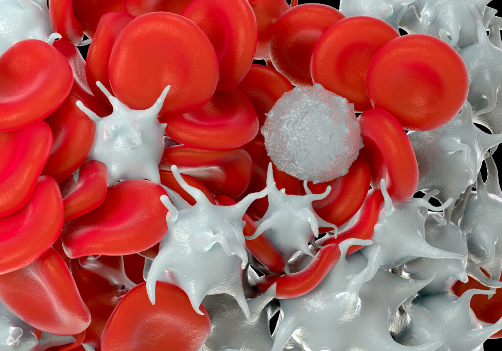
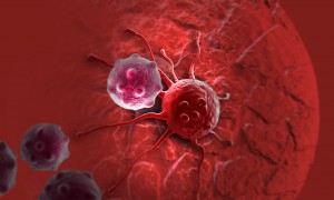
Most cancer-related deaths from solid tumors aren’t caused by the primary tumor itself, but are the result of metastasis. Metastasis is the process by which cancer cells break away from the primary tumor, travel through the bloodstream and establish new tumors in distant tissues. This is a complex, multi-step process and cancer researchers have spent decades trying to understand and disrupt the metastatic cascade. What is known: platelets and calcium play important roles in metastasis. What is unknown: How do these two factors interact?
Here, we explore a recent study published in Scientific Reports by researchers at George Washington University1. They investigate how calcium levels influence platelet-cancer cell interactions and what happens when both factors converge.
Kierkegaard observed that one of humanity’s enduring tensions is that while life can only be understood backwards, it must be lived forwards. It’s a truth medicine knows intimately: in the treatment that worked until it didn’t, the resistance that arrived without warning, the moment a doctor has to tell a patient that the drug that was helping has stopped. Not because anyone made a mistake, but because the critical knowledge that would have mattered arrived too late, if at all.
A recent paper from the National Cancer Institute is, in a small but meaningful way, science’s pursuit of that elusive foresight: an understanding that emerges early enough, for once, to change what happens next.
The Elegant Idea
For decades, chemotherapy has worked by brute force, flooding the body with toxins designed to kill rapidly dividing cells. The problem is that rapid division isn’t unique to cancer. Hair follicle cells, gut lining cells and immune cells also divide rapidly, which is why patients lose hair, lose energy and become susceptible to infection. Chemotherapy targets a behavior, but the drug has no way to tell a healthy cell from a cancerous one.
Antibody-drug conjugates (ADCs) change that. Instead of targeting what cancer cells do, they target what cancer cells are. Cancer cells tend to display certain proteins on their surface in far greater numbers than healthy cells do. The antibody is engineered to seek out those proteins specifically. It navigates to its target, binds and waits for the cell to do what cells routinely do: pull it inside. Once there, the cell’s own digestive machinery (the lysosome) breaks down the chemical tether holding the toxin to the antibody, releasing the toxin to kill the cell from within. More than a dozen ADCs have received FDA approval in recent years, and the field is evolving fast.
What the Cell Does Next
But cancer cells don’t simply accept their fate. Even when an ADC delivers its payload perfectly—the antibody finds its target, the cell pulls it inside, the lysosome cuts the tether—a pump embedded in the cell membrane can grab the released toxin and throw it back out before it causes damage.
The delivery worked. The package got ejected anyway.
These pumps—ATP-binding cassette transporters, or more plainly, efflux pumps—are a normal feature of cell biology. Their job is cellular housekeeping, clearing out unwanted or toxic substances before they cause damage. Under the pressure of drug treatment, cancer cells do what life has always done under pressure: the ones best equipped to survive do. The same mechanism that has shaped living things for billions of years now works against the treatment. Not all cancer cells are identical, and the ones that happen to produce more pumps survive while others don’t, gradually shifting the tumor toward resistance.
The National Cancer Institute’s NCI-60 drug screening panel, comprised of 60 diverse human cancer cell lines, has been a cornerstone in advancing cancer research and drug discovery since its inception in the late 1980s. Developed in response to the need for more predictive and comprehensive preclinical models, the NCI-60 facilitates the screening of thousands of compounds annually, aiming to identify potential anti-cancer drugs across a broad spectrum of human cancers. This article traces the origins, development, and evolution of the NCI-60 panel, highlighting its significant role in advancing our understanding of cancer and therapeutic agents.
At the time of writing this post, no scientist had yet discovered the secret to immortality. In our world, we’ve come to accept that living things are born, grow old and die—the circle of life.
And yet, for many years, life scientists believed that the circle of life did not apply to our constituent cells when cultured in a laboratory. That is, cultured normal human cells were immortal, and they would continue to grow and proliferate forever, as long as they were provided with the necessary nutrients.
Pioneering work published in 1961 by Leonard Hayflick and Paul Moorhead challenged that theory (reviewed in 1). Their research showed that normal cells in culture have a finite capacity to replicate. After they reach a certain number of replicative cycles, cells stop dividing. Hayflick and Moorhead made the important distinction between normal human cells and cultured cancer cells, which are truly immortal. In later years, the limit to the number of replicative cycles normal human cells can undergo became known as the Hayflick limit. Although some scientists still express skepticism about these findings, the Hayflick limit is widely recognized as a fundamental principle of cell biology.
In 1963, Jennifer Harvey was studying Moloney murine leukemia virus (MMLV) at the cancer research department of the London Hospital Research Laboratories. After routine transfers of plasma from MMLV-infected rats to mice, she made an unusual discovery. In addition to the expected leukemia, the mice that received the plasma developed solid tumors (soft-tissue sarcomas), primarily in the spleen (1). A few years later, Werner Kirsten at the University of Chicago observed similar results working with mouse erythroblastosis virus (MEV) (2).
Subsequent research, with the advent of genome sequencing, showed that a cellular rat gene had been incorporated into the viral genome in both cases (3). These genomic sequences contained a mutation later shown to be responsible for the development of sarcomas, and the word “oncogene” became a common part of the vocabulary in cancer publications during the early 1980s (4). Harvey’s discovery led to the naming of the corresponding rat sarcoma oncogene as HRAS, while Kirsten’s related oncogene was named KRAS. Several laboratories, working independently, cloned the human homolog of the viral HRAS gene in 1982 (3). The human KRAS gene was cloned shortly thereafter, as well as a third RAS gene, named NRAS (3). Additional studies showed that a single point mutation in each of these genes led to oncogenic activation, and they have been popular targets for anticancer drug discovery efforts ever since.
In April 2018, a series of 27 papers representing the most comprehensive genomic analysis of human cancers to date was published in Cell Press journals.
The collection constitutes the final outputs from the Cancer Genome Atlas (TCGA) project, a collaboration between the National Cancer Institute (NCI) and the National Human Genome Research Institute (NHGRI) involving analysis of over 11,000 tumors representing 33 different cancers. The many research teams involved analyzed tumor DNA, mRNA, miRNA and chromatin, comparing them to matched normal cellular genomes to perform a complete molecular characterization of cancer-specific changes. The results have been presented with much hope that open access to this type of comprehensive analysis will build on recent advances in understanding tumor biology and spur further progress in developing new approaches to treatment. (See this news item for more detail).
The Pan-Cancer Atlas results are collected on a cell.com portal, where they are presented in three collections grouped by topic: Cell of Origin, Oncogenic Processes and Signaling Pathways. Each collection is accompanied by a “Flagship” paper introducing the topic and summarizing the findings. It seems fitting that these findings have been published in #HumanGenomeMonth. This comprehensive analysis of the genomic and metagenomic profiles of tumors illustrates one powerful application of the type of genomic analysis pioneered by the original Human Genome Project, and shows just how much has been made possible since the initial publication of the human genome fifteen years ago.
At first glance, the biology of magnetic, underwater-dwelling, oxygen-averse bacteria may seem of little relevance to our most pressing human health problems. But science is full of surprises. A paper published in Nature Nanotechnology presents an inspired use of these bacteria to deliver anti-cancer drugs to tumors, specifically targeting the oxygen-starved regions generated by aggressively proliferating cells.
RNA molecules have become a hot topic of research. While I was taught about messenger RNA (mRNA), ribosomal RNA (rRNA) and transfer RNA (tRNA), many more varieties have come into the nomenclature after I graduated with my science degrees. Even more interesting, these RNAs do not code for a protein, but instead have a role in regulating gene expression. From long non-coding RNA (lncRNA) to short interfering RNA (siRNA), microRNA (miRNA) and small nucleolar RNA (snoRNA), these classes of RNAs affect protein translation, whether by hindering ribosomal binding, targeting mRNA for degradation or even modifying DNA (e.g., methylation). This post will cover the topic of microRNAs, explaining what they are, how researchers understand their function and role in metabolism, cancer and cardiovascular disease, and some of the challenges in miRNA research.
What are microRNAs? MicroRNAs (miRNAs) are short noncoding RNAs 19–25 nucleotides long that play a role in protein expression by regulating translation initiation and degrading mRNA. miRNAs are coded as genes in DNA and transcribed by RNA polymerase as a primary transcript (pri-miRNA) that is hundreds or thousands of nucleotides long. After processing with a double-stranded RNA-specific nuclease, a 70–100 nucleotide hairpin RNA precursor (pre-miRNA) is generated and transported from the nucleus into the cytoplasm. Once in the cytoplasm, the pre-miRNA is cleaved into an 18- to 24-nucleotide duplex by ribonuclease III (Dicer). This cleaved duplex associates with the RNA-induced silencing complex (RISC), and one strand of the miRNA duplex remains with RISC to become the mature miRNA.
Over the last few years, human microbiome studies have revealed fascinating connections between our colonizing microorganisms and ourselves—including associations between gut bacterial populations and obesity, disease susceptibility, and even mood. The relationship between us and our microbial colonists—once considered completely benign, is now being revealed as an intricate, complicated partnership with the potential to redefine who “we” are in fundamental ways.
Two papers published back-to-back in the November 27 issue of Science add further to this growing body of knowledge—reporting a new and unexpected connection between gut bacterial species and the effectiveness of cancer immunotherapies in mice. The work suggests one reason why such treatments are effective in some circumstances, but not others. Both papers report that the presence of specific bacterial populations may be required for the efficacy of certain treatments, and raise the intriguing question “Could the composition of bacteria in the gut be manipulated to enhance the effectiveness of cancer treatments?”
Every day scientists apply creative ideas to solve real-world problems. Every so often a paper comes up that highlights the creativity and elegance of this process in a powerful way. The paper “Programmable probiotics for detection of cancer in urine”, published May 27 in Science Translational Medicine, provides one great example of the application of scientific creativity to develop potential new ways for early detection of cancer.
The paper describes use of an engineered strain of E.coli to detect liver tumors in mice. The authors (Danino et al) developed a potential diagnostic assay that uses a simple oral delivery method and provides a readout from urine, all of which is made possible by some seriously complex and elegant science. Continue reading “Designer Bacteria Detect Cancer”
XWe use cookies and similar technologies to make our website work, run analytics, improve our website, and show you personalized content and advertising. Some of these cookies are essential for our website to work. For others, we won’t set them unless you accept them. To learn more about our approach to Privacy we invite you to Read More
By clicking “Accept All”, you consent to the use of ALL the cookies. However you may visit Cookie Settings to provide a controlled consent.
We use cookies and similar technologies to make our website work, run analytics, improve our website, and show you personalized content and advertising. Some of these cookies are essential for our website to work. For others, we won’t set them unless you accept them. To find out more about cookies and how to manage cookies, read our Cookie Policy.
If you are located in the EEA, the United Kingdom, or Switzerland, you can change your settings at any time by clicking Manage Cookie Consent in the footer of our website.
Necessary cookies are absolutely essential for the website to function properly. These cookies ensure basic functionalities and security features of the website, anonymously.
Cookie
Duration
Description
cookielawinfo-checbox-analytics
11 months
This cookie is set by GDPR Cookie Consent plugin. The cookie is used to store the user consent for the cookies in the category "Analytics".
cookielawinfo-checbox-functional
11 months
The cookie is set by GDPR cookie consent to record the user consent for the cookies in the category "Functional".
cookielawinfo-checbox-others
11 months
This cookie is set by GDPR Cookie Consent plugin. The cookie is used to store the user consent for the cookies in the category "Other.
cookielawinfo-checkbox-advertisement
1 year
The cookie is set by GDPR cookie consent to record the user consent for the cookies in the category "Advertisement".
cookielawinfo-checkbox-necessary
11 months
This cookie is set by GDPR Cookie Consent plugin. The cookies is used to store the user consent for the cookies in the category "Necessary".
cookielawinfo-checkbox-performance
11 months
This cookie is set by GDPR Cookie Consent plugin. The cookie is used to store the user consent for the cookies in the category "Performance".
gdpr_status
6 months 2 days
This cookie is set by the provider Media.net. This cookie is used to check the status whether the user has accepted the cookie consent box. It also helps in not showing the cookie consent box upon re-entry to the website.
lang
This cookie is used to store the language preferences of a user to serve up content in that stored language the next time user visit the website.
viewed_cookie_policy
11 months
The cookie is set by the GDPR Cookie Consent plugin and is used to store whether or not user has consented to the use of cookies. It does not store any personal data.
Analytical cookies are used to understand how visitors interact with the website. These cookies help provide information on metrics the number of visitors, bounce rate, traffic source, etc.
Cookie
Duration
Description
SC_ANALYTICS_GLOBAL_COOKIE
10 years
This cookie is associated with Sitecore content and personalization. This cookie is used to identify the repeat visit from a single user. Sitecore will send a persistent session cookie to the web client.
vuid
2 years
This domain of this cookie is owned by Vimeo. This cookie is used by vimeo to collect tracking information. It sets a unique ID to embed videos to the website.
WMF-Last-Access
1 month 18 hours 24 minutes
This cookie is used to calculate unique devices accessing the website.
_ga
2 years
This cookie is installed by Google Analytics. The cookie is used to calculate visitor, session, campaign data and keep track of site usage for the site's analytics report. The cookies store information anonymously and assign a randomly generated number to identify unique visitors.
_gid
1 day
This cookie is installed by Google Analytics. The cookie is used to store information of how visitors use a website and helps in creating an analytics report of how the website is doing. The data collected including the number visitors, the source where they have come from, and the pages visted in an anonymous form.
Advertisement cookies are used to provide visitors with relevant ads and marketing campaigns. These cookies track visitors across websites and collect information to provide customized ads.
Cookie
Duration
Description
IDE
1 year 24 days
Used by Google DoubleClick and stores information about how the user uses the website and any other advertisement before visiting the website. This is used to present users with ads that are relevant to them according to the user profile.
test_cookie
15 minutes
This cookie is set by doubleclick.net. The purpose of the cookie is to determine if the user's browser supports cookies.
VISITOR_INFO1_LIVE
5 months 27 days
This cookie is set by Youtube. Used to track the information of the embedded YouTube videos on a website.
Performance cookies are used to understand and analyze the key performance indexes of the website which helps in delivering a better user experience for the visitors.
Cookie
Duration
Description
YSC
session
This cookies is set by Youtube and is used to track the views of embedded videos.
_gat_UA-62336821-1
1 minute
This is a pattern type cookie set by Google Analytics, where the pattern element on the name contains the unique identity number of the account or website it relates to. It appears to be a variation of the _gat cookie which is used to limit the amount of data recorded by Google on high traffic volume websites.