Trends and Tools Transforming Drug Discovery: Five Takeaways from Discover Glo 2025

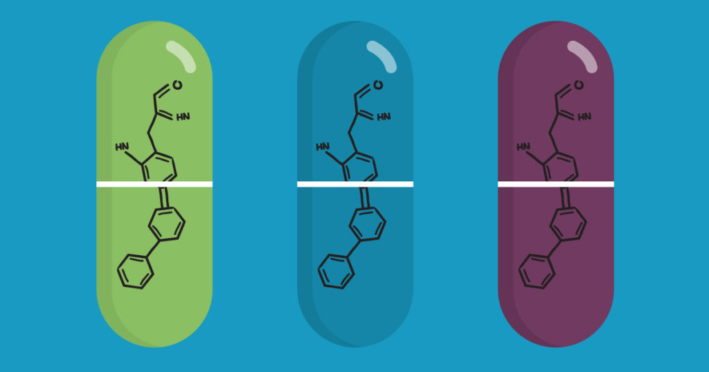
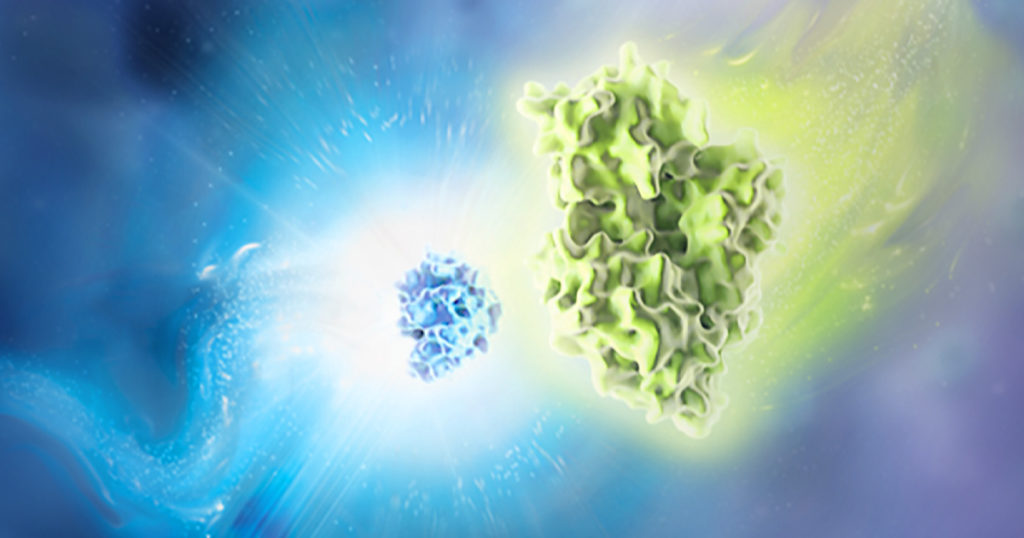
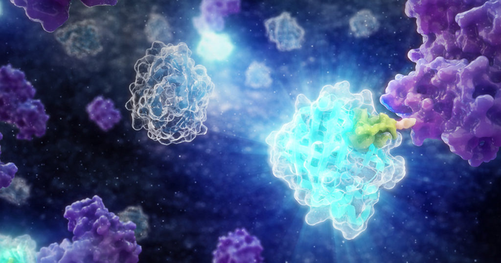
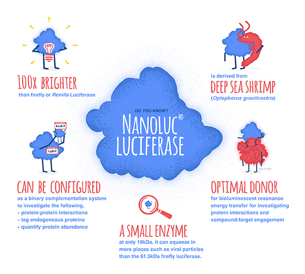
In biologics, cell therapy, and targeted protein degradation, the science is moving fast—and so are the tools. From GPCR-targeted therapies to real-time CAR-T manufacturing tools, new techniques are reshaping how scientists approach drug development, live-cell imaging, and protein degradation.

The “Bringing Light to Science” Discover Glo 2025 speaker series brought together researchers from across academia and industry to share real-world examples of how bioluminescent technologies are helping them advance their research. Now available on demand, these sessions offer fresh perspectives and actionable takeaways on the future of therapeutic development, cellular analysis and assay design.

We’ve distilled five key takeaways from the sessions—practical insights you can apply to your own work or use to stay current with where the field is heading.

Continue reading “Trends and Tools Transforming Drug Discovery: Five Takeaways from Discover Glo 2025”

Understanding Wnt Signaling Through β-Catenin Localization in Live Cells

The Wnt/β-catenin pathway, long studied in the context of developmental biology, has become increasingly recognized for its role in a wide range of human diseases. Its dysregulation has been implicated in cancer, fibrosis, immune modulation, and neurodegenerative conditions—making it a clinically actionable target across diverse therapeutic areas1. In this blog, we cover the fundamentals of Wnt/β-catenin signaling, highlight ongoing research efforts to understand its role in disease, and show how combining live-cell imaging with luminescent assays complements functional studies.

Continue reading “Understanding Wnt Signaling Through β-Catenin Localization in Live Cells”

Drug Repurposing Screens: Redeploying Old Dogs for New Tricks

This blog was written by guest author, Amy Landreman, PhD.

Drug repurposing, identifying new uses for approved or investigational drugs, is an attractive strategy when looking for new disease treatments. Because the compounds have already gone through some level of pre-clinical optimization and safety testing, this approach can reduce risk, reduce cost, and speed up the timeline for further drug development. An additional benefit of this approach is that it can result in new biological insights or a better understanding of disease mechanisms since these compounds usually already have some level of mechanistic characterization. Indeed, there are now a number of compound collections openly available specifically for the purpose of facilitating drug repurposing efforts. For example, the ReFRAME (Repurposing, Focused Rescue, and Accelerated Medchem) library is a collection of 12,000 compounds developed by Scripps Research Center and has been screened to identify novel candidate therapeutics for Cryptosporidium infection (1). The Broad Institute also offers a drug repurposing hub that contains an annotated collection of over 7,000 compounds.

Drug repurposing libraries, although often smaller than novel compound small molecule libraries, are designed for implementation into high-throughput screening workflows in order to efficiently triage compounds for the desired result. Effective compound screens require assays that can be scaled to 384 or 1536-well microplate formats and implemented in batch or continuous processing workflows. The firefly luciferase reaction has been leveraged to create many assays that are well-suited to these types of high-throughput screening approaches. In particular, the generation of “Glow” assays that have stable luminescent signals and homogenous assay design is a good fit. The signal stability allows for multi-plate processing and because the reagent is added directly to cells in culture, pre-processing steps are eliminated allowing for automated workflows. Assay reagents such as the CellTiter-Glo® Cell Viability Assay and the ADP-Glo™ Kinase Assay are commonly used in screening efforts including those done with repurposing libraries.  In addition, there are several firefly luciferase reporter assay reagents such as Steady-Glo® and Bright-Glo™ Luciferase Assays that have been optimized for high-throughput detection of firefly luciferase activity making them well-suited to repurposing screens.

Continue reading “Drug Repurposing Screens: Redeploying Old Dogs for New Tricks”

Bioluminescent Sharks Set the Sea Aglow

Many deep sea creatures are bioluminescent. However, before documenting the luminescence of the kitefin shark, Dalatias licha, there has never been a nearly six-foot long luminous vertebrate creature. In a recent study, Mallefet and colleagues examined three species of sharks: Dalatias licha, Etmopterous lucifer, and Emopterus granulosus and documented their luminescence for the first time. These bioluminescent sharks are the largest bioluminescent creatures known.

Researchers studied three species of bioluminescent sharks near the Chatham Islands, New Zealand
Coastline of one of the Chatham Islands, New Zealand
Continue reading “Bioluminescent Sharks Set the Sea Aglow”

Advancing Understanding of Hypoxic Gene Regulation Using Reporter Genes: Celebrating the Work of Dr. Gregg L. Semenza

This post is written by guest blogger, Amy Landreman, PhD, Sr. Product Manager at Promega Corporation.

Oxygen is necessary for animal life. It’s essential for cellular respiration and the production of energy (ATP) we require to survive. Given the need for oxygen, it isn’t surprising that our bodies have evolved ways to sense and adapt to decreased oxygen conditions (hypoxia). We can increase the production of new blood vessels by producing vascular endothelial growth factor (VEGF) or increase red blood cell (RBC) production by increasing the levels of eythropoietin (EPO), the hormone that plays a key role in the production of RBCs. But how does our body sense low oxygen, increase EPO levels, and kick our RBC production into gear? Nobel laureate Gregg L. Semenza has been honored for his contributions to our understanding of this process, and his research demonstrates the value of reporter genes and bioluminescence for studying gene regulation.

Reporter genes and bioluminescence are important tools for studying gene regulation
Continue reading “Advancing Understanding of Hypoxic Gene Regulation Using Reporter Genes: Celebrating the Work of Dr. Gregg L. Semenza”

A Closer Look at C. difficile Biology with Luminescent Tagging

Clostridium difficile is a bacterium that infects around half a million people per year in the United States. The infection causes symptoms ranging from diarrhea to severe colitis, and it’s one of the most common infections contracted while staying in the hospital. As the global incidence of C. diff infection has risen over the past decade, so has the pressure to develop novel therapeutic strategies. This requires a thorough exploration of all aspects of C. difficile biology.

Two recent papers published by researchers at the University of Leiden have shed light on C. difficile physiology using HiBiT protein tagging. The HiBiT system allows detection of proteins in live cells using an 11 amino acid tag. The HiBiT tag binds to the complementary LgBiT polypeptide to reconstitute the luminescent NanoBiT® enzyme. The resulting luminescence is proportional to the amount of HiBiT-tagged protein that is present.

Continue reading “A Closer Look at C. difficile Biology with Luminescent Tagging”

NanoLuc® Luciferase: Brighter Days Ahead for In Vivo Imaging

nanoluc in vivo imaging

The development of NanoLuc® luciferase technology has provided researchers with new and better tools to study endogenous biology: how proteins behave in their native environments within cells and tissues. This small (~19kDa) luciferase enzyme, derived from the deep-sea shrimp Oplophorus gracilirostris, offers several advantages over firefly or Renilla luciferase. For an overview of NanoLuc® luciferase applications, see: NanoLuc® Luciferase Powers More than Reporter Assays.

The small size of NanoLuc® luciferase, as well the lack of a requirement for ATP to generate a bioluminescent signal, make it particularly attractive as a reporter for in vivo bioluminescent imaging, both in cells and live animals. Expression of a small reporter molecule as a fusion protein is less likely to interfere with the biological function of the target protein. NanoLuc® Binary Technology (NanoBiT®) takes this approach a step further by creating a complementation reporter system where one subunit is just 11 amino acids in length. This video explains how the high-affinity version of NanoBiT® complementation (HiBiT) works:

Continue reading “NanoLuc® Luciferase: Brighter Days Ahead for In Vivo Imaging”

NanoLuc: Tiny Tag with a Big Impact

Synthetic biology—genetically engineering an organism to do or make something useful—is the central goal of the iGEM competition each year. After teams conquer the challenge of cloning their gene, the next hurdle is demonstrating that the engineered gene is expressing the desired protein (and possibly quantifying the level of expression), which they may do using a reporter gene.

Reporters can also play a more significant role in iGEM projects when teams design their organism with reporter genes to detect and signal the presence of specific molecules, like environmental toxins or biomarkers. Three of the iGEM teams Promega sponsored this year opted to incorporate some version of NanoLuc® Luciferase into their projects.

NanoLuc® luciferase is a small monomeric enzyme (19.1kDa, 171 amino acids) based on the luciferase from the deep sea shrimp Oplophorus gracilirostris. This engineered enzyme uses a novel substrate, furimazine, to produce high-intensity, glow-type luminescence in an ATP-independent reaction. Unlike other molecules for tagging and detecting proteins, NanoLuc® luciferase is less likely to interfere with enzyme activity and affect protein production due to its small size.

NanoLuc® Luciferase has also been engineered into a structural complementation reporter system, NanoBiT® Luciferase, that contains a Large subunit (LgBiT) and two small subunit options: low affinity SmBiT and high affinity HiBiT. Together, these NanoLuc® technologies provide a bioluminescent toolbox that was used by the iGEM teams to address a diverse set of biological challenges.

Here is an overview of each team’s project and how they incorporated NanoLuc® technology.

Continue reading “NanoLuc: Tiny Tag with a Big Impact”

WiSciFest 2019: A Retrospective

This past weekend was the 9th Annual Wisconsin Science Festival, and we at Promega were excited to join in the celebration of science throughout the state. We participated in the Discovery Expo on Thursday and Friday, where dozens of demonstrations and exhibits were scattered throughout the Wisconsin Institute for Discovery building. Thousands of children on field trips filled the halls, eager to poke and prod at strange and exciting new things.

At our table, we talked about the science of bioluminescence. With 3D-printed firefly luciferase models in hand, we showed the glow of recombinant luciferase to the incoming children and explained to them how scientists could use bioluminescence like a tiny “flashlight” to look inside of cells and watch what’s happening. Our learners received a nice little reward for their attentiveness in the form of glow-in-the-dark firefly stickers.

Thank you to the tablecloth for providing the darkness needed to grab this pic.
Continue reading “WiSciFest 2019: A Retrospective”

Striking Fear into the Heart of Cardiovascular Disease Using Zebrafish and NanoLuc® Luciferase

Representative images of ApoB-LP localization in zebrafish across developmental, genetic, pharmacological and dietary manipulations.
Credit: Figure 5.D of The LipoGlo reporter system for sensitive and specific monitoring of atherogenic lipoproteins by James Thierer, Stephen C. Ekker and Steven A. Farber.
Article licensed under Creative Commons Attribution 4.0 International License.

Cardiovascular diseases, or CVDs, are collectively the most notorious gang of cold-blooded killers threatening human lives today. These unforgiving villains, including the likes of coronary heart disease, cerebrovascular disease and pulmonary embolisms, are jointly responsible for more deaths per year than any other source, securing their seat as the number one cause of human mortality on a global scale.

One of the trademarks of most CVDs is the thickening and stiffening of the arteries, a condition known as atherosclerosis. Atherosclerosis is characterized by the accumulation of cholesterol, fats and other substances, which together form plaques in and on the artery walls. These plaques clog or narrow your arteries until they completely block the flow of blood, and can no longer supply sufficient blood to your tissues and organs. Or the plaques can burst, setting off a disastrous chain reaction that begins with a blood clot, and often ends with a heart attack or stroke.

Given the global prevalence and magnitude of this problem, there is a significant and urgent demand for better ways to treat CVDs. In a recent study published in Nature Communications, researchers at the Carnegie Institution for Science, Johns Hopkins University and Mayo Clinic are taking the fight to CVDs through the study of low-density lipoproteins (LDLs), the particles responsible for shuttling bad cholesterol throughout the bloodstream.

Continue reading “Striking Fear into the Heart of Cardiovascular Disease Using Zebrafish and NanoLuc® Luciferase”