Protein:DNA Interactions—High-Throughput Analysis

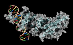
Protein-DNA interactions are fundamental processes in gene regulation in a living cells. These interactions affect a wide variety of cellular processes including DNA replication, repair, and recombination. In vivo methods such as chromatin immunoprecipitation (1) and in vitro electrophoretic mobility shift assays (2) have been used for several years in the characterization of protein-DNA interactions. However, these methods lack the throughput required for answering genome-wide questions and do not measure absolute binding affinities. To address these issues a recent publication (3) presented a high-throughput micro fluidic platform for Quantitative Protein Interaction with DNA (QPID). QPID is an microfluidic-based assay that cam perform up to 4096 parallel measurements on a single device.

The basic elements of each experiment includes oligonucleotides that were synthesized and hybridized to a Cy5-labeled primer and extended using Klenow. All transcription factors that were evaluated contained a 3’HIS and 5’ cMyc tag and were expressed in rabbit reticulocyte coupled transcription and translation reaction (TNT® Coupled Reticulocyte Lysate). Expressed proteins are loaded onto to the QIPD device and immobilized. In the DNA binding assay the fluorescent DNA oligonucleotides are incubated with the immobilized transcription factors and fluorescent images taken. To validate this concept the binding of four different transcription factor complexes to 32 oligonucleotides at 32 different concentrations was characterized in a single experiment. In a second application, the binding of ATF1 and ATF3 to 128 different DNA sequences at different concentrations were analyzed on a single device.

Literature Cited

  1. Ren, B. et al. (2007) Genome-wide mapping of in vivo protein-DNA binding proteins. Science 316, 1497–502.
  2. Garner, M.M. (1981) A gel electrophoresis method for quantifying the binding of proteins to specific DNA regions. Nuc. Acids. Res. 9, 3047-60.
  3. Glick,Y et al. (2016) Integrated microfluidic approach for quantitative high throughput measurements of transcription factor binding affinities. Nuc. Acid Res. 44, e51.

Catching a Child Abuser in Five Days

ishi_27jpg

Next week, forensic analysts from all over the world will gather in Minneapolis for the 27th  International Symposium on Human Identification (ISHI). So today, we’d like to share one story from a forensics lab that highlights the importance of collaboration, knowledge-sharing and technology development–since that is what ISHI is all about. 

Crimes against children are especially heinous, and it is vitally important that the offenders are removed from the streets as soon as possible. In today’s blog, Sarah Chenoweth from the Anne Arundel County Crime Lab in Maryland describes a sexual assault case that was solved in just five days. Key to this speed were the collaboration between lab staff and state police, and the ability to quickly and reliably amplify DNA profiles from low-DNA samples. Thanks to the efforts of the investigators involved, parents in Maryland, and possibly nationwide, can sleep a little easier.

On Friday, February 5th, the Anne Arundel County Crime Lab was notified of a sex offense involving a 7-year-old victim. With our efficient DNA workflow, including use of the Fusion amplification kit with our 3500 Genetic Analyzer, we were able to identify the perpetrator in only five days. Continue reading “Catching a Child Abuser in Five Days”

The Cell Line Identity Crisis

29981164-Whistle

If you work with cell lines you may have paid attention to the dramatic headline published last month in the online journal STAT, Thousands of studies used the wrong cells, and journals are doing nothing.” In their column The Watchdogs (“Keeping an eye on misconduct, fraud, and scientific integrity”), Ivan Oransky and Adam Marcus call out the fact that scientists continue to publish research using cell lines that are contaminated or misidentified. Recent estimates have found that the percentage of misidentified cell lines used by scientists is as high as 20 to 36. The blame here is being placed on the peer reviewed journals for not blowing the whistle. The authors call for journals to put some “kind of disclaimer on the thousands of studies affected.”

This is not a new claim. The continuing problem of cell line misidentification, of lack of authentication, has been covered before in various channels. It’s easy to find news publicizing yet another retracted publication. In May 2015 the journal Nature required authors of all submitted manuscripts to confirm the identity of cell lines used in their studies and provide details about the source and testing of their cell lines.

Continue reading “The Cell Line Identity Crisis”

Digging Up More Clues in the History of the Black Death

Bubonic plague victims in a mass grave in 18th century France. By S. Tzortzis [Public domain], via Wikimedia Commons
My last blog post on the Black Death highlighted research that suggested that the reintroduction of Yersinia pestis, the causative agent of the pandemic, originated in Europe during the 14–18th centuries rather than from Asia, the hypothesized origin. In my post, I wrote about my curiosity regarding what an Asian skeleton positive for Y. pestis from that same time period would reveal about the strain or strains that were circulating. Well, a team of researchers has been exploring the issue of strain circulation and an Asian connection, and recently published what they gleaned from additional historic Y. pestis samples in Cell Host & Microbe.

Teeth from 178 individuals in three different locations (two European, one Asian) were screened for Y. pestis infection using the plasminogen activator (pla) gene. Continue reading “Digging Up More Clues in the History of the Black Death”

Moving Out of the Cell: Advantages of Cell-Free Protein Expression

Cell-free protein expression is a simplified and accelerated avenue for the transcription and/or translation of a specific protein in a quasi cell environment. An alternative to slower, more cumbersome cell-based methods, cell-free protein expression methods are simple and fast and can overcome toxicity and solubility issues sometimes experienced in the traditional E. coli expression systems.

Cell-free protein expression offers significant time savings over cell-based expression methods.

Cell-free protein expression offers a convenient method for generating small amounts of protein for a variety of applications (e.g., protein:protein interactions, protein: nucleic acid interactions, structural analysis, functional assays and toxicity screening). This approach lends itself to specific protein labeling with fluorescence, biotin, radioactivity or heavy atoms, via modified charged tRNA’s or amino acids. Cell-free protein expression systems provide quick access to proteins of interest and remain a staple in the collection of tools available for the elucidation of protein structure and function, understanding cellular pathways and mechanisms and high-throughput screening of compounds for drug discovery. There are a number of different cell-free expressions systems, each with different strengths. Deciding which one is right for you depends upon your research needs and goals.

Continue reading “Moving Out of the Cell: Advantages of Cell-Free Protein Expression”

Magnetic Bacteria Carry Drugs into Tumors

cancer cell

At first glance, the biology of magnetic, underwater-dwelling, oxygen-averse bacteria may seem of little relevance to our most pressing human health problems. But science is full of surprises. A paper published in Nature Nanotechnology presents an inspired use of these bacteria to deliver anti-cancer drugs to tumors, specifically targeting the oxygen-starved regions generated by aggressively proliferating cells.

Continue reading “Magnetic Bacteria Carry Drugs into Tumors”

An Epizootic for the Ages: Revisiting the White-Nose Syndrome Story

Map showing the spread of WNS across North America

In March 2016, two hikers on a trail east of Seattle, WA, found a little brown bat lying on the ground in obviously poor condition. The bat was taken to an animal shelter where it died two days later from White-Nose Syndrome (WNS).

This bat was the first case of WNS found west of the Rocky Mountains. It represented a jump in the spread of WNS, and a troubling one. WNS was first detected in a cave in Albany, New York, and since then it has been moving slowly westward at a rate of about 200 miles per year, according to David Blehert of the United States Geological Survey, the laboratory that confirmed the WNS diagnosis for the Washington bat. Before this year’s discovery outside of Seattle, the westward-most case detected was in eastern Nebraska.

WNS, caused by a cold-loving fungus, Psuedogymnoascus destructans (Pd), can kill 100% of the hibernating bats in a colony, and in the ten years since it has been detected and monitored has killed over 6 million bats in the United States and Canada. As of July 2016, bats infected with the fungus have been found in 29 states and 5 Canadian provinces.

According to Blehert, this is probably the “most significant epizootic of wildlife” ever observed; never before have we seen hibernating mammals specifically affected by a skin fungus. What does that mean? Are we looking at extinction for some bat species? What are the ecological consequences of rapidly losing so many individuals to disease so quickly? And, what, if anything, can be done to combat the disease and help bat populations recover?

Continue reading “An Epizootic for the Ages: Revisiting the White-Nose Syndrome Story”

Easy Automated Genomic DNA Isolation for GMO Testing: From Vision to Reality

29980616-July25-PureFood---Kelly-600x300

The European Union (EU) has a zero tolerance policy for products containing any material from non-authorized genetically modified (GM) crops. Seed entering EU markets may not contain even trace amounts of non-authorized genetically modified material. In 2012, as the global use of GM crops increased, seed testing loads in the EU continued to build. Isolating genomic DNA (gDNA) using traditional manual methods was becoming impractical in the face of increasing amounts of material that required testing. There was a growing need for an automated method to isolate gDNA from seed samples. Working to address this need, a group of scientists from the Bavarian Health and Food Safety Authority collaborated with scientists from Promega Corporation to evaluate the Maxwell® 16 Instrument and the associated chemistry as possible a solution for the testing labs.

Continue reading “Easy Automated Genomic DNA Isolation for GMO Testing: From Vision to Reality”

Optimization of Alternative Proteases for Bottom-Up Proteomics

Alternate Proteases Cover

Bottom-up proteomics focuses on the analysis of protein mixtures after enzymatic digestion of the proteins into peptides. The resulting complex mixture of peptides is analyzed by reverse-phase liquid chromatography (RP-LC) coupled to tandem mass spectrometry (MS/MS). Identification of peptides and subsequently proteins is completed by matching peptide fragment ion spectra to theoretical spectra generated from protein databases.

Trypsin has become the gold standard for protein digestion to peptides for shotgun proteomics. Trypsin is a serine protease. It cleaves proteins into peptides with an average size of 700-1500 daltons, which is in the ideal range for MS (1). It is highly specific, cutting at the carboxyl side of arginine and lysine residues. The C-terminal arginine and lysine peptides are charged, making them detectable by MS. Trypsin is highly active and tolerant of many additives.

Even with these technical features, the use of trypsin in bottom-up proteomics may impose certain limits in the ability to grasp the full proteome, Tightly-folded proteins can resist trypsin digestion. Post-translational modifications (PTMs) present a different challenge for trypsin because glycans often limit trypsin access to cleavage sites, and acetylation makes lysine and arginine residues resistant to trypsin digestion.

Are you looking for proteases to use in your research?
Explore our portfolio of proteases today.

To overcome these problems, the proteomics community has begun to explore alternative proteases to complement trypsin. However, protocols, as well as expected results generated when using these alternative proteases have not been systematically documented.

In a recent reference (2), optimized protocols for six alternative proteases that have already shown promise in their applicability in proteomics, namely chymotrypsin, Lys-C, Lys-N, Asp-N, Glu-C and Arg-C have been created.

Data describe the appropriate MS data analysis methods and the anticipated results in the case of the analysis of a single protein (BSA) and a more complex cellular lysate (Escherichia coli). The digestion protocol presented here is convenient and robust and can be completed in approximately in 2 days.


Try a sample of high-efficiency Trypsin Platinum today!

Visit our website for more on Trypsin Platinum, Mass Spectrometry Grade, with enhanced proteolytic efficiency and superior autoproteolytic resistance.


Reference

  1. Laskay, U. et al. (2013) Proteome Digestion Specificity Analysis for the Rational Design of Extended Bottom-up and middle-down proteomics experiments. J of Proteome Res. 12, 5558–69.
  2. Giansanti, P. et. al. (2016) Six alternative protease for mass spectrometry based proteomics beyond trypsin. Nat. Protocols 11, 993–6

Related Posts

Calling All Red Sharpies—A Lighthearted Commentary on Lab Supply Shortages

Every lab has it—that one thing that is always in short supply no matter how many you order. What is your lab’s “red sharpie”?

eh_July2016_800px

If you like this cartoon, there are more like it in our Cartoon Lab.