Inflammasomes and Pyroptosis

In today’s post, guest blogger,  Martha O’Brien, PhD, provides a preview of her upcoming AAI poster and block symposium talk on the inflammasome, caspase-1 activity and pyroptosis.

Schematic of the Caspase-Glo 1 Inflammasome Assay.

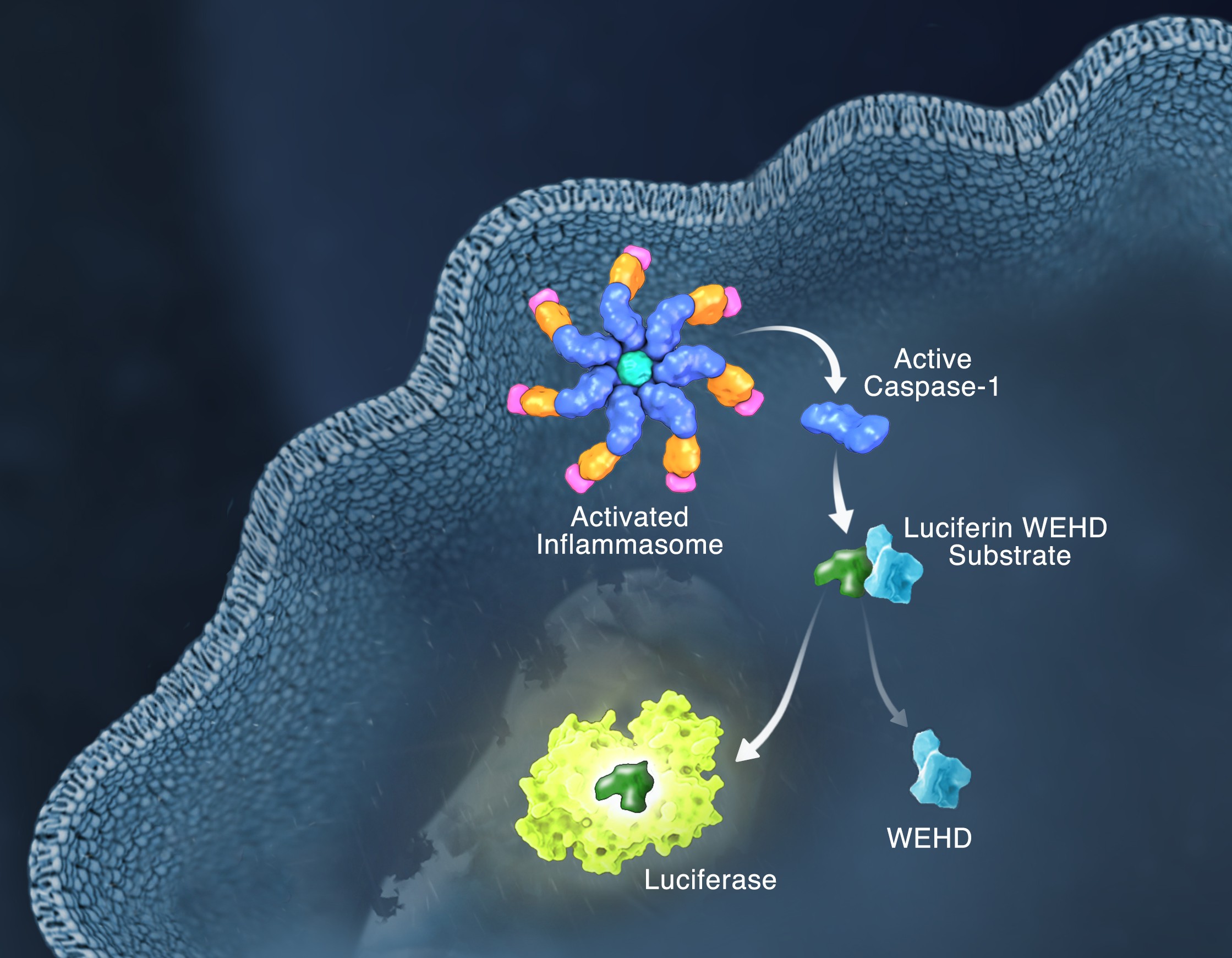
Responding rapidly to microbial pathogens and damage-associated molecular markers is critical to our innate immune system. Caspase-1 is pivotal in this process leading to processing and release of essential cytokines and an immunogenic form of cell death, termed pyroptosis. Upon sensing pathogen-associated and damage-associated molecular patterns (PAMPs and DAMPs), innate immune cells form inflammasome protein complexes that recruit and activate caspase-1 (canonical inflammasomes). In addition, other inflammatory caspases, 4 and 5 in humans and 11 in mice, directly bind bacterial lipopolysaccharides (LPS), triggering pyroptosis (non-canonical inflammasome). LPS-triggered non-canonical inflammasomes in mice and humans ultimately lead to canonical inflammasome engagement and caspase-1 activation (1–3).  Caspase-1 was originally termed interleukin converting enzyme (ICE) for its well-established role in processing IL-1ß and IL-18, two important inflammation cytokines. How caspase-1 mediates pyroptosis is less well understood, but is beginning to be delineated. Recently, a substrate of the inflammatory caspases, gasdermin D, was identified and its processed fragment, gasdermin-N domain, was shown to be required for pyroptosis in non-canonical inflammasome circumstances (4, 5). The precise role of gasdermin D in canonical inflammasome-triggered pyroptosis is still under investigation. Linking inflammatory caspases directly to pyroptosis is a notable step in understanding the mechanism of this important form of cell death.

Pyroptosis is clearly one means of releasing processed IL-1ß and IL-18 from the cell. However depending on the cell type and stimulus, there is evidence for inflammasome engagement, caspase-1 activation, and release of IL-1ß in the absence of cell death (6, 7). On the flip-side there is also evidence for caspase-1 mediated pyroptosis that helps clear bacteria, independent of IL-1ß and IL-18 involvement (8). To enable further studies on the inflammasome and in particular, assessing the connections between caspase-1 activation, pyroptosis, and cytokine release, Promega developed a new tool to conveniently monitor caspase-1 activation, the Caspase-Glo® 1 Inflammasome Assay.  This bioluminescent, plate-based assay is used to measure caspase-1 activity directly in cell cultures or to monitor released caspase-1 activity in culture medium from treated cells. This flexibility allows easy multiplexing to monitor all three outcomes of inflammasome stimulation; caspase-1 activity, pyroptosis, and release of IL-1ß and IL-18. Caspase-1 activation typically is monitored indirectly with western blots of processed caspase-1. Now the activity of the enzyme can be monitored directly, providing accurate information on temporal aspects of the inflammasome. The assay can be readily combined with real-time measures of cell death (e.g., CellTox™ Green Cytotoxicity Assay) and some of the culture medium can be removed for IL-1ß/IL-18 assessment, leaving the cells and remaining culture medium for caspase-1 activity measurements.

References

  1. Schmid-Burgk et al. (2015) Caspase-4 mediates non-canonical activation of the NLRP3 inflammasome in human myeloid cells. J. Immunol. 45, 2911–7.
  2. Baker et al. (2015) NLRP3 inflammasome activation downstream of cytoplasmic LPS recognition by both caspase-4 and caspase-5. J. Immunol. 45, 2918–26.
  3. Ruhl, S. and P. Broz (2015) Caspase-11 activates a canonical NLRP3 inflammasome by promoting K+ Eur. J. Immunol. 45, 2927–36.
  4. Shi et al. (2015) Cleavage of GSDMD by inflammatory caspases determines pyroptotic cell death. Nature 526, 660–5.
  5. Kayagaki et al. (2015) Caspase-11 cleaves gasdermin D for non-canonical inflammasome signaling. Nature 526, 666–71.
  6. Gaidt et al. (2016) Human monocytes engage an alternative inflammasome pathway. Immunity 44, 833–46.
  7. Chen et al. (2014) The neutrophil NLRC4 inflammasome selectively promotes IL-1ß maturation without pyroptosis during acuteSalmonella Cell Reports 8, 570–82.
  8. Miao et al. (2010) Caspase-1-induced pyroptosis is an innate immune effector mechanism against intracellular bacteria. Nature Immunology 11, 1136–42.

Familial DNA Searching for Criminal Forensics: Q&A

When DNA evidence is collected at a crime scene, submitting the sample for a search within a DNA database does not always identify a profile match. There is a way to extend that search and generate leads, called familial searching (FS). FS is used to identify close biological relatives of an unidentified DNA profile obtained as evidence. The basic premise is that DNA profiles of immediate family members, such as siblings, parents, or children, are likely to have more alleles in common than unrelated individuals. These familial profile matches can generate new investigative leads for law enforcement.

Currently, a few states are using FS under their state database laws, although none explicitly permit FS. Many agencies have yet to adopt policies related to FS, even though it has been found to be as effective as CODIS for identifying sources of evidence. The absence of clear ethical guidelines and policy regarding how to properly utilize FS prevents many local and state jurisdictions from adopting this investigational tool.

In order to address concerns and existing policies related to FS and to guide policy decisions by agencies implementing FS, the National Institute of Justice (NIJ) issued the report Familial DNA Searching: Current Approaches in January 2015. The goal of the report was to provide information to policy makers, law enforcement officials, forensic laboratory practitioners, and legal professionals about how FS is being applied within the criminal justice realm.

Mr. Rock Harmon, former prosecutor
Mr. Rockne Harmon, former prosecutor

Answers to the following questions about FS were provided by Mr. Rockne Harmon, a retired former prosecutor and member of the team that produced the report for the National Institute of Justice.

What is familial DNA searching?

Familial searching (FS) is an additional search of a DNA profile in a law enforcement DNA database that is conducted after a routine search fails to identify any profile matches. The FS process attempts to provide investigative leads to agencies engaged in the pursuit of justice by identifying a close biological relative of the source of the unknown forensic profile obtained from crime scene evidence.

Continue reading “Familial DNA Searching for Criminal Forensics: Q&A”

Characterizing Unique Protein: DNA Interactions Using Cell-Free Protein Expression

Molecular model of human telomere DNA

The POT1 protein plays a critical role in telomere protection and telomerase regulation. POT1 binds single-stranded 5′-TTAGGGTTAG-3′ and forms a dimer with the TPP1 protein. Human POT1 contains two Oligonucleotide/Oligosaccharide Binding (OB) fold domains, OB1 and OB2, which make physical contact with the DNA. OB1 recognizes 5′-TTAGGG whereas OB2 binds to the downstream TTAG-3′ (1,2). Several recent studies from other species have shown that some of these proteins are able to recognize a broader variety of DNA ligands than expected (3). A recent reference reexamined the sequence-specificity of the Human POT1 protein (4).
SELEX (Systematic Evolution of Ligands through Exponential Enrichment) was used  to re-examine the DNA-binding specificity of human POT1 (5).

Continue reading “Characterizing Unique Protein: DNA Interactions Using Cell-Free Protein Expression”

Five Interview Responses Recruiters Can See Right Through

20708189_lRecruiters aren’t pessimists, but throughout the years we have become more cautious and maybe a little suspicious. Many of us interviewed enough candidates that we have come to approach each new person with a “trust but verify” mentality. I’m very trusting in my personal life, but at work, my job is to be a detective. I follow clues to dig up the good, the bad, and the ugly.

During a recent talk with fellow recruiters, we realized there are some things many candidates say that perk up our sleuth ears every single time. These answers may be coming from a truthful and benign place, but they raise suspicions in any good recruiter. The average candidate has no idea what other candidates are saying, so I’m here to share. Recruiters hear these answers often and, take it from us, you’ll come off better in your interview if you avoid them. Continue reading “Five Interview Responses Recruiters Can See Right Through”

Finding a Connection Between Glucose Metabolism and Macrophage Activation

Introduction to Glucose Metabolism

Macrophages. By NIAID ) [CC BY 2.0 , via Wikimedia Commons

Many think of glucose as something diabetics have to test each day using a blood monitor, or a quick energy boost for someone exercising intensely. However, the simple sugar glucose, a monosaccharide, fuels most of the cells in our bodies. Disaccharides that contain glucose (e.g., sucrose is comprised of glucose and fructose) and glucose polymers (e.g., starch and glycogen) are carbohydrates that are consumed by organisms from bacteria to humans to produce energy. These carbohydrates are broken down into component monosaccharides like glucose and lactose. The process of glycolysis generates the energy currency of cells, ATP, as well as precursor molecules for nucleotides, lipids and amino acids. Because glucose is the cell fuel source, the uptake of glucose and its subsequent metabolism is increased by cells that divide rapidly like cancer cells. The more energy and precursor molecules the cancer cell can create for itself, the more rapidly the tumor can grow.

Because glucose metabolism is central to cellular functioning, changes that decrease glucose uptake or increase glycolysis have a widespread effect on on both the cells and organism. How does a simple sugar molecule create such broad effects on health? For example, diabetes results from the inability to store glucose because of a lack of insulin, a hormone that draws glucose from the blood and stores it as glycogen in the liver, muscles and adipose tissue. High levels of sugar in the blood negatively affect the body over the long term, damaging blood vessels and eyesight, making the kidneys work harder to excrete the excess sugar and increasing the risk of stroke and coronary artery disease. Because cancer cells have such a high metabolic demand for glucose, many of the mutations in cancers affect pathways that regulate glucose uptake and glucose breakdown, allowing the cancer cells to survive and grow, crowding out nearby normal cells.

Glucose metabolism is altered by processes other than mutations or an reduced production of a hormone. Throughout its life cycle, a cell will vary its requirements for glucose. For example, the cells that comprise our innate immune response are typically in a quiescent or steady state. However, when these immune cells encounter an foreign invader, they become activated and increase their demand for glucose. To respond to a potential pathogen, the activated cells need glucose to fuel cell proliferation and the production of cytokines, chemicals that activate other immune cells and initiate an inflammatory response. The typical signs of inflammation are red inflamed area that may be painful to the touch, such as a cut that becomes infected. Most inflammation resolves when the infection is eliminated, leaving behind whole skin in the instance of a cut, and the activated immune cells become quiescent again.

An Interesting Observation about Glucose Metabolism in M2 Macrophages

Glucose uptake, immunity and metabolism are cellular pathways that are intertwined such that understanding how glucose is utilized in macrophages illuminates gene induction and regulation in activated macrophages. In a recently published eLife article, Covarrubias et al. studied how activation of murine bone marrow-derived macrophages (BMDMs) by interleukin-4 (IL-4), a signaling cytokine, altered glucose metabolism in the cells and regulated a subset of genes involved in macrophage activation.

Continue reading “Finding a Connection Between Glucose Metabolism and Macrophage Activation”

Vitamin D: Power in Cancer Prevention?

This and vitamin D should get your attention.

Have you ever noticed that after a good long day outdoors, maybe hiking, at the beach or even working in the yard, you feel really strong and healthy, maybe even more relaxed than after an indoor session in front of the telly or computer? Maybe a February trip to someplace sunny like Mexico or the Canary Islands has given you renewed zest for your normal tasks?

While rest and a change of scenery is never a bad thing, time outdoors and in the sunshine might have gained for you something more than rest and relaxation. If it included a little UVB irradiation, your time outdoors may have increased your serum vitamin D level. And though it’s been presumed for years, we now have proof that higher serum vitamin D3 levels correlate with a decreased incidence of certain cancers. Continue reading “Vitamin D: Power in Cancer Prevention?”

For Protein Complementation Assays, Design is Everything

Most, if not all, processes within a cell involve protein-protein interactions, and researchers are always looking for better tools to investigate and monitor these interactions. One such tool is the protein complementation assay (PCA). PCAs use  a reporter, like a luciferase or fluorescent protein, separated into two parts (A and B) that form an active reporter (AB) when brought together. Each part of the split reporter is attached to one of a pair of proteins (X and Y) forming X-A and Y-B. If X and Y interact, A and B are brought together to form the active enzyme (AB), creating a luminescent or fluorescent signal that can be measured. The readout from the PCA assay can help identify conditions or factors that drive the interaction together or apart.

A key consideration when splitting a reporter is to find a site that will allow the two parts to reform into an active enzyme, but not be so strongly attracted to each other that they self-associate and cause a signal, even in the absence of interaction between the primary proteins X and Y. This blog will briefly describe how NanoLuc® Luciferase was separated into large and small fragments (LgBiT and SmBiT) that were individually optimized to create the NanoBiT® Assay and show how the design assists in monitoring protein-protein interactions.

Continue reading “For Protein Complementation Assays, Design is Everything”

Increasing Drug Research and Development Efficiency Using a 4-point Screening Method to Determine Molecular Mechanism of Action

Fig 4. Four point MMOA screen for tideglusib and GW8510. Time dependent inhibition was evaluated by preincubation of TbGSK3β with 60 nM tideglusib and 6 nM GW-8510 with 10μM and 100μM ATP. (A). Tideglusib [60 nM] in 10μM ATP. (B). GW8510 [60 nM] in 10μM ATP. (C.) Tideglusib [60 nM] at 100μM ATP. (D.) GW8510 [60 nM] at 100μM ATP. All reactions preincubated or not preincubated with TbGSK3β for 30 min at room temperature. Experiments run with 10μM GSM peptide, 10μM ATP, and buffer. Minute preincubation (30 min) was preincubated with inhibitor, TbGSK3β, GSM peptide, and buffer. ATP was mixed to initiate reaction. No preincubation contained inhibitor, GSM peptide, ATP, and buffer. The reaction was initiated with TbGSK3β. Reactions were run at room temperature for 5 min and stopped at 80°C. ADP formed was measured by ADP-Glo kit. Values are mean +/- standard error. N = 3 for each experiment and experiments were run in duplicates. Control reactions contained DMSO and background was determined using a zero time incubation and subtracted from all reactions. Black = 30 min preincubation Grey = No preincubation.
Four point MMOA screen for tideglusib and GW8510.
Time dependent inhibition was evaluated by preincubation of TbGSK3β with 60 nM tideglusib and 6 nM GW-8510 with 10μM and 100μM ATP. (A). Tideglusib [60 nM] in 10μM ATP. (B). GW8510 [60 nM] in 10μM ATP. (C.) Tideglusib [60 nM] at 100μM ATP. (D.) GW8510 [60 nM] at 100μM ATP. All reactions preincubated or not preincubated with TbGSK3β for 30 min at room temperature.  Black = 30 min preincubation Grey = No preincubation.
The first small-molecule kinase inhibitor approved as a cancer therapeutic, imatinib mesylate (Gleevec® treatment), has been amazingly successful. However, a thorough understanding of its molecular mechanism of action (MMOA) was not truly obtained until more than ten years after the molecule had been identified.

Understanding the MMOA for a small-molecule inhibitor can play a major role in optimizing a drug’s development. The way a drug actually works–the kinetics of binding to the target molecule and how it competes with endogenous substrates of that target–ultimately determines whether or not a a candidate therapeutic can be useful in the clinic. Drugs that fail late in development are extremely costly.

Drug research and discovery for neglected tropical diseases suffer from a lack of a large commercial market to absorb the costs of late-stage drug development failures. It becomes very important to know as much as possible, simply and quickly, about MMOA for candidate molecules for these diseases that are devastating to large populations.

One such neglected topical disease is Human African trypanosomiasis (HAT, also known as sleeping sickness). Continue reading “Increasing Drug Research and Development Efficiency Using a 4-point Screening Method to Determine Molecular Mechanism of Action”

Analyzing the Effects of Yersinia pestis Infection on Gene Expression

Yersinia pestis. See page for author [Public domain], via Wikimedia Commons
While scientists using ancient DNA analysis are learning how Yersinia pestis developed over time into the causative agent of three worldwide pandemics, there is still much to learn about the early hours and days of an organism infected with the plague. Y. pestis still infects humans so any insight into disease progression is useful for determining treatment timing and even developing novel treatments to supplement or replace antibiotics. A 2012 study observed how Y. pestis injected into mice spread throughout the body using bioluminescent imaging to track the infection. More recent research reported in PLOS ONE used a more real-world route of infection by introducing an aerosolized Y. pestis to a nonhuman primate model and tracking the transcripts altered during the first 42 hours of infection. Continue reading “Analyzing the Effects of Yersinia pestis Infection on Gene Expression”

In the Moment with Promega Software Designer, Dave Romanin

26062334-portrait-WEBWhen Dave Romanin came to work for Promega he was fresh out of school with a degree in bacteriology. His plan was to work for a year in manufacturing and then go back to graduate school. But in the end, he didn’t go. There was no incentive, he explains, for him to spend five years in graduate school making little to no money. He didn’t want to write grants or run his own lab, and he enjoyed what he was doing.

Twenty‐four years later, Dave is still here. He’s moved around a bit, first manufacturing, then dispensing, kit packaging and then on to software development with Lou Mezei. Their first software project was a quality control software to capture data from the scales weighing bottles to ensure they were filled correctly. His experience in manufacturing helped him understand what the program needed to do and helped him define the specifications for the software for the programmer. He has been designing software for the last 10 years, and has worked on projects for everyone from marketing to manufacturing.

He describes his job, in part, as a game of cat and mouse. Dave spends hours testing the software, trying to find the weaknesses the developer didn’t anticipate—in essence, trying to break it. When he finds something that throws the software off or causes it to crash, he and the programmer decide on the next steps. Sometimes it is an easy fix, and sometimes they have to decide if it is worth what it would take to fix it. Would a user be likely to ever do what Dave did? Continue reading “In the Moment with Promega Software Designer, Dave Romanin”