Here in the US, as around the world, we’re beginning to come out of COVID-19 hiding, whether mandated or voluntary. We are slowly starting to leave the confines of home and “safer at home” orders. Many of us are donning masks and venturing out as needed, still under social distancing considerations.
We’re looking forward to a time when social distancing won’t be necessary, when we can see our relatives and friends, and give them a hug without concern for their safety or ours.
When will that time come? Many believe that it won’t be completely safe until there is an effective vaccine to protect us from SARS-CoV-2.
How does a vaccine protect us? Effective vaccines cause our immune system to produce antibodies that are specific for SARS-CoV-2, so that if we come into contact with the virus, it will be neutralized, preventing infection.
At this time, many questions remain about whether SARS-CoV-2 virus causes production of antibodies. And if antibodies are produced, are they protective?
In some exciting news this week, scientists studying SARS-CoV-2 have shown that neutralizing antibodies to this virus are made in humans. Here’s a look at their work.
In their report in Science, “A noncompeting pair of human neutralizing antibodies block COVID-19 virus binding to its receptor ACE2”, Wu et al. describe the isolation of four antibodies from a recovered SARS-CoV-2 patient. They demonstrate the ability of these antibodies to neutralize SARS-CoV-2 and to minimize infection in a mouse that is susceptible to SARS-CoV-2. Here are a few of the study details.
Pathogenic Betacoronavirus: What We Know
We know that SARS-CoV-2 is a betacoronavirus, as are two other pathogenic coronaviruses, SARS-CoV and Middle-Eastern respiratory syndrome coronavirus or MERS-CoV.
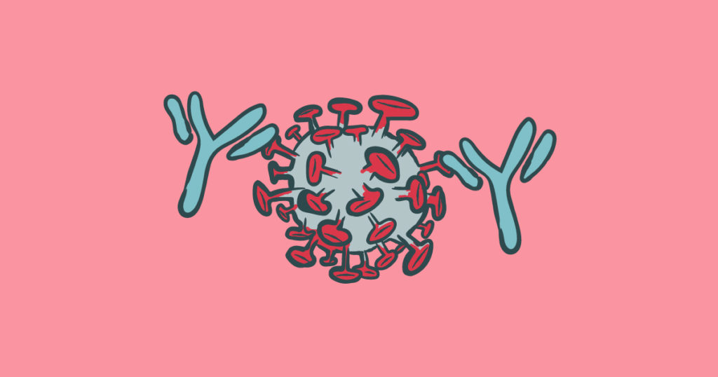
Coronaviruses are characterized by outer surface protein spikes (causing the appearance of a “corona”) and that these spikes are the means by which the virus attaches to human cells. This spike protein attaches to the surface receptor ACE2, which is how the virus gains entry into human cells.
The spike or “S” proteins consist of two protomers, S1 and S2, that comprise the receptor-binding domain or RBD. It is this RBD at the end of the coronavirus spike that connects to the ACE2 receptor.
In both MERS- and SARS-CoV, neutralizing antibodies are produced by infection, and these antibodies bind to the RBD. This information has led researchers to search for neutralizing antibodies to SARS-CoV-2.
Visit our website for resources to support:
SARS-CoV-2 Viral Research
SARS-CoV-2 Serology Testing
SARS-CoV-2: Neutralizing Antibodies Recovered from Human Patient
In their report, Wu et al. describe the isolation and characterization of four antibodies isolated from a convalescing patient.
The researchers used RBD as bait to isolate “specific, single memory” B cells from the blood of a COVID-19 patient. They then amplified the variable heavy and light regions from single B cells and cloned them in a vector with the constant Ig region to produce IgG1 antibodies.
To examine antibody binding abilities, they then used the vector to transfect HEK293T cells, producing monoclonal antibodies (mAb). The supernatants of the HEK293T cells were screened for binding to SARS-CoV-2 RBD, using a non-SARS antibody and a SARS-CoV antibody as controls. Four supernatants bound to the SARS-CoV-2 RBD, and not to SARS-CoV RBD, demonstrating an immunologic distinction between these two SARS viruses.
They next demonstrated the ability of these four mAbs to neutralize SARS-CoV-2 in Vero cells. Cells were treated with SARS-CoV-2 and serial dilutions of the four antibodies. After 5 days, IC50 values for each antibody were calculated by fitting cytopathic effect (CPE) of the antibody dilutions to a dose-response curve. This study further demonstrated that two of the antibodies had an additive effect on virus neutralization, even at higher virus concentrations.
In a competition studies using FACS (flow cytometry cell sorting), results showed that the two antibodies that worked in an additive fashion in virus neutralization, only partially competed in binding to RBD, suggesting these two antibodies bind to different area of the RBD.
Antibodies Neutralize SARS-CoV-2 in Mice
The same two antibodies, B38 and H4, were tested for their ability to inhibit SARS-CoV-2 infection in hACE2 transgenic mice. Twelve hours after intranasal viral infection, mice were given a single dose of either PBS, B38 or H4 antibody intraperitoneally. Body weight of the B38 and H4 mice decreased, then recovered at day 3 after infection, compared to the PBS control.
Viral RNA load was determined from lung tissue by qRT-PCR at day three, and showed significantly higher viral load in the PBS-treated mice, versus the antibody-treated mice, which showed 32.8% and 26% lower RNA copies.
Histopathology of mouse lung tissue also showed severe bronchial and interstitial pneumonia in the PBS-treated mice. Mild bronchopneumonia was observed in the H4-treated mice, while the authors reported no such lesions in the B38 antibody-treated mice.
Structural Analysis of Antibody-RBD Binding
Wu et al. performed a structural analysis of RBD binding by B38 and H4 using crystal structures. The resulting images are available in the paper and corresponding supplemental material. The authors highlight the importance of not only their neutralizing antibody binding studies, but also of the structural analysis, noting:
“Characterization of the epitopes on the COVID-19 virus RBD will provide valuable information for vaccine development. Furthermore, the molecular features of the neutralizing antibody targeting epitopes are helpful for the development of small molecule or peptide drugs/inhibitors.”
Here is the paper:
Wu, Y. et al. (2020) A noncompeting pair of human neutralizing antibodies block COVID-19 virus binding to its receptor ACE2. Science 13 May 2020. DOI: 10.1126/science.abc2241
Promega Corporation has tools and resources available for viral analysis and research. Visit our website to learn more.
Related Posts
Kari Kenefick
Latest posts by Kari Kenefick (see all)
- Base Editing Brilliance: David Liu’s Breakthrough Prize and Its Impact - May 28, 2025
- Neurons’ Role in FBP2 Regulation - October 24, 2024
- Fluorescent Ligands in Biological Research: Where We’ve Been, Where We’re Headed - June 27, 2024