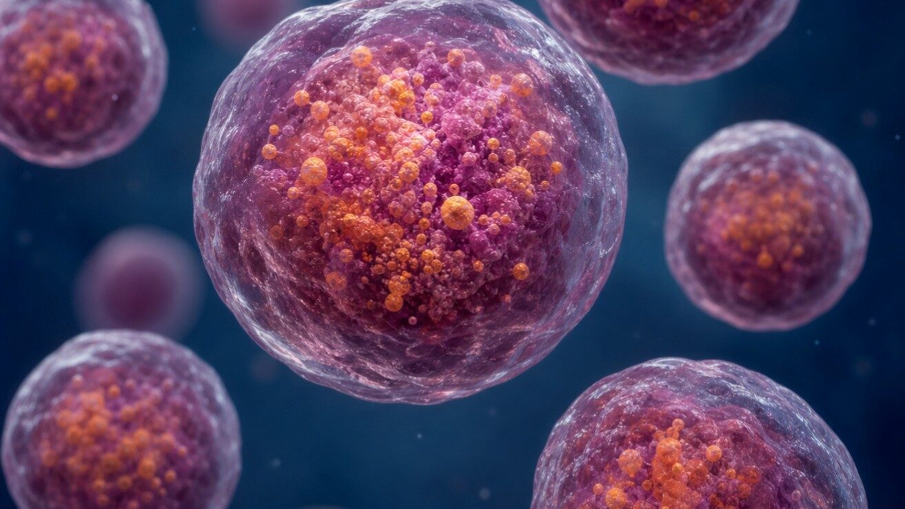
Your cells are constantly juggling two opposing needs: breaking things down and building things up. At the heart of that balancing act are lysosomes—tiny, acid-filled compartments that digest worn-out proteins, recycle cellular debris, and help cells decide whether it’s time to grow or conserve energy.
When lysosomes malfunction, the consequences can be serious. Lysosomal storage diseases, neurodegeneration, and metabolic disorders have all been linked to disrupted lysosome function. A new study published in Nature Communications has uncovered a key part of the control system that keeps lysosomes functioning properly.
What if we could boost crop yields—not by adding foreign genes, but by tweaking the plant’s own DNA in just the right places?
For decades, plant scientists have known that the noncoding DNA flanking a gene—its promoter and regulatory regions—acts like a volume dial, controlling how much protein the gene produces. Adjusting that dial is the premise behind an approach called quantitative trait engineering (QTE), where CRISPR is used to make small, precise changes to these regulatory sequences instead of inserting entire transgenes. The appeal is enormous: nontransgenic edits face fewer regulatory hurdles and are more likely to gain public acceptance.
The problem? We don’t really understand the rules governing plant promoter architecture. Which nucleotides matter? Where can you cut, insert, or swap bases to crank expression up—or dial it down? Previous attempts to answer these questions have been limited in scale and have rarely uncovered gain-of-function mutations that increase gene expression. Now, however, a new study published in Nature Biotechnology suggests we’re closer to that reality than ever before.
This is the third post of three in a series leading up to the 16th annual International Forum on Consciousness, taking place in Madison this May. Hosted by the BTC Institute, Promega and Usona Institute, the forum gathers scientists, philosophers, and practitioners from dozens of different fields to investigate the nature of the mind. This year’s theme, “Unspoken Intelligence,” explores forms of perception and knowing that fall outside conventional cognition.
In 1845, mathematician Urbain Le Verrier calculated where an unseen planet had to be based on irregularities in Uranus’s orbit, wrote a letter to an observatory telling them where to point their telescope, and Neptune was there. He found a planet without ever looking up.
This is what third-person inquiry looks like at its best: observe from the outside, measure what anyone with the right instruments can measure, build a model precise enough to predict what no one has seen yet. Then look. The history of science is full of such moments, equations pointing to phenomena that hadn’t been detected, particles that hadn’t been observed, forces that hadn’t been measured. The method works because it is ruthlessly disciplined about what counts as evidence. The observer is removed, the conditions controlled, and the measurement trusted.
That discipline is not a limitation. It is the engine of over four centuries of extraordinary results. It gave us germ theory, the structure of DNA, and the sequenced human genome. Every time something seemed to resist physical explanation, the method eventually found the mechanism and the method held. The winning streak was long enough that the assumption underneath it stopped looking like an assumption. Outside-in, third-person, measurable evidence stopped looking like one way of knowing. It started looking like the definition of knowing itself.
The assumption felt safe because it had earned its confidence. Digestion, heredity, mental illness, each had seemed to resist physical explanation until it didn’t. The pattern was consistent enough that the method felt inevitable rather than chosen.
Then science turned toward consciousness, and the winning streak entered dangerous territory.
Here is the problem, what philosopher David Chalmers named the “hard problem” of consciousness in 1995.
To understand what Chalmers meant, it helps to start with his own illustration. When you see red, something measurable happens. Light hits the retina. Signals travel along the optic nerve. Specific regions of the visual cortex activate in patterns that neuroscientists can map with increasing precision. All of that is, in principle, fully describable by the third-person, outside-in approach. Given enough time and instruments, you can trace the whole sequence.
What you cannot describe from the outside is what red looks like. The redness of red, that specific quality of experience that exists only in the moment of seeing it, is not in the neural map. No better scanner will find it there, because the felt quality of the experience isn’t a physical thing hiding in the data. It exists only from the inside. The outside measurement, however precise, cannot reach it.
Chalmers used “hard” deliberately, in contrast to what he called the “easy problems” of consciousness: how the brain integrates information, focuses attention, produces behavior. Those are genuinely difficult, but the outside-in approach knows how to go after them. The hard problem is different in kind. It’s the question that remains even after you’ve solved all of the “easy” ones: why does any of it feel like anything at all?
Think of it this way: everything the brain does could, in principle, happen without any felt experience attached. Processing, responding, behaving, all of it could run like a machine in the dark, with no one home. The question Chalmers is asking is why it doesn’t. Philosophers ask it this way: why is there something it’s like to be you, right now, reading this?
No amount of outside-in evidence, however precise, touches that question, not because the science is insufficient but because the method was specifically designed to exclude first-person data. That exclusion was the whole point. It’s what made the outside-in approach so powerful everywhere else.
With consciousness, the method’s central design decision runs into a question it wasn’t built to answer: how do you study first-person experience when your method was built to exclude first-person data?
This is the second post in a series leading up to the 16th annual International Forum on Consciousness, taking place in Madison this May, hosted by the BTC Institute, Promega and Usona Institute. The Forum gathers scientists, philosophers, and practitioners from dozens of different fields to investigate the nature of the mind. This year’s theme, “Unspoken Intelligence,” explores forms of perception and knowing that fall outside conventional cognition.
When she thought about a dog, she saw a dog, more specifically, every dog she had ever encountered, cycling through her mind like a card catalog with pictures attached. She assumed everyone did this. When she discovered they didn’t, that most people access something more like an abstract concept hovering somewhere between language and image, she was genuinely surprised. Temple Grandin had always known her mind didn’t work the way people expected. What she didn’t know, until she was an adult, was the specific shape of the difference.
Most of us know this story, or one like it. We understand that some minds filter experience differently, but the science on this doesn’t stop where the conversation usually does.
For most of its history, the field that mapped minds like Grandin’s looked at those that didn’t fit the available systems and concluded the minds were broken. (It didn’t ask whether the systems were.) More recently, the conversation has been reframing those minds not as deficient but as different.
For many people, that reframing has been transformative, changing how educators teach, how clinicians diagnose, and how workplaces are designed. We are now more familiar with alternative cognitive profiles such as autistic pattern recognition (like that experienced by Grandin), ADHD-associated divergent thinking, and the hyper-focused depth of what researchers call monotropic attention. These are not broken versions of normal cognition. They are different architectures, each with genuine capabilities that other minds aren’t built to produce.
The terms most commonly used to describe these differences, neurotypical and neurdivergent, are useful shorthand but they describe a binary the underlying biology doesn’t support. Cognitive traits distribute across a population the way most biological traits do. “Neurotypical” minds are simply closer to the statistical center. What we call “neurodivergent” can be better understood as the part of that population that differs visibly enough from the statistical center to make the variation impossible to ignore.
This is the first post in a series leading up to the 16th annual International Forum on Consciousness, taking place in Madison this May, hosted by the BTC Institute, Promega and Usona Institute. The Forum gathers scientists, philosophers, and practitioners from dozens of different fields to investigate the nature of the mind. This year’s theme, “Unspoken Intelligence,” explores forms of perception and knowing that fall outside conventional cognition.
There’s a quote that travels well in some intellectual circles:
You don’t have a soul. You are a soul. You have a body.
There’s something genuinely relieving about that idea. It locates the real you somewhere above the fray, untouched by the body’s demands and indignities, the consciousness that thinks and persists while the body handles the inconvenient work of being hungry and tired and sick. The thinking part is what counts.
Plato thought something similar. So did Augustine. As did Descartes. Kant, too. The idea that the thinking self is separate from and superior to the body is Western civilization’s default setting.
It sounds like wisdom. It is also, I’ve come to think, exactly the wrong way to understand what we are.
Here’s a different text, one most millennials can recite from memory. In the opening verse of “Lose Yourself,” Eminem rattles off a visceral catalog of physical symptoms: sweaty palms, weak knees, heavy arms, vomiting. The body staging a complete revolt while the mind tries to execute a plan, until the moment on stage when the mouth opens and nothing comes out. The mind wanted to perform, but the body said no.
Nobody who has memorized those lyrics thinks of them as a description of embodied cognition. They file it under music, or nostalgia, or just a song they played too loud in a car they didn’t own. But the nervous system doesn’t care what you call it, because the body doesn’t catalogue in words.
This is the thing the soul-body quote gets wrong: the body isn’t a vehicle the self rides around in. It’s already thinking, already keeping score, already running a process the mind is only partially aware of. The question is what to do about that.
Despite its many mysteries, a chrysalis is one of the most familiar transformations in nature. We know what goes in. We know what comes out. For a long time, what happened in between was essentially invisible to us. Not because we weren’t curious, but because the mechanism was sealed inside something the size of a thumbnail, and we had no way in.
This same invisibility exists on a much older and much larger scale.
Sometime around two billion years ago, a cell swallowed a bacterium and, instead of digesting it, kept it alive inside itself. This process, called endosymbiosis, is arguably the single most consequential event in the history of complex life. The bacterium became a permanent resident, and over billions of years of co-evolution, it became something else entirely: the mitochondria that power every complex cell on earth. Without it, the living world as we know it doesn’t exist.
Scientists have known for decades that this kind of cellular acquisition had to have occurred. What has proved harder to explain is not that it happened, but how it started. What did the earliest molecular steps actually look like from the inside?
In the ocean, there is a microscopic single-celled organism called Rapaza viridis. It hunts algae by propelling itself through the water on whip-like appendages called flagella. That hunt may be showing us the beginning of a modern endosymbiosis: the same process that gave every complex cell its mitochondria and every plant its chloroplasts.
Kierkegaard observed that one of humanity’s enduring tensions is that while life can only be understood backwards, it must be lived forwards. It’s a truth medicine knows intimately: in the treatment that worked until it didn’t, the resistance that arrived without warning, the moment a doctor has to tell a patient that the drug that was helping has stopped. Not because anyone made a mistake, but because the critical knowledge that would have mattered arrived too late, if at all.
A recent paper from the National Cancer Institute is, in a small but meaningful way, science’s pursuit of that elusive foresight: an understanding that emerges early enough, for once, to change what happens next.
The Elegant Idea
For decades, chemotherapy has worked by brute force, flooding the body with toxins designed to kill rapidly dividing cells. The problem is that rapid division isn’t unique to cancer. Hair follicle cells, gut lining cells and immune cells also divide rapidly, which is why patients lose hair, lose energy and become susceptible to infection. Chemotherapy targets a behavior, but the drug has no way to tell a healthy cell from a cancerous one.
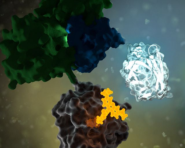
Antibody-drug conjugates (ADCs) change that. Instead of targeting what cancer cells do, they target what cancer cells are. Cancer cells tend to display certain proteins on their surface in far greater numbers than healthy cells do. The antibody is engineered to seek out those proteins specifically. It navigates to its target, binds and waits for the cell to do what cells routinely do: pull it inside. Once there, the cell’s own digestive machinery (the lysosome) breaks down the chemical tether holding the toxin to the antibody, releasing the toxin to kill the cell from within. More than a dozen ADCs have received FDA approval in recent years, and the field is evolving fast.
What the Cell Does Next
But cancer cells don’t simply accept their fate. Even when an ADC delivers its payload perfectly—the antibody finds its target, the cell pulls it inside, the lysosome cuts the tether—a pump embedded in the cell membrane can grab the released toxin and throw it back out before it causes damage.
The delivery worked. The package got ejected anyway.
These pumps—ATP-binding cassette transporters, or more plainly, efflux pumps—are a normal feature of cell biology. Their job is cellular housekeeping, clearing out unwanted or toxic substances before they cause damage. Under the pressure of drug treatment, cancer cells do what life has always done under pressure: the ones best equipped to survive do. The same mechanism that has shaped living things for billions of years now works against the treatment. Not all cancer cells are identical, and the ones that happen to produce more pumps survive while others don’t, gradually shifting the tumor toward resistance.
Last spring, my niece and I made a trip to a home improvement store to put together a Mother’s Day planter for my sister. My niece had a clear vision: my sister’s favorite color is blue, so we were going to buy blue flowers. We walked every aisle of the garden center. We checked the annuals, the perennials, and the hanging baskets then left with purple, red, and a grumpy 7-year-old.
It turns out we were not up against a bad selection. We were up against biology.
The Problem with Blue
Blue is one of the rarest colors in the natural world. The food industry is currently finding that out the hard way. There is a good chance you have eaten something blue today. Maybe it was the frosting on a birthday cake, the coating on some M&M’s® candies, or the sports drink in your refrigerator. That blue almost certainly came from a petroleum-based synthetic dye, and for the first time in decades, the food industry is being asked to find something better.
The FDA banned Red Dye No. 3 in January 2025, and pressure has been building around the remaining synthetic dyes ever since, including Blue No. 1 and Blue No. 2. Major food brands have begun announcing plans to reformulate.
There is just one problem. Blue is genuinely, stubbornly hard to make in nature. It turns out that blue has almost nothing to do with color, and almost everything to do with light.
RNA doesn’t just carry genetic instructions—it also interacts with proteins to regulate nearly every aspect of gene expression, from splicing to translation. When those interactions go awry, the consequences can be devastating. In myotonic dystrophy type 1 (DM1), the most common adult-onset muscular dystrophy, a toxic RNA repeat expansion hijacks a critical protein called MBNL1, trapping it in nuclear clumps called foci. This leads to widespread splicing defects and progressive muscle wasting. But studying these toxic interactions inside living cells—and finding small molecules that can disrupt them—has been a significant challenge.
A recent study led by the Scripps Institute may have a solution. The study introduces a NanoBRET™ assay that can monitor the interaction between the expanded CUG RNA repeats and MBNL1 protein in real time, in live cells. Their findings demonstrate how this platform can be used not only to detect disease-driving RNA–protein complexes but also to identify small molecules that break them apart.
You’re sitting in the dentist’s chair, nodding along to the familiar flossing lecture you’ve been politely ignoring for most of your adult life. Fair enough. It’s hard to get excited about gum health. But it turns out your dentist may have been underselling the pitch.
A study published in January 2026 in Cell Communication and Signaling shows that a common gum disease bacterium can promote breast cancer growth and spread in mice, and the findings hint at a particularly troubling link for people carrying BRCA1 mutations (1). “Floss to help prevent cancer” probably wasn’t on your 2026 bingo card, yet here we are.
XWe use cookies and similar technologies to make our website work, run analytics, improve our website, and show you personalized content and advertising. Some of these cookies are essential for our website to work. For others, we won’t set them unless you accept them. To learn more about our approach to Privacy we invite you to Read More
By clicking “Accept All”, you consent to the use of ALL the cookies. However you may visit Cookie Settings to provide a controlled consent.
We use cookies and similar technologies to make our website work, run analytics, improve our website, and show you personalized content and advertising. Some of these cookies are essential for our website to work. For others, we won’t set them unless you accept them. To find out more about cookies and how to manage cookies, read our Cookie Policy.
If you are located in the EEA, the United Kingdom, or Switzerland, you can change your settings at any time by clicking Manage Cookie Consent in the footer of our website.
Necessary cookies are absolutely essential for the website to function properly. These cookies ensure basic functionalities and security features of the website, anonymously.
Cookie
Duration
Description
cookielawinfo-checbox-analytics
11 months
This cookie is set by GDPR Cookie Consent plugin. The cookie is used to store the user consent for the cookies in the category "Analytics".
cookielawinfo-checbox-functional
11 months
The cookie is set by GDPR cookie consent to record the user consent for the cookies in the category "Functional".
cookielawinfo-checbox-others
11 months
This cookie is set by GDPR Cookie Consent plugin. The cookie is used to store the user consent for the cookies in the category "Other.
cookielawinfo-checkbox-advertisement
1 year
The cookie is set by GDPR cookie consent to record the user consent for the cookies in the category "Advertisement".
cookielawinfo-checkbox-necessary
11 months
This cookie is set by GDPR Cookie Consent plugin. The cookies is used to store the user consent for the cookies in the category "Necessary".
cookielawinfo-checkbox-performance
11 months
This cookie is set by GDPR Cookie Consent plugin. The cookie is used to store the user consent for the cookies in the category "Performance".
gdpr_status
6 months 2 days
This cookie is set by the provider Media.net. This cookie is used to check the status whether the user has accepted the cookie consent box. It also helps in not showing the cookie consent box upon re-entry to the website.
lang
This cookie is used to store the language preferences of a user to serve up content in that stored language the next time user visit the website.
viewed_cookie_policy
11 months
The cookie is set by the GDPR Cookie Consent plugin and is used to store whether or not user has consented to the use of cookies. It does not store any personal data.
Analytical cookies are used to understand how visitors interact with the website. These cookies help provide information on metrics the number of visitors, bounce rate, traffic source, etc.
Cookie
Duration
Description
SC_ANALYTICS_GLOBAL_COOKIE
10 years
This cookie is associated with Sitecore content and personalization. This cookie is used to identify the repeat visit from a single user. Sitecore will send a persistent session cookie to the web client.
vuid
2 years
This domain of this cookie is owned by Vimeo. This cookie is used by vimeo to collect tracking information. It sets a unique ID to embed videos to the website.
WMF-Last-Access
1 month 18 hours 24 minutes
This cookie is used to calculate unique devices accessing the website.
_ga
2 years
This cookie is installed by Google Analytics. The cookie is used to calculate visitor, session, campaign data and keep track of site usage for the site's analytics report. The cookies store information anonymously and assign a randomly generated number to identify unique visitors.
_gid
1 day
This cookie is installed by Google Analytics. The cookie is used to store information of how visitors use a website and helps in creating an analytics report of how the website is doing. The data collected including the number visitors, the source where they have come from, and the pages visted in an anonymous form.
Advertisement cookies are used to provide visitors with relevant ads and marketing campaigns. These cookies track visitors across websites and collect information to provide customized ads.
Cookie
Duration
Description
IDE
1 year 24 days
Used by Google DoubleClick and stores information about how the user uses the website and any other advertisement before visiting the website. This is used to present users with ads that are relevant to them according to the user profile.
test_cookie
15 minutes
This cookie is set by doubleclick.net. The purpose of the cookie is to determine if the user's browser supports cookies.
VISITOR_INFO1_LIVE
5 months 27 days
This cookie is set by Youtube. Used to track the information of the embedded YouTube videos on a website.
Performance cookies are used to understand and analyze the key performance indexes of the website which helps in delivering a better user experience for the visitors.
Cookie
Duration
Description
YSC
session
This cookies is set by Youtube and is used to track the views of embedded videos.
_gat_UA-62336821-1
1 minute
This is a pattern type cookie set by Google Analytics, where the pattern element on the name contains the unique identity number of the account or website it relates to. It appears to be a variation of the _gat cookie which is used to limit the amount of data recorded by Google on high traffic volume websites.