An amazing transformation is taking place, unseen and unnoticed, within the microscopic bits that make you, you.
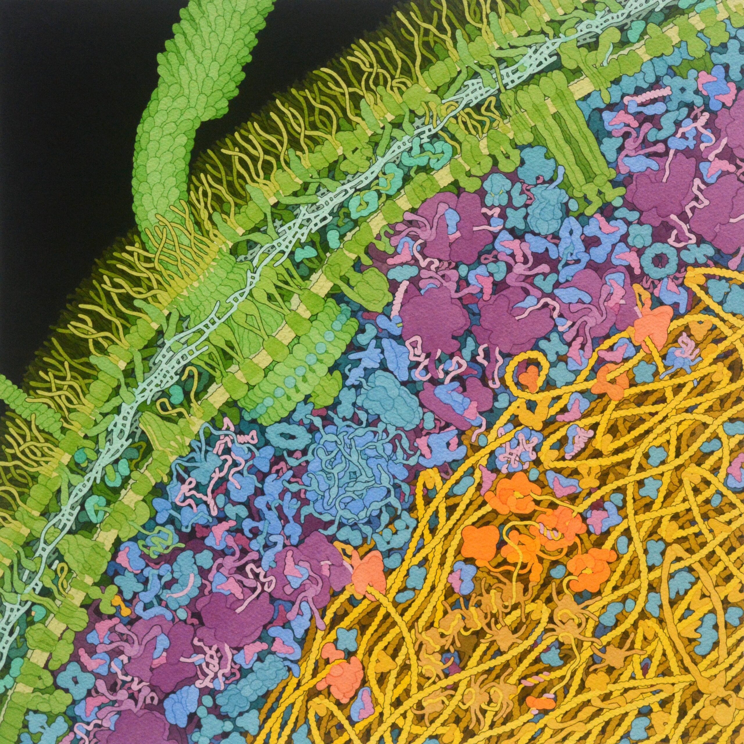
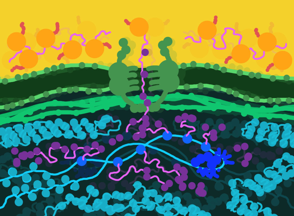
A tightly coiled lattice unspools to reveal a sinuous DNA stand. Along its length, tendrils of RNA sprout, growing bit by genetic bit. Eventually, the signal to stop and break away arrives, yielding a new strand of RNA that faithfully transcribes the DNA strand’s genetic code. Proteins trim and splice this new growth, pruning it so it takes its final form, messenger RNA. More proteins then ferry this mRNA strand through a pore in the nuclear envelope into the open space of the cell’s cytoplasm. Ribosomes and codon-carrying tRNA alight onto the released mRNA strand, reading the instructions it has carried from the DNA in the nuclear nursery. From this trio new forms emerge, bulbous proteins shaped by their destined purpose.
And so it goes, every second of every day, in the tens of trillions of cells in your body…
…And on the tens of thousands of kit packages we deliver to customers across the globe every year.
Our New “Face”
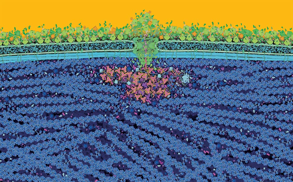
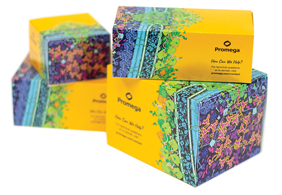
In 2018, Promega unveiled a new kit packaging design. Against a warm yellow horizon, rainbow-hued figures tumble in a seemingly chaotic and undeniably eye-catching mix. This image, however, is far from chaotic. It depicts a fundamental and tightly controlled biological process: the transfer of information from DNA into the structure and function of the proteins that give living beings their own function and form.
Our packaging design is an illustration anchored in the scientific experience and artistry of its creator, David Goodsell, a scientist-artist known for his colorful and painstakingly accurate watercolor paintings of biological “landscapes”. Goodsell, a professor of computational biology at Scripps Research, paints microscopic worlds utterly unlike the simplified infographics or cinematic 3D renderings you might encounter in textbooks or life science marketing materials or research articles. His watercolors uniquely capture the crowdedness and complexities of biological structures and processes.
When the time came to update our kit packaging design, we turned to Goodsell. What he crafted became the new “face” of Promega and is a striking visual representation of our science and our values.
This transformation was the third kit packaging design for Promega. In 1996, we launched our first kit packaging design: a color-blocked illustration of a cell, its bright color palette reminiscent of the decade’s iconic windbreaker jackets. Four years later, we introduced a new, pared-back look that featured the warm yellow color we call “sol”. While we remember our previous kit packaging designs fondly, our current packaging is uniquely able to communicate our identity.
Below, we share the story behind this illustration and how a single piece of art manages to capture the creativity, commitment to quality and science that form the core of Promega.

Illustrating a Living Cell
The scientific community first encountered Goodsell’s scientific artistry in a 1991 feature article in Trends in Biochemical Sciences (1). In the article, Goodsell sets out to illustrate an Escherichia coli bacterium at 1,000,000x magnification. While the greyscale drawings lack the vibrancy of his later work, the simplified forms and crowded cellular compartments unmistakably come from Goodsell’s hand.
Goodsell opens the article explaining why he took on this project:
“A clear picture of the interior of a living cell that shows the average distribution of molecules at the proper scale, the proper concentration, and with no missing parts, seems to me to be central to the understanding of the workings of life. However, this type of picture is virtually absent from the popular and technical literature.”
He goes on to explain the careful calculations and research required to not only capture which biomolecules are in an E. coli cell, but how many proteins are present and what their shape and relative sizes are:
“The typical cell is an E. coli strain B/r in balanced growth at 37°C in glucose minimal medium. The cell is assumed to be 70% water, yielding a volume of 0.88µm3. This volume corresponds to a cell about 2.95µm long and 0.64µm wide. The volume of the envelope is 0.14µm3, assuming a width of 7.5nm for the two membranes and a width of 10nm for the periplasmic space. The nuclear material occupies another 0.14µm3, leaving 0.6µm3 of cytoplasm.”
With his meticulous attention to known details and “a liberal dose of artistic license and scientific intuition” where there are unknowns, Goodsell managed, in his own words, to “almost” capture the complexity of a living cell (2).
Since that 1991 publication, Goodsell has updated his E. coli painting and has created many other molecular landscapes, including one of a SARS-CoV-2 vaccine, a casein micelle in milk and the protective myelin layer that surrounds and protects nerve cells. More examples of his artwork can be found at the PDB-101 website.
Collaborating with David Goodsell
In 2016, we reached out to David Goodsell about the possibility of creating illustrations that would highlight some of our flagship product groups. For genomics, we proposed the idea of illustrating how information encoded in DNA flows to proteins’ structure and function, which is often described as the Central Dogma of molecular biology (more on that later).
One challenge to taking on this assignment, however, is that there are a lot of biological processes wrapped up in the Central Dogma.
“I couldn’t figure out a composition that included DNA replication along with transcription and translation,” Goodsell wrote in an email. “I often show all of this for prokaryotic cells, but I haven’t found a way to do it for eukaryotes. We settled on showing transcription and translation.”
During transcription, a strand of DNA is “unzipped” and serves as a template for enzymes to create a strand of RNA. In other words, the genetic code built into DNA is transcribed to a strand of RNA. This happens in a cell’s nucleus. Then, during translation, RNA serves as a template, this time providing the information needed to create proteins. Here, a strand of nucleotides is translated into a fundamentally different biomolecule made up of linked amino acids.
After the team agreed with this approach and approved a concept sketch, Goodsell began creating his illustration. During January 2017, he worked during the evenings to put the pieces together and create a series of draft images.
Though the illustration was originally intended to represent one product group, we recognized the opportunity to use the Central Dogma image for all of our kit packaging because of how it actually encompasses all of our product areas.
“The Central Dogma is the foundation of life and what we do as a life science company. It encompasses all our products whether it’s genomics, protein analysis and expression, cellular analysis, drug discovery or genetic identity,” said Jennifer Mook, now a senior applications scientist at Promega. Mook was one of the team members who collaborated with Goodsell.
To that end, Goodsell’s final image shows a larger portion of the nucleus, extending the boundaries of the image so it could wrap around our kit packaging.
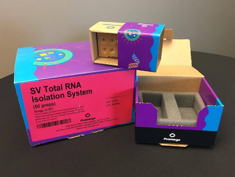
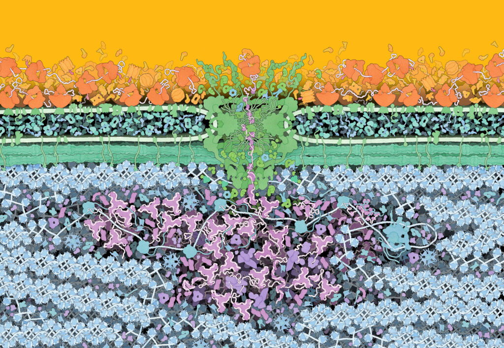
After Goodsell provided his final illustration, Promega graphic designer Jenna Stormberg adjusted the color palette and translated the 2D image onto the 3D surface of our kit packages. Packaging wrapped in this imagery began showing up in customer labs in 2018.
The Central Dogma
In a 1957 lecture, Francis Crick introduced what he termed a “Central Dogma” (3). Most well-known today for his role in solving the double helix structure of DNA, Crick’s dogma provided a hypothesis of how information could transfer from genetic biomolecules to and from proteins.
“I shall…argue that the main function of the genetic material is to control (not necessarily directly) the synthesis of proteins. There is a little direct evidence to support this, but to my mind the psychological drive behind this hypothesis is at the moment independent of such evidence.”
At the time, scientists knew little about how DNA’s structure and its sequence of base pairs might inform the amino acid sequence and structure of proteins. Crick’s dogma doesn’t deal with details about the machinery or mechanisms that enable this transfer and interconversion of these sequences. Instead, it simply asserts in which directions sequence information can and cannot be passed.
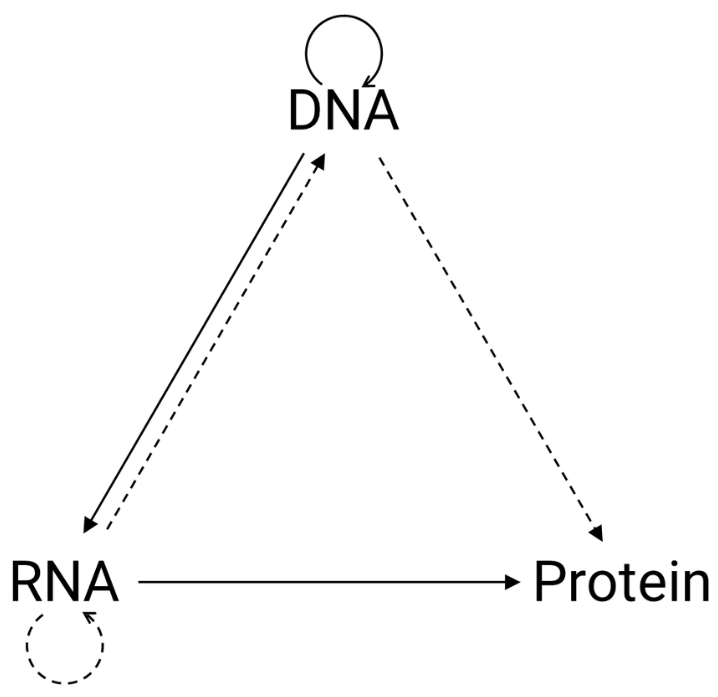
In short, DNA’s sequence can be transcribed into RNA, whose base pair sequence can then be translated to a protein’s amino acid sequence. The amino acid sequence of a protein, however, does not inform or impact the sequence encoded into DNA. Crick later captured this concept visually in a simple, triangular diagram (right).
Solid lines communicate what were known information transfers. Dashed lines show transfers that Crick deemed possible, but had no, or little evidence to confirm. More striking to his audience would be the absence of arrows pointing from “protein” to “DNA” or “RNA”, which captures this key assertion:
“Once information has got into a protein it can’t get out again. Information here means the sequence of amino acids residues, or other sequences related to it.”
Amazingly, Crick’s assertions have stood the test of time, even as the details have been uncovered.
The Central Dogma of Molecular Biology painting on our kit packaging benefits from decades of experimentation and discovery in genetics and molecular biology. Though it’s certainly a more dramatic and detailed illustration, it’s also simply an alternative way of communicating what Crick was attempting with his solid arrows:
- The transcription of DNA’s nucleotide sequence to its RNA mirror image.
- The translation of RNA’s nucleotide sequence into a protein’s amino acid sequence.
Capturing the Details
More of the details and science behind what’s shown in Goodsell’s Central Dogma illustration are captured in his book The Machinery of Life and a 2011 paper (5,6). Beyond transcription and translation, the image also includes mRNA splicing, RNA transport through the nuclear pore and membrane-bound ribosomes.
While the illustration’s details are largely grounded in what’s known to scientists about cellular structures, there are still unanswered questions and unknowns. As with his 1991 illustration of E. coli, Goodsell needed to take some artistic liberties in our Central Dogma image:
“There still aren’t good structures for the cone-shaped basket at the bottom of the nuclear pore,” Goodsell wrote in an email. Furthermore, “there’s much controversy about the 30nm filament that is depicted here for the chromatin, and I chose to draw the traditional filament.”
Beyond Boxes
In the years since launching our new kit packaging design, we’ve explored other ways we can use our Central Dogma design (a.k.a. our Goodsell Imagery) to introduce customers and others to Promega.
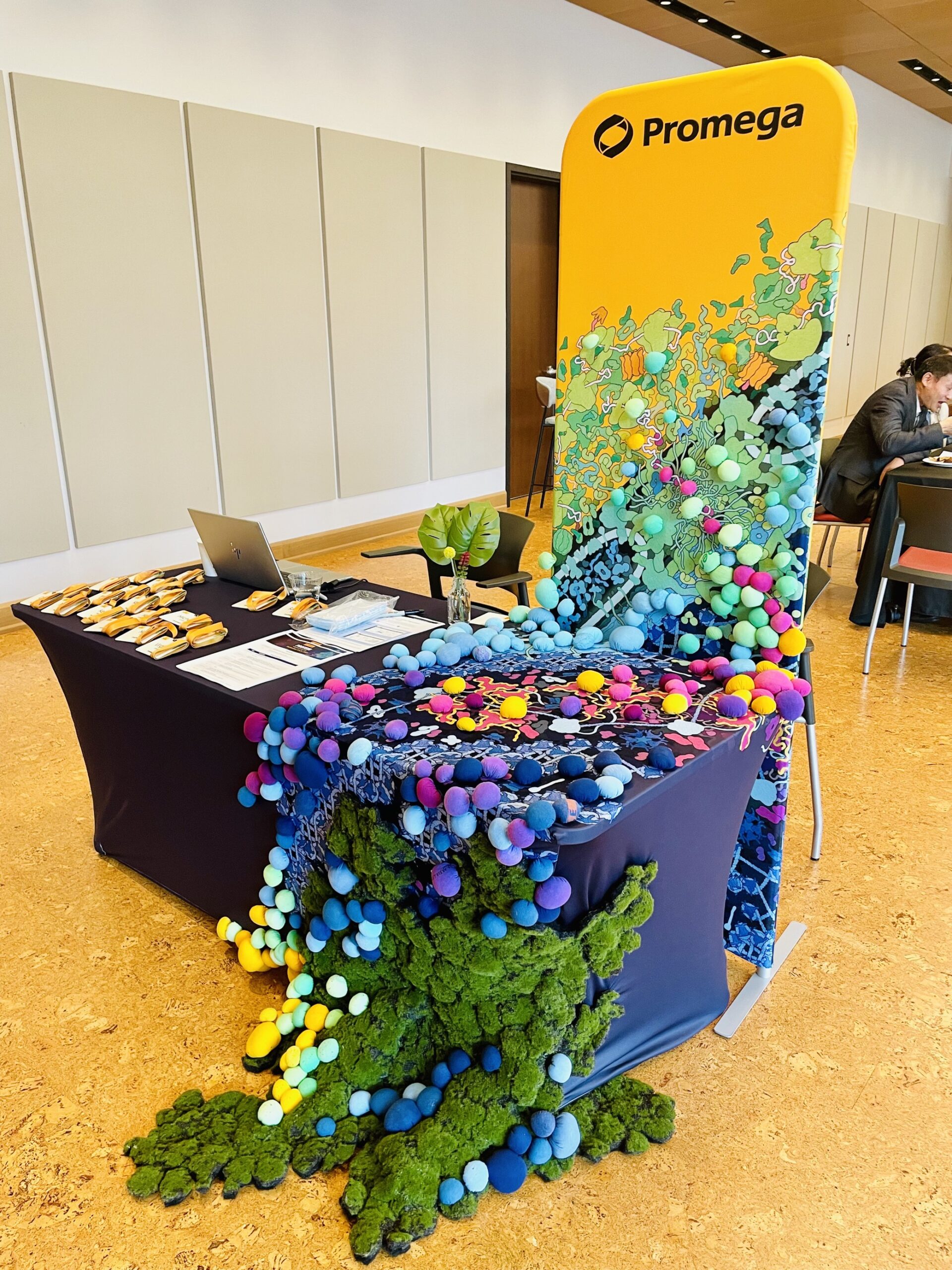
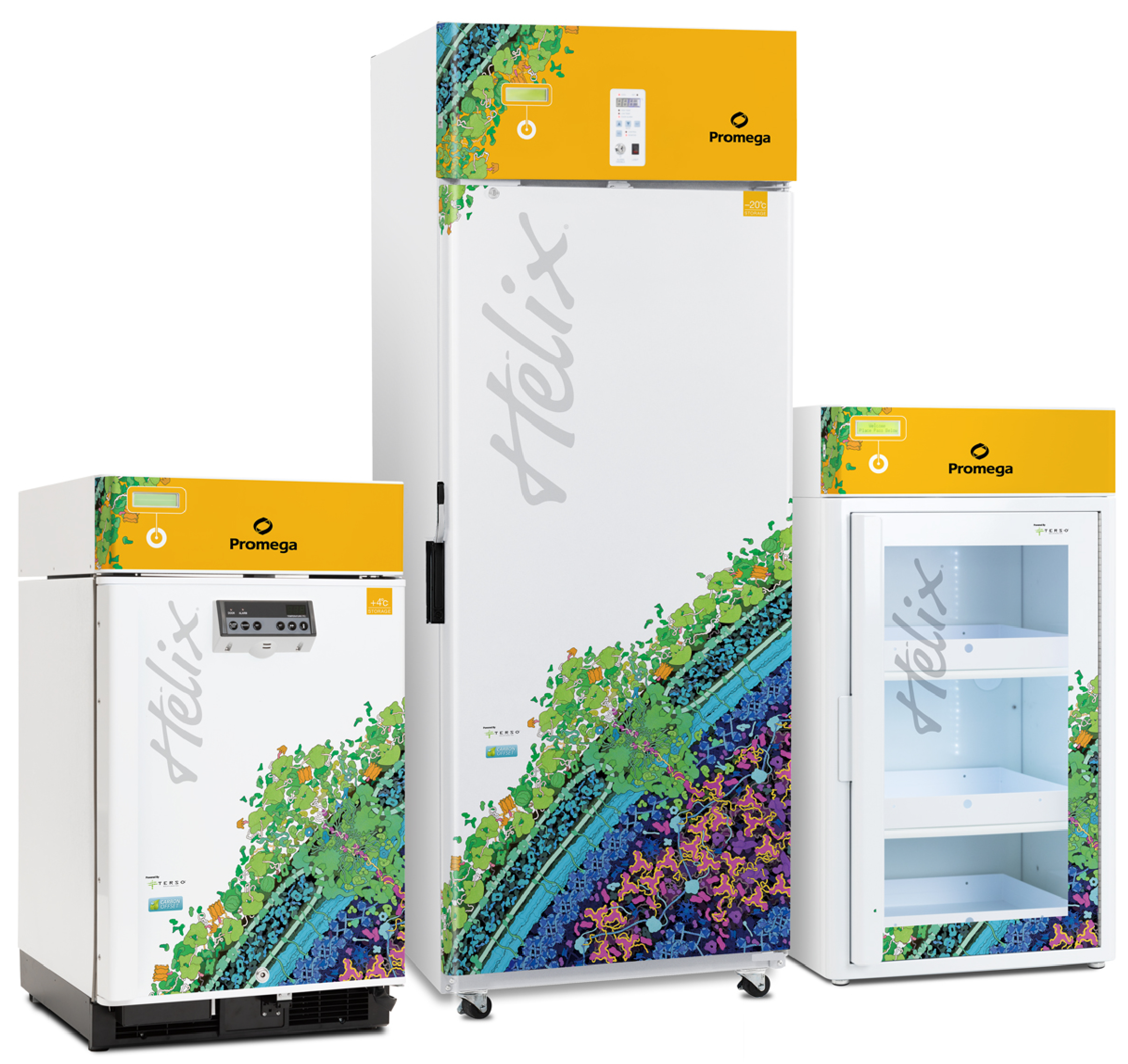
You’ll find it on our Helix™ freezers, Lab Sets made with LEGO elements and swirling across our home web page. And continues to find new life and new interpretations elsewhere, like in our Student Resource Center and new tradeshow booth designs.


Art Fuels Our Science
There’s an incredible power in communicating concepts and knowledge visually. Visual communication allows us to describe the same phenomenon in vastly different ways with vastly different outcomes. For example, Crick’s triangle does away with the details so viewers can absorb a paradigm-shifting framework while Goodsell’s landscape invites viewers to stand in awe of life’s complexities and the monumental efforts in uncovering those complexities.
The ability to hold up a concept and inspect it at any one of an infinite number of angles and then communicate what you’ve learnt and what you still don’t know is at the heart of science and of art.
At Promega, we believe that creating, considering and cultivating art helps us all to ask better questions, listen more deeply and understand each other and our world a little bit better.

Did you know that in addition to reflecting our love of art and science, we designed our kit packaging to reduce carbon emissions? It’s part of our commitment to sustainability and corporate responsibility efforts that benefit our planet, people, communities and products. Learn more about our award-winning sustainable packaging design.
References
- Goodsell, D.S. (1991) Inside a living cell. Trend. in Biochem. Sci. 16, 203. doi: 10.1016/0968-0004(91)90083-8
- Goodsell, D.S. (2021) Art as a tool for science. Nat. Struct. Mol. Biol. 28, 402. doi: 10.1038/s41594-021-00587-5
- Cobb, M. (2017) 60 years ago, Francis Crick changed the logic of biology. PLOS Biol. 15, e2003243. doi: 10.1371/journal.pbio.2003243
- Crick, F. (1970) Central Dogma of Molecular Biology. Nature 227, 561. doi: 10.1038/227561a0
- Goodsell, D.S. (2009) The Machinery of Life. Springer-Verlag, New York. doi: 10.1007/978-0-387-84925-6
- Goodsell, D.S. (2011) Eukaryotic cell panorama. Biochem. Mol. Biol. Ed. 39. 91. doi: 10.1002/bmb.20494
Latest posts by Jordan Nutting (see all)
- The Central Dogma of Promega: The Story and Science Behind Our Kit Packaging Design - May 7, 2024
- Silencing the Immunogenicity of AAV Vectors - April 4, 2024
- Discovering Cyclic Peptides with a “One-Pot” Synthesis and Screening Method - February 29, 2024