Illustration showing NanoLuc and firefly luciferase reporters.
The luciferase immunoprecipitation system (LIPS) assay is a liquid phase immunoassay allowing high-throughput serological screening of antigen-specific antibodies. The immunoassay involves quantitating serum antibodies by measuring luminescence emitted by the reporter enzyme Renilla luciferase (Rluc) fused to an antigen of interest. The Rluc-antigen fusion protein is recognized by antigen-specific antibodies, and antigen-antibody complexes are captured by protein A/G beads that recognize the Fc region of the IgG antibody (1).
Ubiquitin modification of a protein directs events such as targeting for proteasomal degradation. Targeting a protein for degradation through ubiquitin modification is one way to regulate the amount of time a signaling protein, such as a kinase or other enzyme, is available to participate in cell signaling events. Deubiquitinases (DUBs) are enzymes that cleave the ubiquitin tags from proteins, and they have been implicated in several diseases, including cancer.
With their roles in the stabilization of proteins involved in cell cycle progression and other critical processes, DUBs are promising targets for small molecule inhibitors, particularly since they may provide a “back door” for targeting otherwise intractable, undruggable proteins by modulating their half lives. However, finding small molecule inhibitors of the ubiquitin proteases to date has not been trivial. Here we highlight two papers describing the identification and characterization of small molecule inhibitors against the DUB USP7. Continue reading “Deubiquitinases: A Backdoor into Undruggable Targets?”
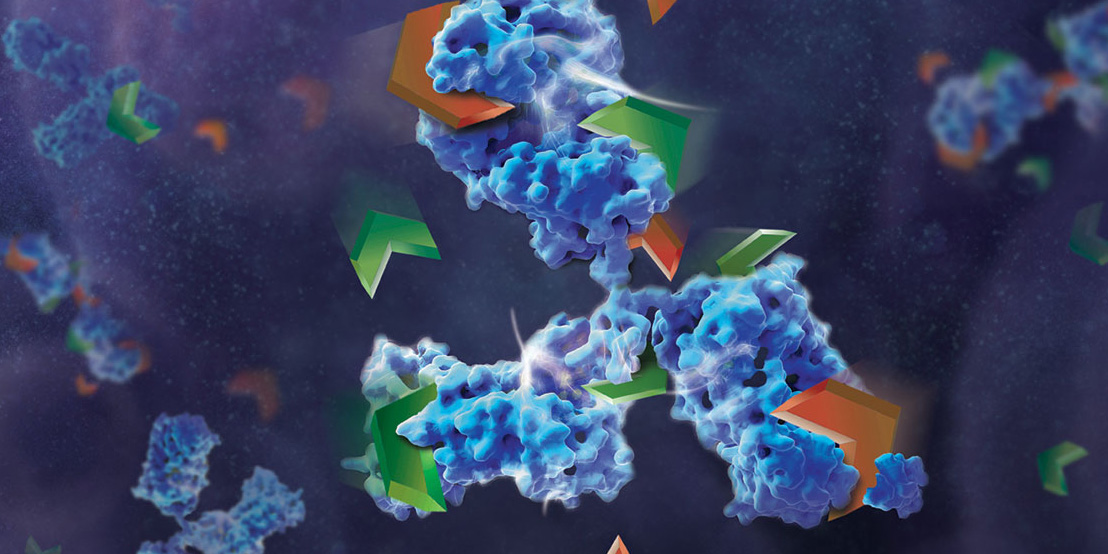
Monoclonal antibodies (mAbs) have been widely used to eliminate undesired cells via various mechanisms, including antibody-dependent cell-mediated cytotoxicity (ADCC), complement-dependent cytotoxicity (CDC) and programmed cell death (PCD). Unlike the Fc-dependent mechanism of ADCC and CDC, certain antibody–antigen interactions can evoke direct PCD via apoptosis or oncosis. Previously, researchers have reported the specific killing of undifferentiated human embryonic stem cells (hESC) by mAb84 (IgM) via oncosis (1)
In a recent publication (2), a monoclonal antibody (mAb), TAG-A1 (A1), was generated to selectively kill residual undifferentiated human embryonic stem cells (hESC). One of the many experimental tools used to characterize the mechanism of oncosis was the fragmention of the A1 antibody with IdeS and papain.
Papain digestion of IgG produces Fab fragments in the presence of reducing agent. F(ab)2 fragments of A1 were produced using IdeS Protease.
The results indicate that both Fab_A1 and F(ab)2_A1 bind to hESC but only F(ab)2_A1 retained hESC killing. Hence bivalency, but not Fc-domain, is essential for A1 killing on hESC.
Choo, A.B. et al. (2008) Selection against undifferentiated human embryonic stem cells by a cytotoxic antibody recognizing podocalyxin-like protein-1. Stem Cells26, 1454.
Zheng, J.Y. et al. (2017) Excess reactive oxygen species production mediates monoclonal antibody-induced human embryonic stem cell death via oncosis. Cell Death and Differentiation24, 546–58.
Are you looking for proteases to use in your research? Explore our portfolio of proteases today.
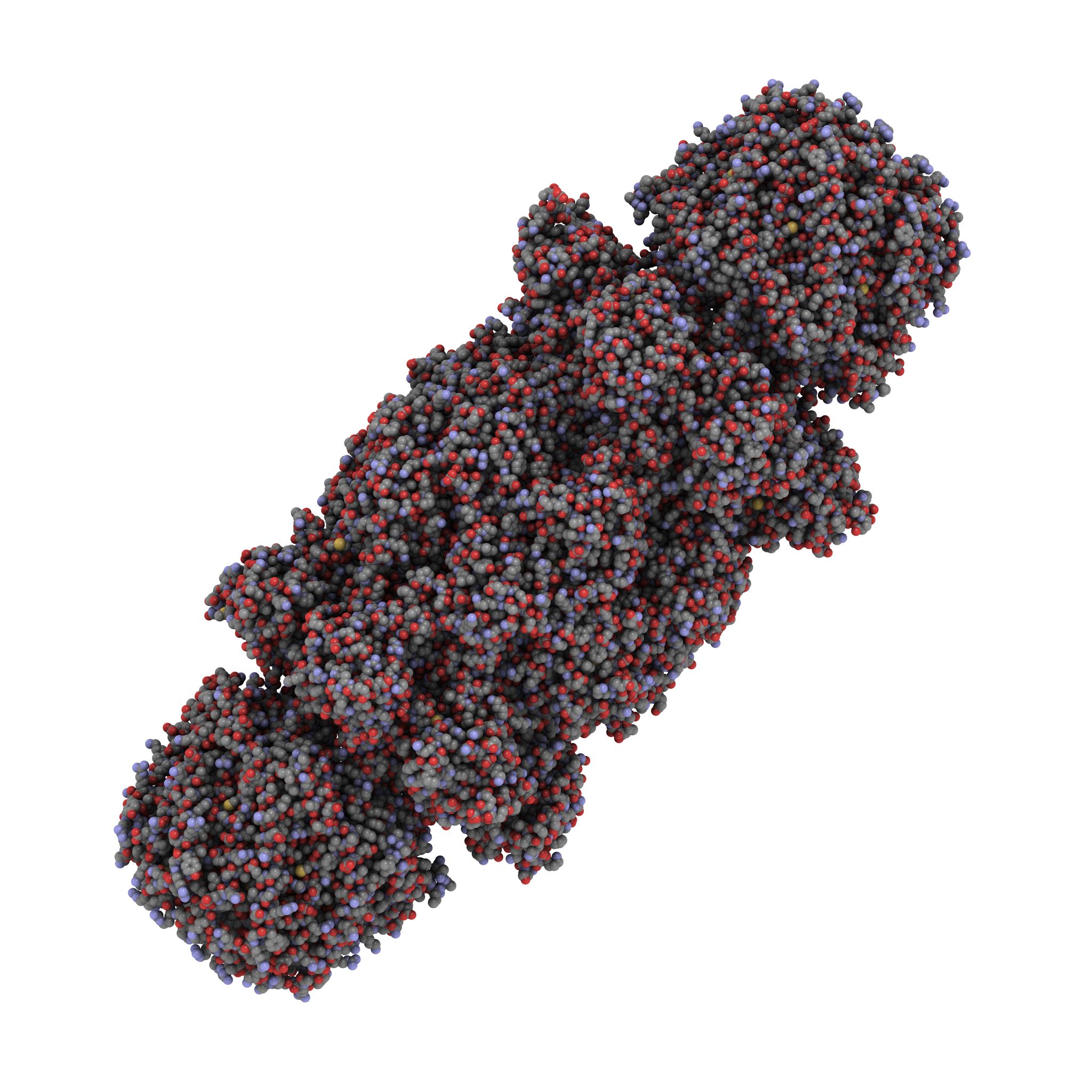
Several pharmaceutical companies have biosimilar versions of therapeutic mAbs in development. Biosimilars can promise significant cost savings for patients, but the unavoidable differences between the original and thencopycat biologic raise questions regarding product interchangeability. Both innovator mAbs and biosimilars are heterogeneous populations of variants characterized by differences in glycosylation,oxidation, deamidation, glycation, and aggregation state. Their heterogeneity could potentially affect target protein binding through the F´ab domain, receptor binding through the Fc domain, and protein aggregation.
As more biosimilar mAbs gain regulatory approval, having clear framework for a rapid characterization of innovator and biosimilar products to identify clinically relevant differences is important. A recent reference (1) applied a comprehensive mass spectrometry (MS)-based strategy using bottom-up, middle-down, and intact strategies. These data were then integrated with ion mobility mass spectrometry (IM-MS) and collision-induced unfolding (CIU) analyses, as well as data from select biophysical techniques and receptor binding assays to comprehensively evaluate biosimilarity between Remicade and Remsima.
The authors observed that the levels of oxidation, deamidation, and mutation of individual amino acids were remarkably similar. they found different levels of C-terminal truncation, soluble protein aggregates, and glycation that all likely have a limited clinical impact. Importantly, they identified more than 25 glycoforms for each product and observed glycoform population differences.
Overall the use of mass spectrometry-based analysis provides rapid and robust analytical information vital for biosimilar development. They demonstrated the utility of our multiple-attribute monitoring workflow using the model mAbs Remicade and Remsima and have provided a template for analysis of future mAb biosimilars.
Research surrounding drug discovery has historically been highly competitive and expensive. Unfortunately, many late-stage drug failures have occurred over recent years, often due to lack of efficacy. These failures have left the industry searching for new means by which to improve early drug discovery efforts aimed at understanding the drug target and its role in disease. One idea that is gaining traction is partnerships to openly share information at the early, precompetitive stages of drug discovery.
I used to think of open access only in terms of publishing data and information—online sites where you could freely access data without a subscription or membership, and without payment.
Structural Genomics Consortium logo.
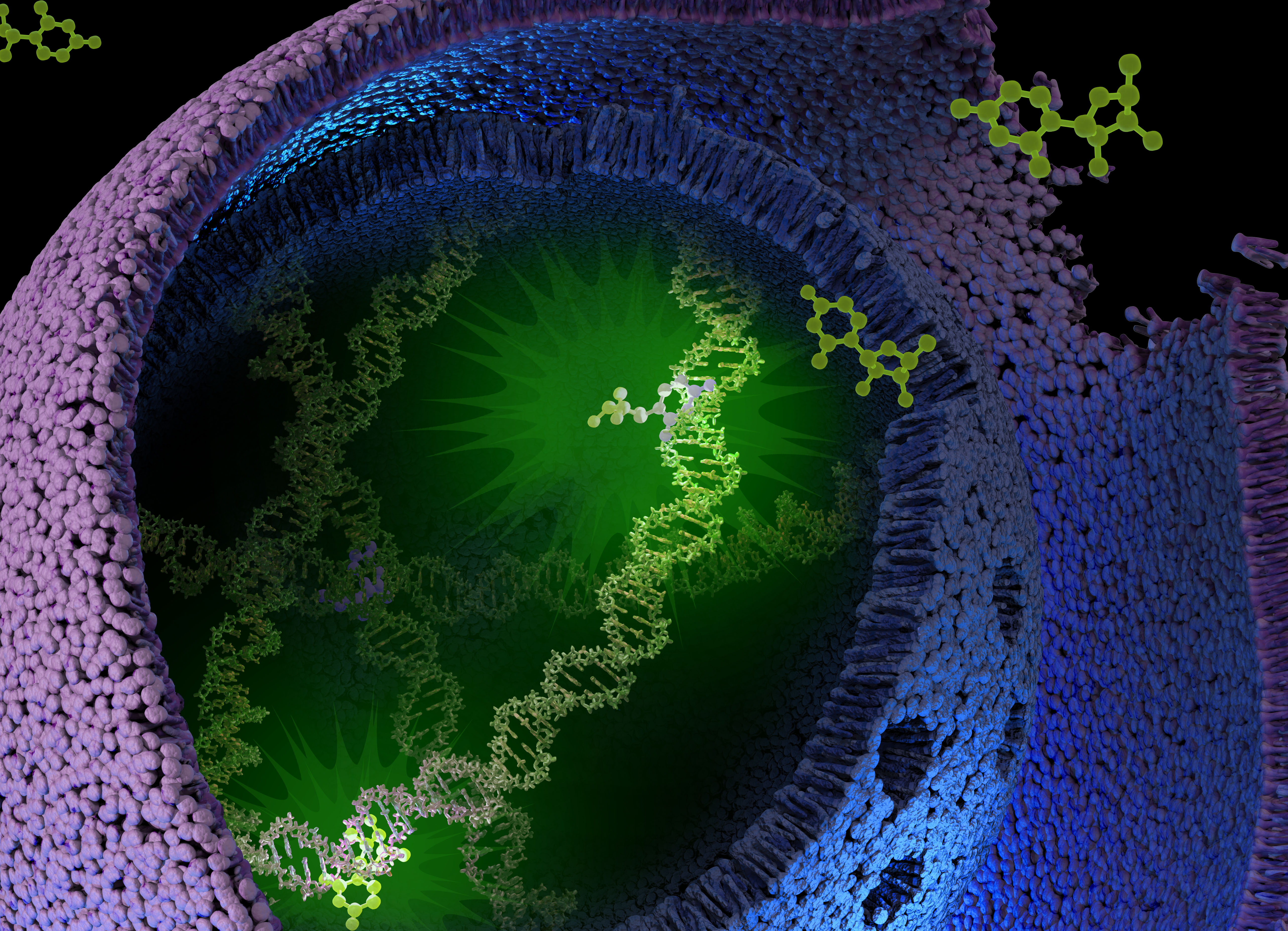
Meet the Structural Genomics Consortium (SGC), the international partnership that’s taking open access to a new level in order to advance scientific research for scientists working in a variety of disciplines—structural genomics and beyond. The SGC might just become your new, best laboratory research partner. Continue reading “Your New Best Research Partner: The Structural Genomics Consortium”
What if you could uncover a small but significant cellular response as your population of cells move toward apoptosis or necrosis? What if you could view the full picture of cellular changes rather than a single snapshot at one point? You can! There are real-time assays that can look at the kinetics of changes in cell viability, apoptosis, necrosis and cytotoxicity—all in a plate-based format. Seeking more information? Multiplex a real-time assay with endpoint analysis. From molecular profiling to complementary assays (e.g., an endpoint cell viability assay paired with a real-time apoptosis assay), you can discover more information hidden in the same cells during the same experiment.
Whether your research involves screening a panel of compounds or perturbing a regulatory pathway, a more complete picture of cellular changes gives you the benefit of more data points for better decision making. Rather than assessing the results of your experiment using a single time point, such as 48 hours, you could monitor cellular changes at regular intervals. For instance, a nonlytic live-cell reagent can be added to cultured cells and measurements taken repeatedly over time. Pairing a real-time cell health reagent with a detection instrument that can maintain the cells at the correct temperature means you can automate the measurements. These repeated measurements over time reveal the kinetic changes in the cells you are testing, giving a real-time status update of the cellular changes from the beginning to the end of your experiment. Continue reading “Reveal More Biology: How Real-Time Kinetic Cell Health Assays Prove Their Worth”
Dr. Walter Blum wins trip to Promega headquarters as part of Promega Switzerland’s 25th Anniversary celebration.
Walter Blum knew how normal cells worked. He had studied and read about the pathways that regulated cell cycles, growth and development; he saw the cell as an amazingly well programmed, intricate machine. What he wanted to understand was: “Why does a cell become crazy? How does it escape immune system surveillance?”
Last week I had the opportunity to sit down with Dr. Blum, a customer of our Promega Switzerland branch. Dr. Blum won a trip to visit our campus in Madison for a week as part of an anniversary celebration for our Switzerland branch. While here, he got an inside peek at research and manufacturing operations, chatted with our scientists, met with our marketing teams and saw the sights in Madison. We talked about his work and what he learned and is taking back with him from his trip to Madison. Continue reading “Genes to Cells to Genomes: Where Will Your Research Questions Take You?”
Welcome to the emerging frontier of immunometabolism. A decade ago, immunology and metabolism were seen as two distinct areas of study. However, we now know that specific metabolic activities are required for proper immune cell differentiation and function. In tumor microenvironments, immune cells may even alter their metabolism to compete with tumor cells for limiting nutrients.
Glucose metabolism in Naïve vs Effector T cells
What does your car and T cells have in common? They both shift gears! You can shift gears on your car to change the way the engine’s power is used to match driving conditions; when you’re going uphill, you switch to a higher gear. Similarly, when T cells are activated, they change the way they generate energy to match functional needs. This makes sense because activated T cells (known as effector T cells) require more energy and biomass to support growth, proliferation and effector functions.
While cars run on gas, the main fuel for T cells is glucose. Each glucose molecule is broken down into pyruvate while generating 2 ATP molecules. Naïve T cells completely oxidize pyruvate through oxidative phosphorylation to generate 36 ATPs per glucose molecule. However, when T cells are activated and become effector T cells, glycolysis is used to produce 2 ATPs per glucose molecule. Continue reading “Measuring Metabolic Changes in T cells with the Lactate-Glo Assay”
A recent paper in PLOS One demonstrated real-time cytotoxicity profiling of approximately 10,000 chemical compounds in the Tox21 compound library, using two Promega assays, RealTime-Glo™ MT Cell Viability Assay and CellTox™ Green Cytotoxicity Assay. This is exciting to me, a science writer working at Promega; exciting because it’s tricky figuring out how to write about the utility of our products without sounding like an evangelist.
I don’t know about you, but I tend to shut out evangelists and their messages.
Instead of me telling you about real-time viability and cytotoxicity assays from Promega, here is an example of their use in Tox21 chemical compound library research.
Fascinating bioluminescent organisms floating on dark waters of the ocean. Polychaete tomopteris.
Today’s blog comes to you from the Promega North America Branch Office.
In nature, the ability to “glow” is actually quite common. Bioluminescence, the chemical reaction involving the molecule luciferin, is a useful adaptation for many lifeforms. Fireflies, mushrooms and creatures of the ocean deep use their internal lightshows to cope with a variety of situations. Used for hunting, communicating, ridding cells of oxygen, and simply surviving in the darkness of the ocean depths, bioluminescence is one of nature’s more flashy, and advantageous traits.
In new research published in April in the journal Scientific Reports, MBARI researchers Séverine Martini and Steve Haddock found that three-quarters of all sea animals make their own light. The study reviewed 17 years of video from Monterey Bay, Calif in oceans that descended to 2.5 miles, to determine the commonality of bioluminescence in the deep waters.
Martini and Haddock’s observations concluded that 76 percent off all observed animals produced some light, including 97 to 99.7 cnidarians (jellyfish), half of fish, and most polychaetes (worms), cephalopods (squid), and crustaceans (shrimp).
Most of us are familiar with the fabled anglerfish, the menacing deep-sea creature known for attracting ignorant prey with a glowing lure attached to their head. As you descend below 200 meters, where light no longer penetrates, you will be surprised at the unexpected color display of the oceans’ sea life. Bioluminescence is not simply an exotic phenomenon, but an important ecological trait that the oceans’ sea creatures have wholeheartedly adopted to cope with complete darkness.
XWe use cookies and similar technologies to make our website work, run analytics, improve our website, and show you personalized content and advertising. Some of these cookies are essential for our website to work. For others, we won’t set them unless you accept them. To learn more about our approach to Privacy we invite you to Read More
By clicking “Accept All”, you consent to the use of ALL the cookies. However you may visit Cookie Settings to provide a controlled consent.
We use cookies and similar technologies to make our website work, run analytics, improve our website, and show you personalized content and advertising. Some of these cookies are essential for our website to work. For others, we won’t set them unless you accept them. To find out more about cookies and how to manage cookies, read our Cookie Policy.
If you are located in the EEA, the United Kingdom, or Switzerland, you can change your settings at any time by clicking Manage Cookie Consent in the footer of our website.
Necessary cookies are absolutely essential for the website to function properly. These cookies ensure basic functionalities and security features of the website, anonymously.
Cookie
Duration
Description
cookielawinfo-checbox-analytics
11 months
This cookie is set by GDPR Cookie Consent plugin. The cookie is used to store the user consent for the cookies in the category "Analytics".
cookielawinfo-checbox-functional
11 months
The cookie is set by GDPR cookie consent to record the user consent for the cookies in the category "Functional".
cookielawinfo-checbox-others
11 months
This cookie is set by GDPR Cookie Consent plugin. The cookie is used to store the user consent for the cookies in the category "Other.
cookielawinfo-checkbox-advertisement
1 year
The cookie is set by GDPR cookie consent to record the user consent for the cookies in the category "Advertisement".
cookielawinfo-checkbox-necessary
11 months
This cookie is set by GDPR Cookie Consent plugin. The cookies is used to store the user consent for the cookies in the category "Necessary".
cookielawinfo-checkbox-performance
11 months
This cookie is set by GDPR Cookie Consent plugin. The cookie is used to store the user consent for the cookies in the category "Performance".
gdpr_status
6 months 2 days
This cookie is set by the provider Media.net. This cookie is used to check the status whether the user has accepted the cookie consent box. It also helps in not showing the cookie consent box upon re-entry to the website.
lang
This cookie is used to store the language preferences of a user to serve up content in that stored language the next time user visit the website.
viewed_cookie_policy
11 months
The cookie is set by the GDPR Cookie Consent plugin and is used to store whether or not user has consented to the use of cookies. It does not store any personal data.
Analytical cookies are used to understand how visitors interact with the website. These cookies help provide information on metrics the number of visitors, bounce rate, traffic source, etc.
Cookie
Duration
Description
SC_ANALYTICS_GLOBAL_COOKIE
10 years
This cookie is associated with Sitecore content and personalization. This cookie is used to identify the repeat visit from a single user. Sitecore will send a persistent session cookie to the web client.
vuid
2 years
This domain of this cookie is owned by Vimeo. This cookie is used by vimeo to collect tracking information. It sets a unique ID to embed videos to the website.
WMF-Last-Access
1 month 18 hours 24 minutes
This cookie is used to calculate unique devices accessing the website.
_ga
2 years
This cookie is installed by Google Analytics. The cookie is used to calculate visitor, session, campaign data and keep track of site usage for the site's analytics report. The cookies store information anonymously and assign a randomly generated number to identify unique visitors.
_gid
1 day
This cookie is installed by Google Analytics. The cookie is used to store information of how visitors use a website and helps in creating an analytics report of how the website is doing. The data collected including the number visitors, the source where they have come from, and the pages visted in an anonymous form.
Advertisement cookies are used to provide visitors with relevant ads and marketing campaigns. These cookies track visitors across websites and collect information to provide customized ads.
Cookie
Duration
Description
IDE
1 year 24 days
Used by Google DoubleClick and stores information about how the user uses the website and any other advertisement before visiting the website. This is used to present users with ads that are relevant to them according to the user profile.
test_cookie
15 minutes
This cookie is set by doubleclick.net. The purpose of the cookie is to determine if the user's browser supports cookies.
VISITOR_INFO1_LIVE
5 months 27 days
This cookie is set by Youtube. Used to track the information of the embedded YouTube videos on a website.
Performance cookies are used to understand and analyze the key performance indexes of the website which helps in delivering a better user experience for the visitors.
Cookie
Duration
Description
YSC
session
This cookies is set by Youtube and is used to track the views of embedded videos.
_gat_UA-62336821-1
1 minute
This is a pattern type cookie set by Google Analytics, where the pattern element on the name contains the unique identity number of the account or website it relates to. It appears to be a variation of the _gat cookie which is used to limit the amount of data recorded by Google on high traffic volume websites.


 What if you could uncover a small but significant cellular response as your population of cells move toward apoptosis or necrosis? What if you could view the full picture of cellular changes rather than a single snapshot at one point? You can! There are real-time assays that can look at the kinetics of changes in cell viability, apoptosis, necrosis and cytotoxicity—all in a plate-based format. Seeking more information? Multiplex a real-time assay with endpoint analysis. From molecular profiling to complementary assays (e.g., an endpoint cell viability assay paired with a real-time apoptosis assay), you can discover more information hidden in the same cells during the same experiment.