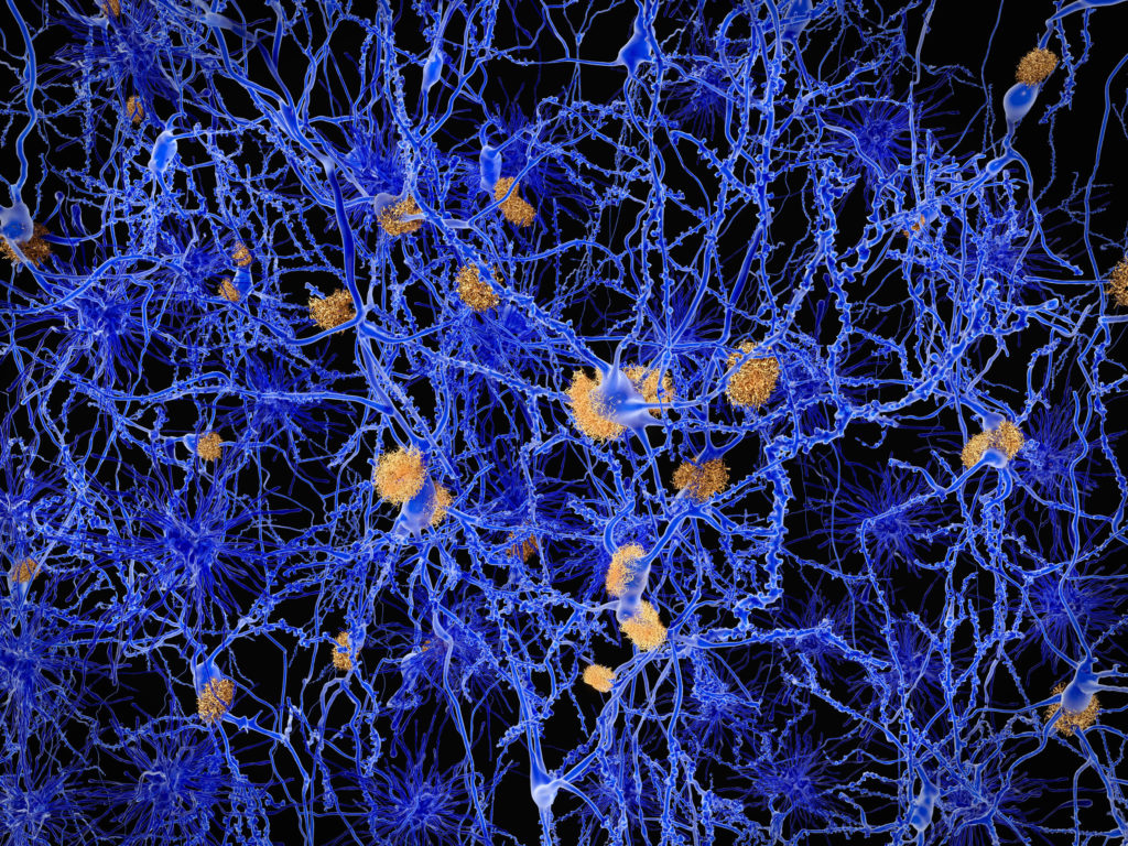
Every neuron relies on a protein called tau to keep its internal skeleton, the microtubule network, working properly. In a group of brain diseases known as “tauopathies,” tau stops doing its job. It misfolds, clumps together, and eventually contributes to the neuron loss that causes memory changes and shifts in behavior. Scientists studying these diseases have long wanted to watch this process unfold from its earliest moments in living human neurons. However, this has been surprisingly difficult.
Why Tau Disease Is Hard to Model in a Dish
Human tau is encoded by the MAPT gene and comes in six isoforms that differ mainly in whether they carry three or four copies of a microtubule-binding segment, called 3R or 4R tau. A healthy adult brain keeps these two forms in rough balance. In several tauopathies, however, that balance tips toward 4R tau.
Neurons grown from induced pluripotent stem cells (iPSCs) are one of the best tools available for modeling this kind of disease in a personalized, physiologically relevant way. But there’s a catch: these neurons tend to behave like fetal neurons and produce almost exclusively 3R tau, regardless of which disease mutation they carry. Researchers have worked around this by combining multiple mutations or adding tau “seeds” from outside the cell, but these approaches do not reflect true disease biology.
Developing a Physiologically Relevant 4R Tau Model
A recent study tested whether disease-relevant splice-site mutations could shift the balance without stacked coding mutations or added tau seeds. The researchers used CRISPR-Cas9 to introduce a mutation (S305N), found in families with frontotemporal dementia (FTD), into human iPSCs. They then differentiated the cells into cortical neurons using a rapid, transcription-factor-based protocol.
S305N sits right at the sequence that determines whether the MAPT messenger RNA includes or skips exon 10, the exon that adds the extra microtubule-binding repeat unique to 4R tau. Surprisingly, that one coding change was enough. Within a week, the edited neurons were making more than 80% 4R tau, climbing above 90% by day 28. This suggests that a single coding change had shifted the neurons from a fetal isoform profile to the 4R-dominant one seen in disease.
A Cascade That Mirrors Human Disease
As 4R tau built up, so did hallmarks of tau pathology. The neurons developed hyperphosphorylated tau and lost tau from their axons as it accumulated around the cell body instead. By day 28, they had begun forming seed-competent tau assemblies capable of templating misfolding in a separate biosensor cell line.
Live imaging over several days showed these assemblies appearing first in axons before spreading toward the cell body, providing a rare, real-time view of tau pathology as it develops. Taken together, these findings suggest the model reflects several of the key pathogenic events seen in human tauopathies.
Making the Model Screening-Ready Using HiBiT
A disease model becomes more valuable when it can also be used to test potential treatments. To track endogenous tau levels at scale, the team used CRISPR to fuse an 11–amino acid HiBiT® tag onto the tau protein at its native genomic location. Due to its small size, the HiBiT tag is unlikely to disturb how tau folds and where it localizes in the cell.
On its own, the tag produces no signal. The Nano-Glo® HiBiT Lytic Detection Reagent supplies the complementary LgBiT® protein, which binds HiBiT tightly enough to reconstitute an active NanoLuc® luciferase enzyme. The luciferase then converts the reagent’s substrate into light in proportion to how much tagged tau is present. Tau abundance therefore reads out as a luminescent signal, with no antibody staining required.
The assay responded as expected to known tau-modulating compounds. Blocking autophagy raised tau levels, while promoting protein clearance or inhibiting tau aggregation lowered them. That kind of predictable, quantifiable readout makes it ideal for high-throughput drug screening.
Why This Model Matters
This study presents the first iPSC-derived neurons carrying a single coding mutation in which endogenous tau assemblies form and can be followed over time. The model isn’t perfect; it does not yet reproduce the mature, thread-like tangles seen in advanced diseases.
Despite its limitations, this cell model is able to capture the sequence of early events—tau misfolding, phosphorylation, and seeding—in a genetically simple, physiologically relevant system. This provides a new scalable approach to study how 4R tauopathies begin, and a platform for testing what might stop them. In the future, it could be used to test potential therapies against various tauopathies.
Reference: Tsefou, E. et al. (2026) Scalable human neuronal models of tauopathy producing endogenous seed-competent 4R tau. Sci Adv. 12(31):eaeg1445
Learn more about HiBiT® protein tagging technology

Latest posts by Johanna Lee (see all)
- A New Human Neuron Model for Tau-Targeted Drug Screening - August 27, 2026
- The Hidden Switch That Controls Lysosome Function - June 11, 2026
- Tiny Edits, Big Harvests: How a Massive Mutation Screen Could Transform Crop Engineering - May 21, 2026
