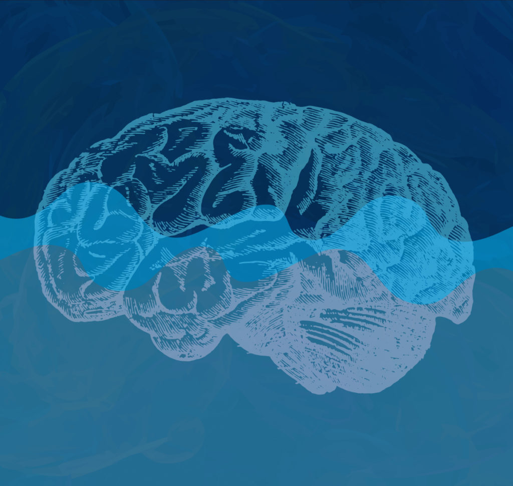
While you and I are getting some shut eye each night, things are happening in our brains. Good things. Therapeutic things.
Think of it as brainwashing of a sort. There is a multiplicity of brain activities going on during sleep, and a November 1 paper in Science shows for the first time when and where in the brain these activities occur, and how they are connected.
Here’s a bit of backstory.
To assess both the progression and pathogenesis of Alzheimer’s disease (AD), as well as the efficacy of AD drugs in clinical trials, there has been interest in the concentrations of amyloid-beta (Aβ) and tau protein in cerebral spinal fluid (CSF).
Early studies of CSF collected by lumbar puncture in human patients showed wide variations in Aβ levels. More recently, studies involving CSF collection by indwelling catheters (allowing more frequent sampling), also revealed significant variability in Aβ levels in human subjects over 24- to 48-hour collection periods (1).
In 2005, reporting in Neuron, Cirrito et al. showed that “Abeta levels in the brain interstitial fluid are dynamically and directly influenced by synaptic activity on a timescale of minutes to hours”, demonstrating a connection between neuron activity and Aβ concentrations (2).
Kang et al. published studies on a connection in variations in Aβ concentration with the sleep-wake cycle, noting that chronically restricted sleep significantly increased Aβ plaque formation in transgenic mice (3).
In the latest studies, published in Science November 1, Fultz et al. simultaneously measured neuron activity, sleep cycle and CSF movement in humans, demonstrating a previously unknown relationship among these brain activities.
Fultz et al. studied human subjects during non-REM sleep, which occurs early in the sleep cycle. Non-REM sleep is recognized as the sleep phase during which memory consolidation occurs.
The study subjects went to sleep in an MRI scanner, allowing researchers to note electrical activity in the brain and thus identify the sleep stage subjects were in, while monitoring the flow of CSF through the brain. At the same time, subjects’ EEGs were tracked to determine brain oxygen levels.
This trifecta of measurements allowed Fultz et al. to see that during non-REM sleep, neuron firing appeared to synchronize, with neurons turning on and off together. As neuron activity decreased, the researchers noted decreased blood flow and thus oxygen levels. At this point large, slow washes of CSF were seen to move through the brain.
The flow of CSF was rhythmic and continued in cycles, along with changes in oxygenation and neuron firing, during non-REM sleep.
This work is fascinating in its explanation of both neural activity during sleep vs. wake cycles, not to mention contribution to how CSF flow and oxygenation occurs during non-REM sleep.
In their Perspective article in the Nov. 1 issue of Science, Grubb and Lauritzen note the contributions of this work to understanding brain communication and a role for CSF washes, noting “The CSF oscillations in human sleep described by Fultz et al. may contribute to the disposal of waste products, such as toxic brain proteins that cause neurodegeneration.”
Here is the paper: Fultz et al. (2019) Coupled electrophysiological, hemodynamic, and cerebrospinal fluid oscillations in human sleep. Science 366, 628–31.
Related Posts
Kari Kenefick
Latest posts by Kari Kenefick (see all)
- What Drives Muscle Fiber Shifts in Obesity and Type ll Diabetes? - October 9, 2025
- How Thalidomide and Molecular Glues Are Redefining Drug Discovery - August 21, 2025
- ATP-Powered Proteins Beyond Kinases – and Why Helicases Are Stealing the Spotlight - August 7, 2025