Did you see the movie where Spider-Man files his taxes? Or the one where Wonder Woman sits on hold with her insurance company while her pasta water boils over? Or where Captain America finds blight on his tomato plants and drives to the county extension office where he spends fifty minutes with a seventy-four-year-old master gardener named Marlys then leaves with a handwritten note covering his soil composition, his watering schedule, and what Marlys calls “the mulching situation”?
No. Because the ordinary day-to-day doesn’t stand a chance next to the saving of the world.
We spend most of our lives in the ordinary. Not because we’re failing to reach the extraordinary, but because the ordinary is what holds everything together while we get there. It’s not the backdrop but the foundation. It’s what the story depends on, whether or not it gets any credit.
Drug Discovery Has a Storytelling Problem
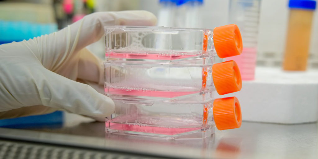
Drug discovery runs almost entirely on ordinary days, punctuated by the moments that make the news: a new target gets identified, a compound shows promise, a trial produces results. Those moments get the headlines, press releases and keynote slots. What doesn’t get the same attention is the years of work behind those moments: the assays, the failed experiments, the redesigns, the slow accumulation of evidence that either holds up or doesn’t. That work has always been the majority of drug discovery.
Some of the most important work in drug discovery ends in a result nobody publishes, but a dead end isn’t a failure of the program. It’s the program working. The researcher who rules something out has learned something true. That knowledge travels forward even when it doesn’t make the headline because it can redirect the next hypothesis, narrow the next experiment or just quietly move things along. That work moves research forward without anyone announcing it.
The Shiniest Thing in the Room
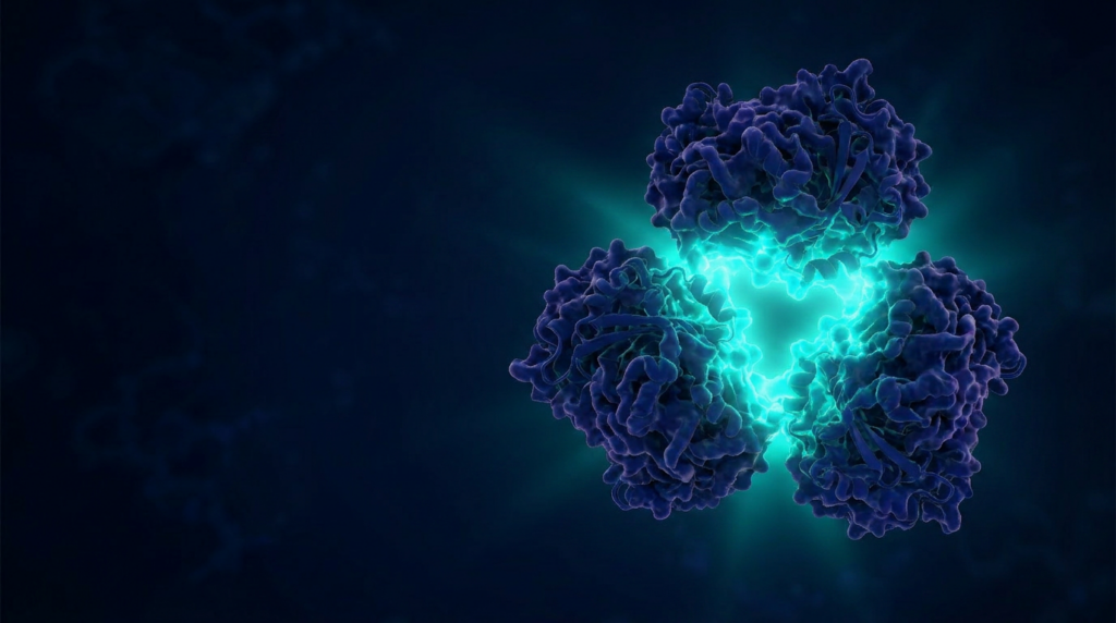
Artificial intelligence is drug discovery’s latest extraordinary announcement, and the fanfare is legitimate. Most of the druggable proteome has never been touched. Of approximately 4,500 human proteins considered druggable, all approved drugs to date work through only 716 distinct targets. Drug hunters knew there was more biology to address but lacked a way to find and prioritize candidates at scale. AI is changing that. By scanning genetic evidence, biological networks and scientific literature at a scale no human team can match, AI is surfacing targets that were previously out of reach and ranking them by the strength of the evidence behind them.
Continue reading “AI Identifies the Target. Someone Still Has to Validate It.”