Virus-Like Particles: All the Bark, None of the Bite

Globally, there have been over 5 million deaths attributed to COVID-19 since the start of the pandemic. Throughout the ongoing battle against SARS-CoV-2, researchers have been studying the viral lineage and the variants that are emerging as the virus evolves over time. The more opportunities that the virus has to replicate (i.e., the more people it infects), the greater the likelihood that a new variant will emerge.

This short video from the World Health Organization explains how viral variants develop.

The US Centers for Disease Control and Prevention (CDC) classify SARS-CoV-2 variants into four groups: Variants Being Monitored (VBM), Variants of Interest (VOI), Variants of Concern (VOC) and Variants of High Consequence (VOHC). So far, no variants in the US have been identified as VOHC or VOI. Currently, the most common variant in the US is the Delta variant (which includes the B.1.617.2 and AY viral lineages), and it is classified as a VOC.

The Delta variant originated in India and spread rapidly across the UK before making its way into the US (1). Current vaccines, including mRNA and adenoviral vector vaccines, have demonstrated effectiveness against the Delta variant. However, it is a VOC because it is more than twice as contagious as previous variants, and some studies have shown that it is associated with more severe symptoms.

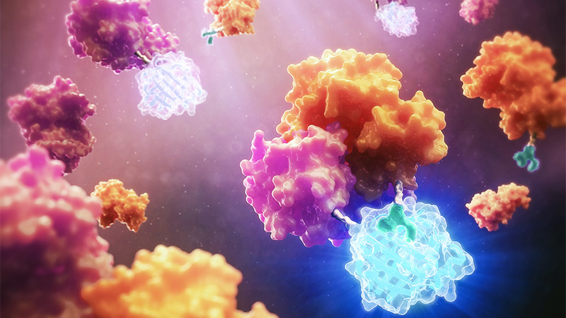
A recent study (2) provides one explanation for the higher infectivity of the Delta variant, using an approach based on virus-like particles (VLPs). The research team was led by Dr. Jennifer Doudna, 2020 Nobel Prize winner for her work on CRISPR-Cas9 gene editing, and Dr. Melanie Ott, director of the Gladstone Institute of Virology at the University of California–Berkeley.

Continue reading “Virus-Like Particles: All the Bark, None of the Bite”
Bringing Cutting Edge Technologies to Academic Researchers Through the Academic Access Program

This post was written by guest blogger Iain Ronald, Director Academic/Government Market Segment at Promega.

My back story is similar to most of you reading this blog, high school education, undergraduate degree then onto a postgraduate degree. However, over 25 years ago during my undergraduate study, I was fortunate enough to work in the lab of Professor Ray Waters studying DNA damage in S. cerevisiae as a model organism and at the time PCR was cutting-edge technology and the PCR license was in full effect. However, there was one company that was fighting the good fight to democratize PCR for the good of the scientific community, Promega.

I became enamored with Promega then, and the next steps in my career were taken with a view to working at this company who, for all intents and purposes, seemed to really care about the progression of science beyond self-aggrandizement.

Now that I am working at Promega in a position where I can bring benefit to our academic community, I have pondered what I can do to equal the disruptive attitude I observed in this company all those years ago when they were fighting the then “big tech” for the enablement of the scientific community. 

Reporter bioassays are one of hte many offerings of the academic access program.
Continue reading “Bringing Cutting Edge Technologies to Academic Researchers Through the Academic Access Program”

A Closer Look at C. difficile Biology with Luminescent Tagging

Clostridium difficile is a bacterium that infects around half a million people per year in the United States. The infection causes symptoms ranging from diarrhea to severe colitis, and it’s one of the most common infections contracted while staying in the hospital. As the global incidence of C. diff infection has risen over the past decade, so has the pressure to develop novel therapeutic strategies. This requires a thorough exploration of all aspects of C. difficile biology.

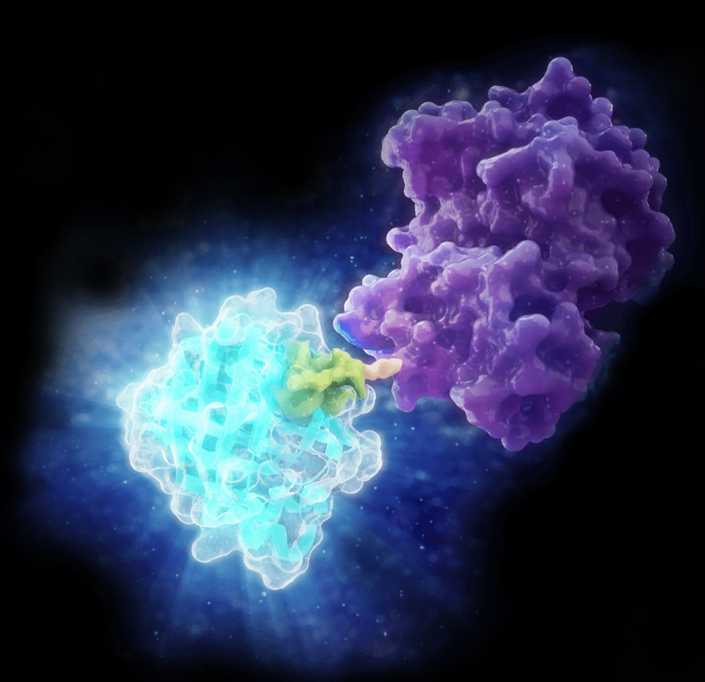
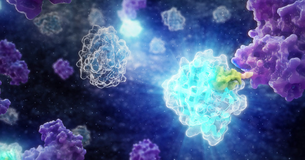
Two recent papers published by researchers at the University of Leiden have shed light on C. difficile physiology using HiBiT protein tagging. The HiBiT system allows detection of proteins in live cells using an 11 amino acid tag. The HiBiT tag binds to the complementary LgBiT polypeptide to reconstitute the luminescent NanoBiT® enzyme. The resulting luminescence is proportional to the amount of HiBiT-tagged protein that is present.

Continue reading “A Closer Look at C. difficile Biology with Luminescent Tagging”

NanoLuc® Luciferase: Brighter Days Ahead for In Vivo Imaging

nanoluc in vivo imaging

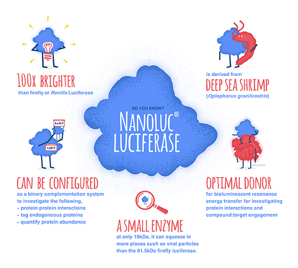
The development of NanoLuc® luciferase technology has provided researchers with new and better tools to study endogenous biology: how proteins behave in their native environments within cells and tissues. This small (~19kDa) luciferase enzyme, derived from the deep-sea shrimp Oplophorus gracilirostris, offers several advantages over firefly or Renilla luciferase. For an overview of NanoLuc® luciferase applications, see: NanoLuc® Luciferase Powers More than Reporter Assays.

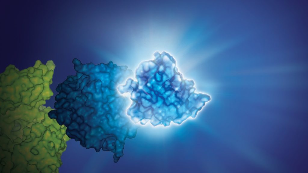
The small size of NanoLuc® luciferase, as well the lack of a requirement for ATP to generate a bioluminescent signal, make it particularly attractive as a reporter for in vivo bioluminescent imaging, both in cells and live animals. Expression of a small reporter molecule as a fusion protein is less likely to interfere with the biological function of the target protein. NanoLuc® Binary Technology (NanoBiT®) takes this approach a step further by creating a complementation reporter system where one subunit is just 11 amino acids in length. This video explains how the high-affinity version of NanoBiT® complementation (HiBiT) works:

Continue reading “NanoLuc® Luciferase: Brighter Days Ahead for In Vivo Imaging”

CRISPR/Cas9 HiBiT Knock-In: A Scalable Approach for Studying Endogenous Protein Dynamics

Studying protein function in live cells is limited by the tools available to analyze the expression and interactions of those proteins. Although mass spectrometry and antibody-based protein detection are valuable technologies for protein analysis, both methods have drawbacks that limit the range of targets and contexts in which proteins can be investigated.

Mass spectrometry is often poor at detecting low-abundance proteins. Antibody-based techniques require high quality, specific antibodies, which can be difficult to impossible to acquire. Both methods require cell lysis, preventing real-time analysis and limiting the physiological relevance, and both methods can be limiting for higher-throughput analysis. While plasmid-based overexpression of tagged target proteins simplifies detection and can allow for real time analysis, protein levels don’t typically resemble endogenous levels. Overexpression also has the potential to create experimental artifacts or limit the dynamic range of an observed response.

In 2018, Promega R&D scientists published a paper in ACS Chemical Biology demonstrating the use of CRISPR/Cas9 to integrate the 11 amino acid, bioluminescent HiBiT tag directly into the genome to serve as an easily measured reporter for endogenous proteins. This provides a highly quantitative method for investigating cellular protein dynamics that sidesteps the need for cloning and other drawbacks to conventional methods, including the ability to measure changing protein dynamics in real-time. (For more details about CRISPR/Cas9 knock-in tagging and other applications, read this blog.)

While their findings showed that this method provides efficient and specific tagging of endogenous proteins, the research was limited to just five different proteins within a single signaling pathway in two cell lines. This left unanswered questions about whether this approach was scalable, had broader applications and how accurately the natural biology of the cells was represented.

Continue reading “CRISPR/Cas9 HiBiT Knock-In: A Scalable Approach for Studying Endogenous Protein Dynamics”

NanoLuc® Luciferase Powers More than Reporter Assays

Bright NanoLuc® Luciferase

What can you do with a small, super bright luciferase? Amazing things. We’ve highlighted many of the papers and new applications that NanoLuc® luciferase has enabled on this blog. While NanoLuc® luciferase was first introduced as a reporter enzyme to assess promoter activity, its capabilities have expanded far beyond a genetic reporter, creating bioluminescent tools used to study endogeneous protein dynamics, target engagement, protein degradation, immunodetection and more. So where did the NanoLuc luciferase come from and how does one enzyme power so many research capabilities? Read further for a primer on the various technologies and applications developed from this enzyme over the last 10 years.

Continue reading “NanoLuc® Luciferase Powers More than Reporter Assays”

Popular Papers from Promega Authors

Promega is a chemistry and instrument supplier to scientists in diverse industries and research labs around the world. True. But we are more than just a supply company; we are scientists dedicated to supporting the work of other scientists. We want the science behind the technologies we develop to be both vetted and valued by the scientific community at large, which is one reason our scientists take the time to prepare and submit manuscripts to peer-reviewed journals. Here we call out some of our published research papers that were highly read in 2019. In the journal ACS Chemical Biology alone, five Promega-authored papers were among the top 10 most read papers in 2019. Here’s a quick review of the highlights from these ACS papers.

Continue reading “Popular Papers from Promega Authors”

CRISPR/Cas9 Knock-In Tagging: Simplifying the Study of Endogenous Biology

Understanding the expression, function and dynamics of proteins in their native environment is a fundamental goal that’s common to diverse aspects of molecular and cell biology. To study a protein, it must first be labeled—either directly or indirectly—with a “tag” that allows specific and sensitive detection.

Using a labeled antibody to the protein of interest is a common method to study native proteins. However, antibody-based assays, such as ELISAs and Western blots, are not suitable for use in live cells. These techniques are also limited by throughput and sensitivity. Further, suitable antibodies may not be available for the target protein of interest.

Continue reading “CRISPR/Cas9 Knock-In Tagging: Simplifying the Study of Endogenous Biology”

NanoLuc: Tiny Tag with a Big Impact

Synthetic biology—genetically engineering an organism to do or make something useful—is the central goal of the iGEM competition each year. After teams conquer the challenge of cloning their gene, the next hurdle is demonstrating that the engineered gene is expressing the desired protein (and possibly quantifying the level of expression), which they may do using a reporter gene.

Reporters can also play a more significant role in iGEM projects when teams design their organism with reporter genes to detect and signal the presence of specific molecules, like environmental toxins or biomarkers. Three of the iGEM teams Promega sponsored this year opted to incorporate some version of NanoLuc® Luciferase into their projects.

NanoLuc® luciferase is a small monomeric enzyme (19.1kDa, 171 amino acids) based on the luciferase from the deep sea shrimp Oplophorus gracilirostris. This engineered enzyme uses a novel substrate, furimazine, to produce high-intensity, glow-type luminescence in an ATP-independent reaction. Unlike other molecules for tagging and detecting proteins, NanoLuc® luciferase is less likely to interfere with enzyme activity and affect protein production due to its small size.

NanoLuc® Luciferase has also been engineered into a structural complementation reporter system, NanoBiT® Luciferase, that contains a Large subunit (LgBiT) and two small subunit options: low affinity SmBiT and high affinity HiBiT. Together, these NanoLuc® technologies provide a bioluminescent toolbox that was used by the iGEM teams to address a diverse set of biological challenges.

Here is an overview of each team’s project and how they incorporated NanoLuc® technology.

Continue reading “NanoLuc: Tiny Tag with a Big Impact”

Choosing a Tag for Your Protein

You have identified and cloned your protein of interest, but you want to explore its function. A protein fusion tag might help with your investigation. However, choosing a tag for your protein depends on what experiments you are planning. Do you want to purify the protein? Would you like to identify interacting proteins by performing pull-down assays? Are you interested in examining the endogenous biology of the protein? Here we cover the advantages and disadvantages of some protein tags to help you select the one that best suits your needs.

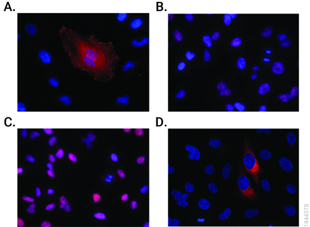
Immunofluorescent detection of HiBiT-tagged proteins in CRISPR-edited cell pools and clones using the Anti-HiBiT Monoclonal Antibody.
CRISPR-Cas9 editing knocked-in HiBiT at the endogenous locus of proteins with varying subcellular localization. Fixed CRISPR-modified clones or pools of cells were imaged by immunofluorescent staining using the Anti-HiBiT Monoclonal Antibody (red) and Hoechst dye (blue). Panel A. VCL-HiBiT pool. Panel B. SMARCA4-HiBiT clone. Panel C. HDAC2-HiBiT clone. Panel D. HSP90B1-HiBiT pool.

Affinity Tags

The most commonly used protein tags fall under the category of affinity tags. This means that the tag binds to another molecule or metal ion, making it easy to purify or pull down your protein of interest. In all cases, the tag will be fused to your protein of interest at either the amino (N) or carboxy (C) terminus by cloning into an expression vector. This protein fusion can then be expressed in cells or cell-free systems, depending on the promoter the vector contains.

Continue reading “Choosing a Tag for Your Protein”