NanoLuc: Tiny Tag with a Big Impact

Synthetic biology—genetically engineering an organism to do or make something useful—is the central goal of the iGEM competition each year. After teams conquer the challenge of cloning their gene, the next hurdle is demonstrating that the engineered gene is expressing the desired protein (and possibly quantifying the level of expression), which they may do using a reporter gene.

Reporters can also play a more significant role in iGEM projects when teams design their organism with reporter genes to detect and signal the presence of specific molecules, like environmental toxins or biomarkers. Three of the iGEM teams Promega sponsored this year opted to incorporate some version of NanoLuc® Luciferase into their projects.
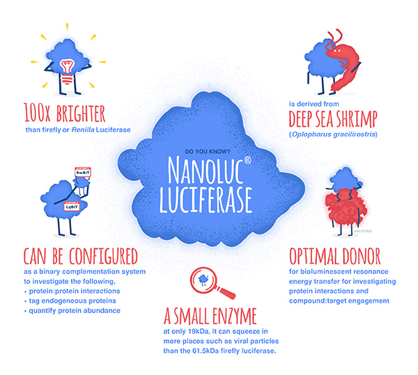
NanoLuc® luciferase is a small monomeric enzyme (19.1kDa, 171 amino acids) based on the luciferase from the deep sea shrimp Oplophorus gracilirostris. This engineered enzyme uses a novel substrate, furimazine, to produce high-intensity, glow-type luminescence in an ATP-independent reaction. Unlike other molecules for tagging and detecting proteins, NanoLuc® luciferase is less likely to interfere with enzyme activity and affect protein production due to its small size.

NanoLuc® Luciferase has also been engineered into a structural complementation reporter system, NanoBiT® Luciferase, that contains a Large subunit (LgBiT) and two small subunit options: low affinity SmBiT and high affinity HiBiT. Together, these NanoLuc® technologies provide a bioluminescent toolbox that was used by the iGEM teams to address a diverse set of biological challenges.

Here is an overview of each team’s project and how they incorporated NanoLuc® technology.

Continue reading “NanoLuc: Tiny Tag with a Big Impact”
Choosing a Tag for Your Protein

You have identified and cloned your protein of interest, but you want to explore its function. A protein fusion tag might help with your investigation. However, choosing a tag for your protein depends on what experiments you are planning. Do you want to purify the protein? Would you like to identify interacting proteins by performing pull-down assays? Are you interested in examining the endogenous biology of the protein? Here we cover the advantages and disadvantages of some protein tags to help you select the one that best suits your needs.

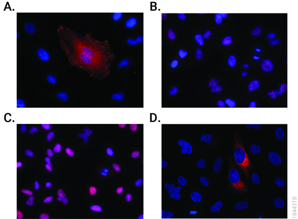
Immunofluorescent detection of HiBiT-tagged proteins in CRISPR-edited cell pools and clones using the Anti-HiBiT Monoclonal Antibody.
CRISPR-Cas9 editing knocked-in HiBiT at the endogenous locus of proteins with varying subcellular localization. Fixed CRISPR-modified clones or pools of cells were imaged by immunofluorescent staining using the Anti-HiBiT Monoclonal Antibody (red) and Hoechst dye (blue). Panel A. VCL-HiBiT pool. Panel B. SMARCA4-HiBiT clone. Panel C. HDAC2-HiBiT clone. Panel D. HSP90B1-HiBiT pool.

Affinity Tags

The most commonly used protein tags fall under the category of affinity tags. This means that the tag binds to another molecule or metal ion, making it easy to purify or pull down your protein of interest. In all cases, the tag will be fused to your protein of interest at either the amino (N) or carboxy (C) terminus by cloning into an expression vector. This protein fusion can then be expressed in cells or cell-free systems, depending on the promoter the vector contains.

Continue reading “Choosing a Tag for Your Protein”

All You Need is a Tether: Improving Repair Efficiency for CRISPR-Cas9 Gene Editing

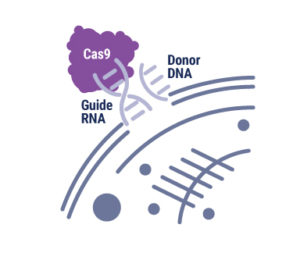
Ribonucleoprotein complex with Cas9, guide RNA and donor ssDNA. Copyright Promega Corporation.

With the advent of genome editing using CRISPR-Cas9, researchers have been excited by the possibilities of precisely placed edits in cellular DNA. Any double-stranded break in DNA, like that induced by CRISPR-Cas9, is repaired by one of two pathways: Non-homologous end joining (NHEJ) or homology-directed repair (HDR). Using the NHEJ pathway results in short insertions or deletions (indels) at the break site, so the HDR pathway is preferred. However, the low efficiency of HDR recombination to insert exogenous sequences into the genome hampers its use. There have been many attempts at boosting HDR frequency, but the methods compromise cell growth and behave differently when used with various cell types and gene targets. The strategy employed by the authors of an article in Communications Biology tethered the DNA donor template to Cas9 complexed with the ribonucleoprotein and guide RNA, increasing the local concentration of the donor template at the break site and enhancing homology-directed repair. Continue reading “All You Need is a Tether: Improving Repair Efficiency for CRISPR-Cas9 Gene Editing”

Lighting Up GPCR Research with Bioluminescent Tagging

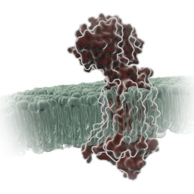
G Protein-Coupled Receptors (GPCRs) are a very large, diverse family of transmembrane receptors in eukaryotes. These receptors detect molecules outside the cell and activate internal signaling pathways by coupling with G proteins. Once a GPCR is activated, β-arrestins translocate to the cell membrane and bind to the occupied receptor, uncoupling it from G proteins and promoting its internalization.

Reporter tags are useful for studying the dynamics of GPCRs and associated proteins, but large tags can disrupt the receptors’ native functioning, and often overexpression of the tagged protein is required to obtain sufficient signal. Here is one example of how researchers have used the small, bright NanoLuc® luciferase to overcome these common challenges and answer questions about GPCRs.

Continue reading “Lighting Up GPCR Research with Bioluminescent Tagging”