Psilocybin as Antidepressant: Quick Acting, Lasting Benefits

Depression is not simply a mood disorder, a feeling of sadness, or being ill at ease. Depression can completely shut a person down, manifesting as an inability to make decisions, to take action, to think. Even sleep is affected by depression.

Researchers and clinicians who treat depression are learning that the physical manifestations can be mirrored by internal, cellular changes. Some people with depression have decreases in their gray matter volume, particularly in areas like the hippocampus (important to memory, learning, and emotions) and prefrontal cortex (where higher-level thought and planning abilities are based).

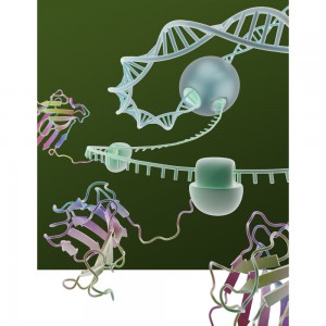
Additionally, imaging has shown a decrease in the number of synapses—the structures through which electrical or chemical signals are passed between neurons and other cells—in persons with chronic depression. Without the signals that synapses transmit, brain function is disrupted.

And without intervention in depression, synapse decrease can continue.

While there are drugs and behavioral therapies to treat depression, these therapies can be slow to act and sometimes ineffective. In addition, once synaptic loss has occurred, these therapies are less effective.

In their August 2021 paper, “Psilocybin induces rapid and persistent growth of dendritic spines in frontal cortex in vivo” (1), Shao et al. state,

“It has long been recognized that these compounds (serotonergic psychedelics like psilocybin) may have therapeutic potential for neuropsychiatric disorders, including depression, obsessive-compulsive disorder and addiction”.

Continue reading “Psilocybin as Antidepressant: Quick Acting, Lasting Benefits”

How Does DNA Template Length Influence Gene Expression in Cell Free Systems?

3D sketch of coupled transcription/translation in a cell free system. A recent paper looks at the effect of DNA Template Length  on gene expression.

Cell-free gene expression systems are a staple tool for the researcher seeking to understand the regulation of transcription and translation. Many factors can affect the efficiency of cell-free gene expression including vector sequence, reaction components and the template DNA concentration. One factor that has not been extensively studied is how DNA template length influences gene expression.

Continue reading “How Does DNA Template Length Influence Gene Expression in Cell Free Systems?”

I Have My Luciferase Vector, Now What?

Choosing and Optimizing Transfection Methods

Here in Technical Services we often talk with researchers at the beginning of their project about how to carefully design and get started with their experiments. It is exciting when you have selected the Luciferase Reporter Vector(s) that will best suit your needs; you are going to make luminescent cells! But, how do you pick the best way to get the vector into your cells to express the reporter? What transfection reagent/method will work best for your cell type and experiment? Do you want to do transient (short-term) transfections, or are you going to establish a stable cell line?

Continue reading “I Have My Luciferase Vector, Now What?”

Why You Don’t Need to Select a Wavelength for a Luciferase Assay

Promega kit depicted; test involves wavelength for a luciferase assay.

It’s a question I’m asked probably once a week. “What wavelength do I select on my luminometer when performing a luciferase assay?” The question is a good and not altogether unexpected one, especially for those new to bioluminescent assays. The answer is that in most cases, you don’t and in fact shouldn’t select a wavelength (the exception to this rule is if you’re measuring light emitted in two simultaneous luciferase reactions). To understand why requires a bit of an explanation of absorbance, fluorescence, and luminescence assays, and the differences among them.

Absorbance, fluorescence, and luminescence assays are all means to quantify something of interest, be that a genetic reporter, cell viability, cytotoxicity, apoptosis, or other markers. In principle, they are all similar. For example, a genetic reporter assay is an indicator of gene expression. The promoter of a gene of interest can be cloned upstream of a reporter such as β-galactosidase, GFP, or firefly luciferase. The amount of each of these reporters that is transcribed into mRNA and translated into protein by the cell is indicative of the endogenous expression of the gene of interest.

Continue reading “Why You Don’t Need to Select a Wavelength for a Luciferase Assay”

Working with RNA doesn’t have to be a nightmare

We’re all familiar with the Central Dogma of Molecular Biology: DNA is transcribed into RNA, which is translated into proteins. It’s drilled into our heads from the early days of biology classes, and it’s surprisingly useful when we start exploring in our own research projects. For example, if you’re interested in gene expression, you’ll most likely be working with RNA, specifically mRNA. Messenger RNA (mRNA) is transcribed from DNA and is used by ribosomes as a “template” for a specific protein. The total mRNA in a cell represents all of the genes that are actively being transcribed. So, if you want to know whether or not a gene is being transcribed, RNA purification is a great place to start.

When preparing your RNA samples for a downstream assay, there are several roadblocks and pitfalls that could give you quite a headache. Let’s tackle two of the most common.

Continue reading “Working with RNA doesn’t have to be a nightmare”

Shining Light on a Superbug

Antibiotic-resistant bacteria and their potential to cause epidemics with no viable treatment options have been in the news a lot. These “superbugs,” which have acquired genes giving them resistance to common and so-called “last resort” antibiotics, are a huge concern as effective treatment options dwindle. Less attention has been given to an infection that is not just impervious to antibiotics, but is actually enabled by them.

Clostridium diffic33553646_lile Infection (CDI) is one of the most common healthcare-associated infections and a significant global healthcare problem. Clostridium difficile (C. diff), a Gram-positive anaerobic bacterium, is the source of the infection. C. diff spores are very resilient to environmental stressors, such as pH, temperature and even antibiotics, and can be found pretty much everywhere around us, including on most of the food we eat. Ingesting the spores does not usually lead to infection inside the body without also being exposed to antibiotics.

Individuals taking antibiotics are 7-10 times more likely to acquire a CDI. Antibiotics disrupt the normal flora of the intestine, allowing C. diff to compete for resources and flourish. Once exposed to the anaerobic conditions of the human gut, these spores germinate into active cells that embed into the tissue lining the colon. The bacteria are then able to produce the toxins that can cause disease and result in severe damage, or even death. Continue reading “Shining Light on a Superbug”

A Crash Course in CRISPR

CRISPR is a hot topic right now, and rightly so—it is revolutionizing research that relies on editing genes. But what exactly is CRISPR? How does it work? Why is everyone so interested in using it? Today’s blog is a beginner’s guide on how CRISPR works with an overview of some new applications of this technology for those familiar with CRISPR.

Introduction to CRISPR/Cas9

27850283-June-13-CRISPR-image-WEB

Clustered Regularly Interspaced Short Palindromic Repeats (CRISPR) were discovered in 1987, but it took 30 years before scientists identified their function. CRISPRs are a special kind of repeating DNA sequence that bacteria have as part of their “immune” system against invading nucleic acids from viruses and other bacteria. Over time, the genetic material from these invaders can be incorporated into the bacterial genome as a CRISPR and used to target specific sequences found in foreign genomes.

CRISPRs are part of a system within a bacterium that requires a nuclease (e.g. Cas9), a single guide RNA (sgRNA) and a tracrRNA. The tracrRNA recruits Cas9, while sgRNA binds to Cas9 and guides it to the corresponding DNA sequence of the invading genome. Cas9 then cuts the DNA, creating a double-stranded break that disables its function. Bacteria use a Protospacer Adjacent Motif, or PAM, sequence near the target sequence to distinguish between self and non-self and protect their own DNA.

While this system is an effective method of protection for bacteria, CRISPR/Cas9 has been manipulated in order to perform gene editing in a lab (click here for a video about CRISPR). First, the tracrRNA and sgRNA are combined into a single molecule. Then the sequence of the guide portion of this RNA is changed to match the target sequence. Using this engineered sgRNA along with Cas9 will result in a double-stranded break (DSB) in the target DNA sequence, provided the target sequence is adjacent to a compatible PAM sequence.

Continue reading “A Crash Course in CRISPR”

Analyzing the Effects of Yersinia pestis Infection on Gene Expression

Yersinia pestis. See page for author [Public domain], via Wikimedia Commons
While scientists using ancient DNA analysis are learning how Yersinia pestis developed over time into the causative agent of three worldwide pandemics, there is still much to learn about the early hours and days of an organism infected with the plague. Y. pestis still infects humans so any insight into disease progression is useful for determining treatment timing and even developing novel treatments to supplement or replace antibiotics. A 2012 study observed how Y. pestis injected into mice spread throughout the body using bioluminescent imaging to track the infection. More recent research reported in PLOS ONE used a more real-world route of infection by introducing an aerosolized Y. pestis to a nonhuman primate model and tracking the transcripts altered during the first 42 hours of infection. Continue reading “Analyzing the Effects of Yersinia pestis Infection on Gene Expression”

Cancer Detection on a Chip?

Figure 5 shows typical scanned images of bead-array for analyzing adjacent normal tissue and tumor tissue. Huang et al. (2015) Digital Detection of Multiple Minority Mutants and Expression Levels of Multiple Colorectal Cancer-Related Genes Using Digital-PCR Coupled with Bead-Array. PLOS ONE 10(4):e0123420. doi:10.1371/journal.pone.0123420.g005
The ideal cancer detection method would involve giving a sample of blood or tissue and using DNA or RNA analysis to determine if there were any gene sequence or gene expression changes that are known hallmarks of cancer. Unfortunately, most current screening methods used are not so precise and in some cases are invasive. However useful tests for colon cancer may be, many people do not subject themselves to the standard colonoscopy. What if there was an easier, noninvasive method that could be used to screen for cancer and detect changes at the early, easily treatable stages of cancer? A recent article in PLOS ONE describes just such a mutation detection method for colorectal cancer using purified nucleic acid with a method that involves emulsion PCR, bead arrays and fluorescent probes. Continue reading “Cancer Detection on a Chip?”

Riboprobes: RNA Probes Are Still Valuable Research Tools

9613ca[1]Riboprobes are RNA probes that can be produced by in vitro transcription of cloned DNA inserted in a suitable plasmid downstream of a viral promoter.
Viruses code for their own RNA polymerases, which are highly specific for the viral promoters. Using these enzymes, labeled NTPs, and inserts in both forward and reverse orientations, both sense and antisense riboprobes can be generated from a cloned gene.
Transcription of RNA is performed with the appropriate RNA polymerase (T3, T7 or SP6), depending on the RNA polymerase promoter sites present in the chosen vector. Because these polymerases are extremely promoter-specific (i.e., there is almost no transcriptional cross talk), virtually homogeneous RNA can be obtained using plasmid DNA as the template in a transcription reaction. When it is desirable to copy only insert DNA sequences, the plasmid is linearized at an appropriate restriction site before the transcription reaction and only discrete “run-off” transcripts are obtained, virtually free of vector sequences. RNA transcripts may be used to generate radioactive probes for hybridization to Northern and Southern blots, plaque and colony lifts as well as non-radioactive probes (i.e, labeled with digoxgenin)for in situ hybridization.

Recent references using riboprobes include: Continue reading “Riboprobes: RNA Probes Are Still Valuable Research Tools”