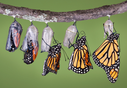
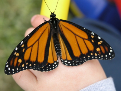
Metamorphosis. In the case of butterflies, it is nature’s version of the great makeover. A plump and slow caterpillar transforms itself into a chrysalis and, tucked snuggly away from the curious prying eyes of the world, metamorphoses into a colorful, graceful butterfly.
Growing up in Iowa, my sister and I had a summer tradition of stalking the milkweed plants in search of Monarch caterpillars. Once we captured our prey, we brought our new acquisitions home and placed them in lovingly crated Mason jar containers filled with milkweed leaves and sticks. Over the next few days these lucky caterpillars lived in the lap of luxury with a constant supply of milkweed leaves. Once the time came for the caterpillar to transform into a chrysalis, we waited with baited breath for the butterfly to emerge.
As a child, those ten or so days I spent watching the unchanging chrysalis were filled with breathless speculation about what must be happening inside. Years later in biology class I learned all about the stages of butterfly development and what was really happening inside the shell of the chrysalis. This knowledge came with a little kernel of sadness though, because there was only one way for science to have figured out what was happening inside the chrysalis, and that way did not end well for the butterfly-to-be.
Wouldn’t it have been nice if we could take pictures of what was happening inside the chrysalis without disturbing nature’s makeover mid cycle?
This is just what researchers from the University of Manchester and the Natural History Museum in London have done (1). Using high-resolution X-ray computed tomography (CT), they developed three-dimensional images of what was happening inside the chrysalis of a painted lady butterfly.
This approach allowed the researchers to follow a single individual pupa through its development, something that previous approaches couldn’t do. They were able to take measurements that really couldn’t be done during dissection, such as measuring tracheal and gut volume. They were also able to reveal new developmental details such as the surprising early development of the adult respiratory system.
This new approach could open up a whole new way to study the development of any number insects. For example, it could help us understand the impact of environmental factors such as pesticide use on honey bee populations. It also means that future biology students might be able to learn about insect development in more detail and without the twinge of sadness that I experienced. In all this is a good thing for us and the butterflies.
Reference
- Lowe, T., Garwood, R.J., Simonsen, .TJ., Bradley, R.S. and Withers, P.J. ( 2013) Metamorphosis revealed: time-lapse three dimensional imaging inside a living chrysalis. J. R. Soc. Interface 10, 20130304. http://dx.doi.org/10.1098/rsif.2013.0304
Kelly Grooms
Latest posts by Kelly Grooms (see all)
- Growing Our Understanding of Triple-Negative Breast Cancer in Sub-Saharan Africa: Why Comprehensive Population Data Matters - June 5, 2025
- Measles and Immunosuppression—When Getting Well Means You Can Still Get Sick - April 17, 2025
- IC50, EC50 and Kd: What is the Difference and Why Do They matter? - March 6, 2025
Very cool. Thanks for sharing!