If you’re familiar with bioluminescence, you’ve probably used it in plate-based experiments to track various biological processes. You understand it provides distinct advantages over traditional fluorescence assays, particularly when it comes to sensitivity. However, there’s always that one nagging question: how representative is the signal on a cell-to-cell level?
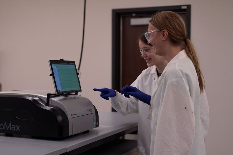
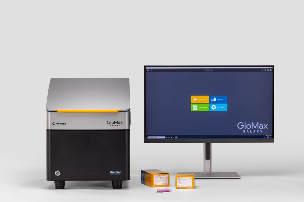
Traditional approaches to decipher cell-to-cell signal rely on complex, time-intensive measures that only approximated the findings acquired through bioluminescence. That’s where the GloMax® Galaxy Bioluminescence Imager comes in. Built to visualize the NanoLuc®-based assays we engineered, the Galaxy goes beyond simple numeric outputs to reveal what’s happening in individual cells.
NanoLuc® luciferase is up to 150X brighter than firefly or Renilla luciferase, giving it the sensitivity to detect subtle protein interactions in complex biological systems. This bright luminescent enzyme enables a much broader linear range than fluorescence, improving detection of small changes in protein activity, such as proteins interacting. Microplate readers measuring NanoLuc® assays rely on signal generated from many cells. This results in an approximation of what is occurring biologically. Truly validating those luminescent readings within a cell population has been challenging—until now. The GloMax® Galaxy allows you to perform bioluminescence imaging, moving beyond the numbers, offering the power to visualize protein interactions directly.
Continue reading “Visualize Protein:Protein Interactions with Bioluminescence Imaging”