Liver disease is a global health challenge, affecting millions each year. The liver has a remarkable ability to regenerate; however, chronic damage arising from obesity, alcohol, or metabolic dysfunction can lead to irreversible failure. At the University of Edinburgh’s Centre for Regenerative Medicine, Professor David Hay’s lab is developing innovative ways to study liver function and disease using a lab-grown mini-organ. In this blog, we highlight how Dr. Hay’s lab is redefining liver disease research through 3D models that reveal how hormones influence metabolic health.
Continue reading “Insights from 3D Liver Models: Rethinking Fatty Liver Disease with Hormone Correction”Tips and Tools
Exploring How NEAT1 Shapes Granulosa Cell Function

Granulosa cells (GCs), which surround and support developing oocytes, play a critical role in estrogen production, follicle maturation and overall ovarian health (3). Their ability to regulate hormone production and cell survival makes them a central focus in studies of ovarian biology.
A recent study investigated how the long non-coding RNA (lncRNA) NEAT1 regulates GC function and mapped a pathway that links NEAT1 expression to cell proliferation, apoptosis and hormone production (1).
Continue reading “Exploring How NEAT1 Shapes Granulosa Cell Function”Cellular Selectivity Profiling: Unveiling Novel Interactions and More Accurate Compound Specificity
This blog was written by guest contributor Tian Yang, Associate Product Manager, Promega, in collaboration with Kristin Huwiler, Manager, Small Molecule Drug Discovery, Promega.
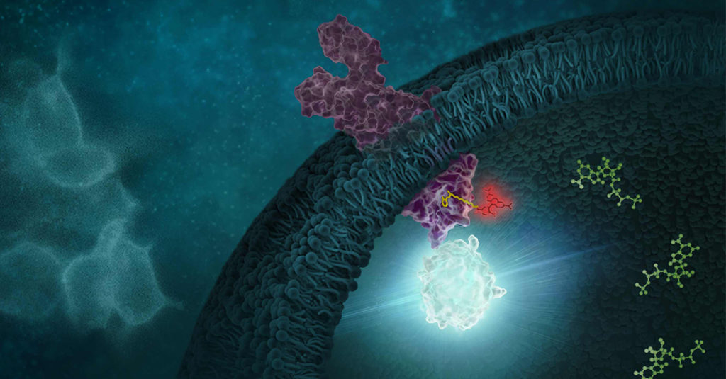
Determining the selectivity of a compound is critical during chemical probe or drug development. In the case of chemical probes, having a clearly defined mechanism of action and specific on-target activity are needed for a chemical probe to be useful in delineating the function of a biological target of interest in cells. Similarly, optimizing a drug candidate for on-target potency and reducing off-target interactions is important in the drug development process (1,2). A thorough understanding of the selectivity profile of a drug can facilitate drug repurposing, by enabling approved therapeutics to be applied to new indications (3). Interestingly, small molecule drugs do not necessarily require the same selectivity as a chemical probe, since some drugs may benefit from polypharmacology to achieve their desired clinical outcome.
Selectivity profiling panels based on biochemical methods have commonly been used to assess compound specificity for established target classes in drug discovery and chemical probe development. Biochemical assays are target-specific and often quantitative, enabling direct measurements of compound affinities for targets of interest and facilitate comparison of compound engagement to a panel of targets. As an example, several providers offer kinase selectivity profiling services using different assay formats and kinase panels comprised of 100 to 400 kinases (4). However, just as biochemical target engagement does not always translate to cellular activity, selectivity profiles based on biochemical platforms may not reflect compound selectivity in live cells (5).
Continue reading “Cellular Selectivity Profiling: Unveiling Novel Interactions and More Accurate Compound Specificity”Unlocking the Power of Live-Cell Kinetics in Degrader Development
In targeted protein degradation (TPD), timing is everything. Understanding not just whether a degrader works—but how fast, how thoroughly and how sustainably—can dramatically influence early discovery decisions. Dr. Kristin Riching (Promega) dove into the real-time world of degradation kinetics in the webinar: Degradation in Motion: How Live-Cell Kinetics Drive Degrader Optimization, sharing how dynamic data provides a clearer view of degrader performance than traditional endpoint assays.
Whether you’re exploring your first PROTAC or optimizing a molecular glue series, the expertise offered in Dr. Riching’s presentation gives you actionable insights that will help you connect kinetic data to better therapeutic design.
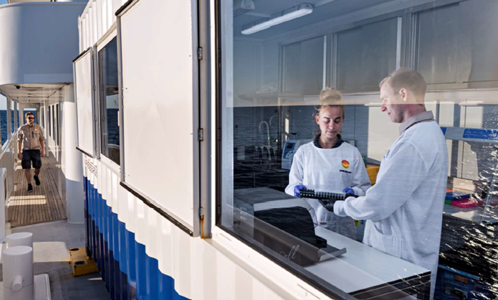
How The OceanOmics Centre is Using the Maxwell RSC to Scale eDNA Biodiversity Monitoring
This blog is written by guest blogger Ben Rushton, Application Specialist/Territory Manager at Promega Australia.
When you’re monitoring marine biodiversity at scale, every drop of seawater tells a story. At Minderoo OceanOmics Centre at the University of Western Australia, scientists are uncovering that story through environmental DNA (eDNA)—and automation is helping them listen more clearly.
Laura Missen, a Scientific Officer at OceanOmics Centre, shares how automating their DNA extraction workflow with the Maxwell® RSC 48 system has transformed how they gather and interpret data from marine ecosystems.
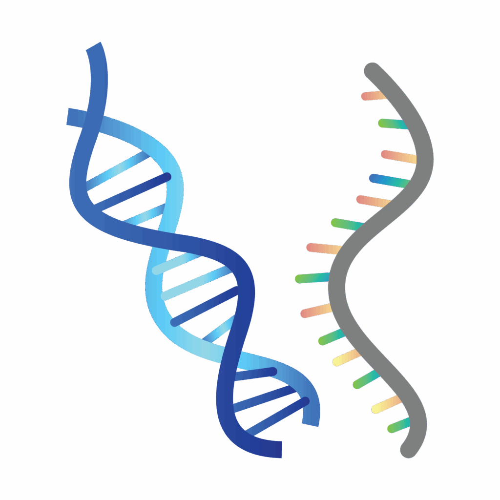
Exploring the Relationship Between IC50 and Kd in Pharmacology
This guest blog post is written by Tian Yang, Associate Product Manager at Promega.
In the realm of chemical probe development and drug discovery, understanding the interactions between drugs/compounds and their targets is crucial. Two frequently used metrics to characterize these interactions are IC50 and Kd, which guide researchers in evaluating the potential of compounds in effecting changes in target function. IC50 offers insights into a compound’s potency by quantifying its ability to inhibit a specific biological activity. Kd provides a measure of the affinity between a ligand and its receptor, reflecting how tightly a compound binds to its target (1). Together, these parameters are instrumental in the early stages of drug development, helping to identify promising candidates by assessing a compounds’s binding characteristics and its observed efficacy.
Continue reading “Exploring the Relationship Between IC50 and Kd in Pharmacology”Why mRNA Transfection Is Transforming Transient Expression Workflows
Transfection is a core technique in molecular biology used to introduce foreign nucleic acids—such as DNA, RNA, or small RNAs like siRNA, shRNA, and miRNA—into eukaryotic cells. This enables researchers to manipulate gene expression and study cellular processes, disease mechanisms and therapeutic strategies (1).
Advances in transfection technology now support a range of nucleic acid types and cell models. Researchers can pursue transient or stable expression to achieve specific goals: knocking down transcripts, expressing proteins, or probing promoter activity in systems from immortalized lines to stem cells (1).
Continue reading “Why mRNA Transfection Is Transforming Transient Expression Workflows”5 Questions to Ask When Your RT-qPCR Isn’t Working

RT-qPCR (reverse transcription quantitative PCR) is a powerful technique for quantifying RNA expression—but it doesn’t always cooperate. Even when you’ve followed the protocol carefully, unexpected results can appear: flat curves, unexpected Cq values, or inconsistent replicates. When that happens, you’re left wondering… what went wrong?
In this blog, we’ll walk through five key questions to help you troubleshoot RT-qPCR issues with confidence. From common errors to more stubborn challenges, we’ll also explore what to consider when technique isn’t fully the problem—and when it might be time to rethink your reagents.
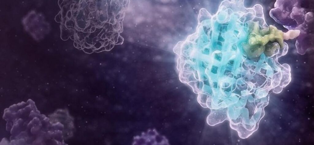
Continue reading “5 Questions to Ask When Your RT-qPCR Isn’t Working”Drug Target Confirmed? Tivantinib’s Lesson on the Importance of Cellular Target Engagement
This guest blog post is written by Tian Yang, Associate Product Manager at Promega.
There are often challenges with translating results from a test tube into a living system, demanding more physiologically relevant assays. In drug discovery, demonstrating a compound’s ability to modulate its target protein in live cells is a critical step in the hit-to-lead workflow. A variety of cell-based assays can be used to assess a compound’s activity in live cells. Take kinase inhibitors as an example, these assays can range from substrate phosphorylation assays that more directly report on the activity of target kinases, to genetic reporter assays or cell viability assays that assess the downstream effects of target modulation.
In the case of Tivantinib, several pieces of data from its development were used to establish its role as an inhibitor of MET kinase. MET Kinase is a prominent target for anti-cancer therapeutics due to frequent MET dysregulation in a wide range of tumors. For example, over-activation of MET drives cancer proliferation and metastasis. In the initial report on Tivantinib, in addition to biochemical activity assays performed with purified MET, the activity of Tivantinib in cells was verified by several methods, including: 1) inhibition of phosphorylation of MET and downstream signaling pathways, 2) cytotoxicity in cancer cell lines expressing MET, and 3) antitumor activity in xenograft mouse models (1). Additionally, a co-crystal structure of the MET-Tivantinib complex was solved, seemingly confirming that Tivantinib is a bona fide MET inhibitor capable of engaging MET in live cells (2). Based on these observations and other pre-clinical data, Tivantinib appeared to be a promising drug candidate and was taken through phase 3 clinical trials targeting cancers with MET overexpression. However, Tivantinib ultimately was not approved as a new therapeutic, failing to show efficacy in these phase 3 clinical trials (3,4).
Within three years of the initial publication on Tivantinib, two separate articles challenged the mechanism of action in Tivantinib-induced cytotoxicity of tumor cells (5,6). Authors for both articles showed that Tivantinib can kill both MET-addicted and nonaddicted cells with similar potency. Both articles also concluded that perturbation of microtubule dynamics, instead of MET inhibition, is likely responsible for the cytotoxicity observed with Tivantinib. Considering the failed clinical trials and uncertainties regarding the mechanism of action, one may wonder if the original pre-clinical work adequately determined if Tivantinib effectively binds and inhibits MET in cells? If Tivantinib’s cellular engagement to MET was assessed directly rather than by MET phosphorylation analysis, would a different pre-clinical recommendation have been made?
Continue reading “Drug Target Confirmed? Tivantinib’s Lesson on the Importance of Cellular Target Engagement”Base Editing Brilliance: David Liu’s Breakthrough Prize and Its Impact
On April 5, 2025, Dr. David R. Liu stood in the spotlight at the Barker Hangar in Santa Monica, California, to receive the Breakthrough Prize in Life Sciences—one of the most prestigious honors in science.
Dubbed the “Oscars of Science,” the Breakthrough Prizes were launched in 2012 by tech philanthropists including Sergey Brin, Mark Zuckerberg and Priscilla Chan, Yuri and Julia Milner and Anne Wojcicki. These prizes recognize groundbreaking achievements in life sciences, physics, and mathematics, with each laureate receiving a $3 million award—more than twice the amount of a Nobel Prize.
The winners are selected by panels of previous Breakthrough Prize recipients, ensuring peer-driven recognition. The annual ceremony brings together not only the best minds in science but also celebrities, filmmakers, and tech industry leaders, creating an uncommon crossover between pop culture and research, in an effort to bring more public attention as well as funding to scientific achievement.
Dr. Liu was honored for inventing base editing and prime editing, technologies that allow precise, programmable rewriting of DNA to correct mutations linked to genetic disease—without introducing double-stranded breaks. These tools have rapidly transitioned from the bench to the clinic, with at least 15 clinical trials currently underway worldwide targeting diseases like sickle cell anemia, T-cell leukemia, and others.
Continue reading “Base Editing Brilliance: David Liu’s Breakthrough Prize and Its Impact”