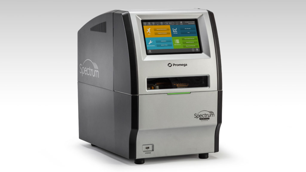
Here’s the good news: The Spectrum Compact CE System is now available from Promega.
Here’s the better news: Labs of all sizes now have the opportunity to perform Sanger sequencing and fragment analysis with a personal, benchtop instrument.
Lynch Syndrome is the autosomal dominant hereditary predisposition to develop colorectal cancer and certain other cancers. This simple, one sentence definition seems woefully inadequate considering the human toll this condition has inflicted on the families that have it in their genetic pedigree.
They Called it a Curse
To one family, perhaps the family when it comes to this condition, Lynch Syndrome has meant heartache and hope; grief and joy; death and life. Their story is told by Ami McKay in her book Daughter of Family G, and it is at once both a memoir of a Lynch Syndrome previvor (someone with a Lynch Syndrome genomic mutation who has not yet developed cancer) and a poignant and honest account of the family that helped science put name to a curse.
“The doctors called it cancer. I say it’s a curse. I wish I knew what we did to deserve it.”
Anna Haab from Daughter of Family G (1)
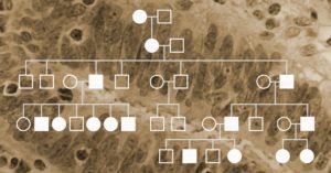
The scientific community first met “Family G” as the meticulously created family tree, filled with the stunted branches that mark early deaths by cancer. The pedigree was first published in 1913 in Archives of Internal Medicine (2). In the article, Dr. Alderd Warthin wrote: “A marked susceptibility to carcinoma exists in the case of certain family generations and family groups.” In 1925, an expanded pedigree of circles and squares was published in Dr. Warthin’s follow up study in the Journal of Cancer Research (3). But each circle and square in that pedigree denotes a person. Each line represents their dreams together for the future, and Ms. McKay wants us to know their names: Johannes and Anna, Kathrina, Elmer, Tillie, Sarah Anne (Sally); and—most importantly—Pauline. Because without Pauline there would be no story.
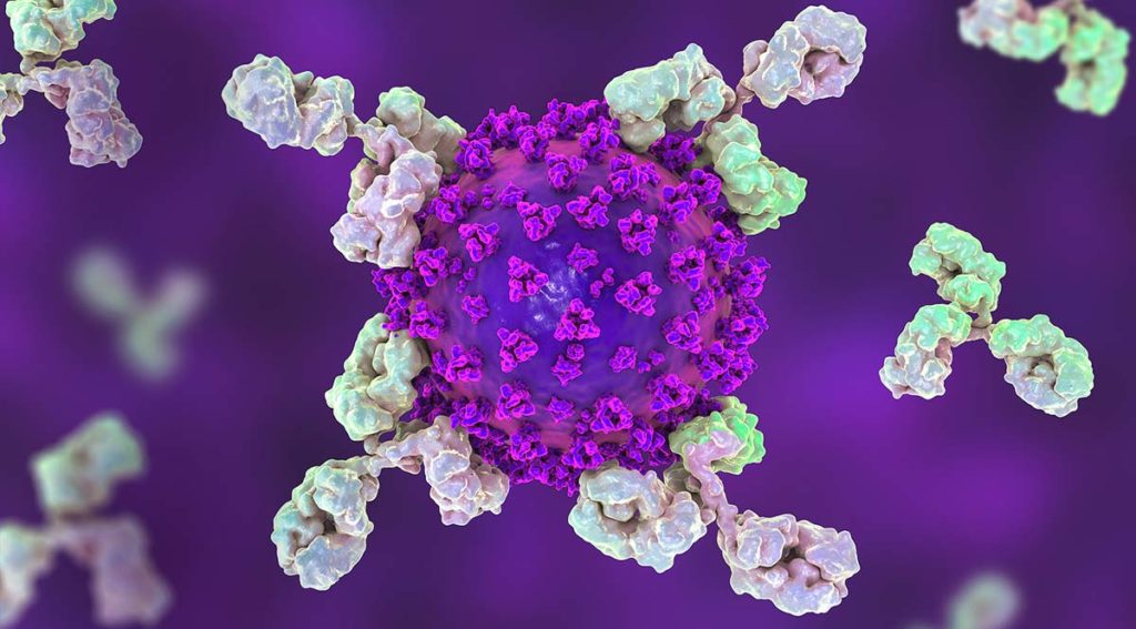
In the nine months since the first cases of COVID-19 were noticed in Wuhan, China, the virus has spread around the globe and infected over 22 million people. As with all emerging infectious diseases, we often find ourselves with more questions than answers. However, through the tireless work of researchers, doctors and public health officials worldwide, we have learned a lot about the virus, how it spreads and how to contain it.
This blog is written by guest blogger, Heather Tomlinson, former Director of Clinical Diagnostics at Promega.
Finding safe and effective treatments for human diseases takes time. Medication and diagnostic tests can take decades to discover, develop and prove safe and effective. In the United States, the FDA stands as the gold-standard gatekeeper to ensure that treatments and tests are reliable and safe. The time we wait in review and clearance means less risk of ineffective or unsafe treatments.
And yet, in a pandemic, we are behind before we even start the race to develop diagnostic tests, so critical for understanding how an infectious disease is spreading. That is when processes like the FDA’s fast track of Emergency Use Authorization (EUA) are critical. Such authorization allows scientists and clinicians to be nimble and provide the best possible test protocol as quickly as possible, with the understanding that these protocols will continue to be evaluated and improved as new information becomes available. The EUA focuses resources and accelerates reviews that keep science at the fore and gets us our best chance at staying safe and healing.
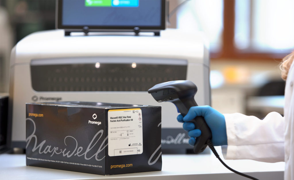
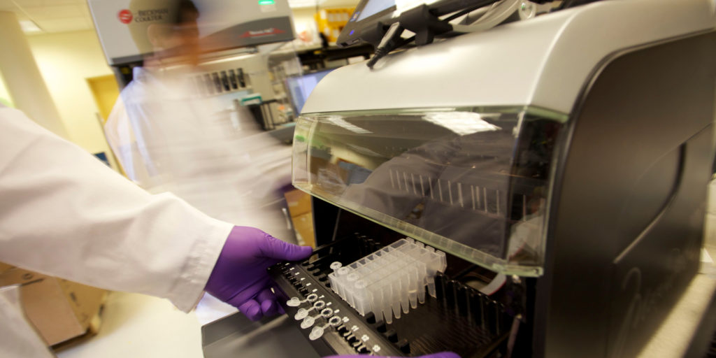
The Maxwell 48 RSC Instrument and the Maxwell RSC Total Viral Nucleic Acid Isolation Kit are now listed as options within the CDC EUA protocol.
For scientists working around the clock, the FDA’s EUA process is ready to review and respond. Getting an EUA gives clinical labs a very specific and tested resource to guide them to the tools and tests to use in a crisis.
Typically the Centers for Disease Control (CDC) will develop the first test or protocol that receives FDA EUA in response to a crisis like a pandemic. For COVID-19 the CDC 2019-Novel Coronavirus Real-Time RT-PCR Diagnostic Panel received FDA EUA clearance in early February. This is the test protocol used by the public health labs that work with the CDC and test manufacturers around the world.
Throughout a crisis such as the current pandemic, scientists continually work to improve the testing protocols and add options to the EUA protocols. This enables more flexibility in the test protocols. Promega is fortunate to play a part of the CDC EUA equation for diagnostic testing. Our GoTaq® Probe 1-Step PRT-qPCR System is one of a few approved options for master mixes in the CDC qPCR diagnostic test, and now our medium-throughput Maxwell 48 Instrument and Maxwell Viral Total Nucleic Acid Purification Kit were added to the CDC protocol as an option for the RNA isolation step as well. These additions to the CDC EUA means that laboratories have more resources at their disposal for the diagnostic testing which is so critical to effective pandemic response.
The Emergency Use Authorization provides the FDA guidance to strengthen our nation’s public health during emergencies, such as the current COVID-19 pandemic. The EUA allows continual improvement of an authorized protocol through the collaborative efforts scientists in all academia, government and industry to identify and qualify the most reliable technologies and systems, giving labs more flexibility as new products are added as options.
Dr. Tomlinson was the Director for the Global Clinical Diagnostics Strategic Business Unit at Promega Corporation bringing over 15 years of experience in clinical diagnostic test development. She was responsible for leading the team that drives strategy in the clinical market for Promega. Her background was in infectious disease diagnostic testing, with a focus on HIV drug resistance and evolution. Her last work focused on oncology companion diagnostic test development. Heather was an accomplished international presenter, delivering conference presentations in the United States, Europe, Asia, and Africa. Heather passed away in 2023.
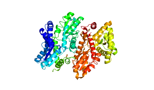
Human Ferrochelatase 2 angstrom crystal structure. Generated from 1HRK (RCSB PDB) using Pymol. Copyright: Sarah Wilson / CC BY-SA
Understanding how disease states arise from genetic variants is important for understanding disease resistance and progression. What can complicate our understanding of disease development is when two people have the same genetic variant, but only one has the disease. To investigate what might be happening with ferrochelatase (FECH) variant alleles that result in erythropoietic protoporphyria (EPP), scientists used next-generation sequencing (NGS) along with RNA analysis and DNA methylation testing to assess the FECH locus in 72 individuals from 24 unrelated families with EPP.
What is FECH and its relationship to EPP?
FECH is the gene for ferrochelatase, the last enzyme in the pathway that synthesizes heme. The inherited metabolic disorder, EPP, is caused when the activity of FECH is reduced to less than a third of normal levels thus, increasing the levels of protoporphyrin (PPIX) without metal in erythrocytes. The consequences of the low-metal PPIX include severe phototoxic skin reactions and hepatic injury due to PPIX accumulation in the liver.
How does FECH expression affect EPP?
The EPP disease state is not simply the lack of two functional FECH genes. Disease occurs with a hypomorphic allele, mutations in FECH that reduce its function, in trans to a null FECH allele. Researchers focused on three common variants called the GTC haplotype that are associated with expression quantitative trait loci (eQTL) that reduce FECH activity. Interestingly, these three variants have been found in trans, but researchers wanted to learn if there were individuals who were homozygous for the GTC allele and how EPP manifested for them.
Loss of smell (olfaction) is a commonly reported symptom of COVID-19 infection. Recently, Bilinska, et al. set out to better understand the underlying mechanisms for loss of smell resulting from SARS-CoV-2 infection. In their research, they used in situ hybridization to investigate the expression of TMPRSS2, a SARS-CoV-2 viral entry protein in olfactory epithelium tissues of mice.
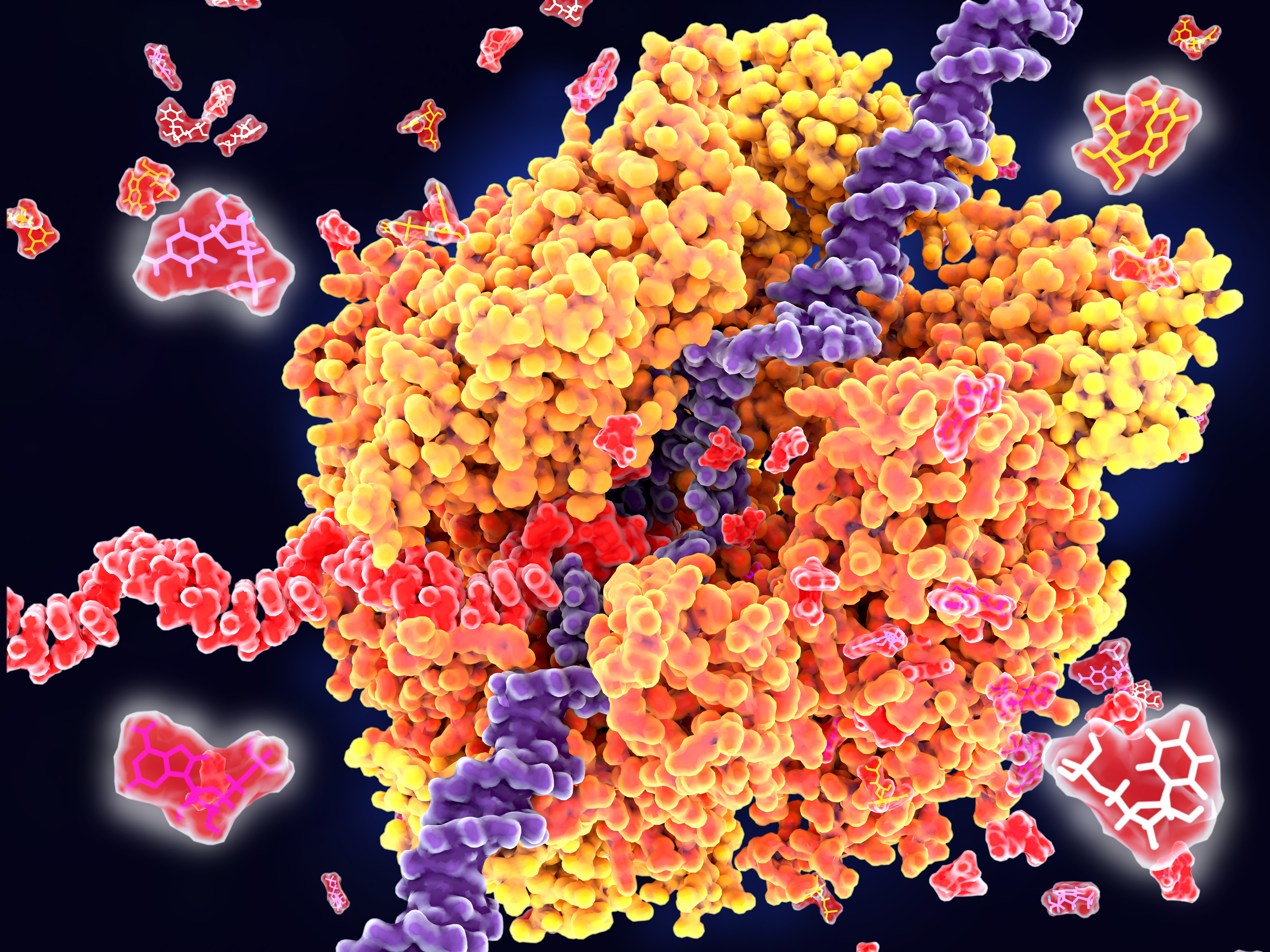
RNA polymerase unwinds DNA strands for transcription.
Transcription is the production of RNA from a DNA sequence. It’s a necessary life process in most cells. Transcription performed in vitro is also a valuable technique for research applications—from gene expression studies to the development of RNA virus vaccines.
During transcription, the DNA sequence is read by RNA polymerase to produce a complimentary, antiparallel RNA strand. This RNA strand is called a primary transcript, often referred to as an RNA transcript. In vitro transcription is a convenient method for generating RNA in a controlled environment outside of a cell.
In vitro transcription offers flexibility when choosing a DNA template, with a few requirements. The template must be purified, linear, and include a double stranded promoter region. Acceptable template types are plasmids or cloning vectors, PCR products, synthetic oligos (oligonucleotides), and cDNA (complimentary DNA).
In vitro transcription is used for production of large amounts of RNA transcripts for use in many applications including gene expression studies, RNA interference studies (RNAi), generation of guide RNA (gRNA) for use in CRISPR, creation of RNA standards for quantification of results in reverse-transcription quantitative PCR (RT-qPCR), studies of RNA structure and function, labeling of RNA probes for blotting and hybridization or for RNA:protein interaction studies, and preparation of specific cDNA libraries, just to name a few!
In vitro transcription can also be applied in general virology to study the effects of an RNA virus on a cell or an organism, and in development and production of RNA therapeutics and RNA virus vaccines. The large quantity of viral RNA produced through in vitro transcription can be used as inoculation material for viral infection studies. Viral mRNA transcripts, typically coding for a disease-specific antigen, can be quickly created through in vitro transcription, and used in the production of vaccines and therapeutics.
Monitoring the use of performance-enhancing substances among athletes is complex and the requirements for tests and assays that detect use of such substances have changed significantly over the last few decades.
The haematological (blood) module of Athlete Biological Passport was adopted December 1, 2009 (ABP) by the World Anti-Doping Agency. The module sets out standard protocols to monitor doping of professional athletes by looking at changes in biological parameters, without relying on the detection of illegal compounds in body fluids. Such biological methods eliminate the need to develop and validate a test to detect every new compound that can be used for doping. The current version of the ABP, adopted in 2014, also adds monitoring of certain steroid use indicators from urine samples.
Blood doping which aims at increasing red blood cells so that more oxygen can be transported to muscles to increase stamina or performance is particularly difficult to detect. There are typically three ways that it is accomplished: use of erythropoietin (EPO) or synthetic oxygen carriers and blood transfusions. While transfusions of large volumes of blood or use of EPO can be detected, microdosing EPO or transfusing smaller volumes of packed red blood cells is much harder to detect.
Nicolas Leuenberger and colleagues at the Swiss Laboratory for Doping Analysis have developed a method to detect blood doping. In addition to addressing the detection of blood doping, his laboratory is also concerned about easing the transport and storage requirements for samples and ensuring that sample collection does not adversely affect athlete performance.
Improving Collection and Storage of Blood Samples
Because sample collection and storage are so critical to accurate test results, any new assays developed to detect blood doping benefit from ease of collection and storage. The Leuenberger laboratory investigated the use of the TAP™ Push Button collection device, which is billed as a simple method for blood collection that is easy to use and eliminates the need for painful needle sticks or finger pricks that can affect athlete performance. After TAP collection, 20µl of blood from the device was placed on to filter paper and dried (dried blood samples; DBS), which are much easier to store and transport from collection site to laboratory.
An RNA Biomarker for Blood Doping
Blood withdrawal and autologous transfusion or recombinant human EPO injection stimulate erythropoiesis and immature red blood cells can be distinguished based on their gene expression profiles. One of the genes that is expressed by immature red blood cells is aminoleuvulinate synthase 2, a gene that encodes an enzyme ALAS2 involved in the synthesis of heme, a pathway active during RBC maturation. RNA transcripts are unstable and tend to degrade rapidly, so isolating linear RNA transcripts from a collected sample can be difficult. However circular RNAs (circRNAs) are a class of RNA molecule produced by the backsplicing of pre-mRNAs that are high in abundance, quite stable and maintain cell-type specific expression. The Leuenberger laboratory developed a method for measuring the linear and circular forms of ALAS2 RNA in DBS to monitor erythropoiesis.
One of the greatest challenges in developing this protocol was achieving efficient RNA extraction from only 20ul of dried blood. Leuenberger and his colleagues adopted a two-step purification; beginning with a phenol:chloroform extraction on the DBS followed by a further purification on the Maxwell® RSC automated instrument, using the Maxwell RSC miRNA Serum and Plasma kit. Switching from a manual to an automated method for the second step was crucial. It reduced chances of contamination as well reduced pipetting errors, without compromising good quality and yield of RNA therefore contributing to assay reproducibility. To normalize volumes within the blood spot, the protocol uses RNA produced by housekeeping genes. The work to automate the assay has been published in Bioanalysis.
What’s Next
This protocol is being tested to see if microdosing of EPO or small transfusions can also be detected by monitoring ALAS2 RNA expression in DBS. The Swiss laboratory of Doping Analysis is also in the process of developing a method to detect gene doping by isolating plasmid DNA from whole blood samples, using the Maxwell® RSC.
Additionally, the collection and storage methods used have implications for the clinic, especially for patients that need routine blood monitoring. The ability to isolate circular RNAs shows promise in forensic applications to identify body fluids.
Want to know more about how the Maxwell® RSC can give you the freedom to focus on the work that interests you the most? To learn more, click here.
The genetic abnormality called microsatellite instability, or MSI, has been linked to cancer since its discovery in 1993 (1). MSI is the accumulation of insertion or deletion errors at microsatellite repeat sequences in cancer cells and results from a functional deficiency within one or more major DNA mismatch repair proteins (dMMR). This deficiency, and the resulting genetic instability, is closely related to the carcinogenicity of tumors (2).
Historically MSI has been used to screen for Lynch Syndrome, a dominant hereditary cancer propensity. More recently, tumors with deficient MMR function have been identified as being more likely to respond to immune checkpoint inhibitor (ICI) therapies (3.). Because MSI can be the first evidence of an MMR deficiency, MSI-High status is predictive of a positive response to immunotherapies such as ICI therapies. (3).
A protein first purified and sold by Promega almost four decades ago has emerged as a crucial tool in many COVID-19 testing workflows. RNasin® Ribonuclease Inhibitor was first released in 1982, only four years after the company was started. At that time, the entire Promega catalog fit on a single sheet of 8.5 × 11” paper, and RNasin was one of the first products to draw widespread attention to Promega. Today, the demand for this foundational product has skyrocketed as it supports labs responding to the COVID-19 pandemic.
What is RNasin® Ribonuclease Inhibitor?
RNA is notoriously vulnerable to contamination by RNases. These enzymes degrade RNA by breaking the phosphodiester bonds forming the backbone of the molecule. To say that RNases are everywhere is barely an exaggeration – almost every known organism produces some form of RNase, and they’re commonly found in all kinds of biological samples. They’re easily introduced into experimental systems, since even human skin secretes a form of RNase. Once they’re present, it’s very hard to get rid of them. Even an autoclave can’t inactivate RNases; the enzymes will refold and retain much of their original activity.
RNasin® Ribonuclease Inhibitor is a protein that has been shown to inhibit many common contaminating RNases, but without disrupting the activity of enzymes like reverse transcriptase that may be essential to an experiment. It works by binding to the RNase enzyme, prevent it from acting on RNA molecules. This is important for ensuring that RNA samples are intact before performing a complex assay.
XWe use cookies and similar technologies to make our website work, run analytics, improve our website, and show you personalized content and advertising. Some of these cookies are essential for our website to work. For others, we won’t set them unless you accept them. To learn more about our approach to Privacy we invite you to Read More
By clicking “Accept All”, you consent to the use of ALL the cookies. However you may visit Cookie Settings to provide a controlled consent.
We use cookies and similar technologies to make our website work, run analytics, improve our website, and show you personalized content and advertising. Some of these cookies are essential for our website to work. For others, we won’t set them unless you accept them. To find out more about cookies and how to manage cookies, read our Cookie Policy.
If you are located in the EEA, the United Kingdom, or Switzerland, you can change your settings at any time by clicking Manage Cookie Consent in the footer of our website.
Necessary cookies are absolutely essential for the website to function properly. These cookies ensure basic functionalities and security features of the website, anonymously.
Cookie
Duration
Description
cookielawinfo-checbox-analytics
11 months
This cookie is set by GDPR Cookie Consent plugin. The cookie is used to store the user consent for the cookies in the category "Analytics".
cookielawinfo-checbox-functional
11 months
The cookie is set by GDPR cookie consent to record the user consent for the cookies in the category "Functional".
cookielawinfo-checbox-others
11 months
This cookie is set by GDPR Cookie Consent plugin. The cookie is used to store the user consent for the cookies in the category "Other.
cookielawinfo-checkbox-advertisement
1 year
The cookie is set by GDPR cookie consent to record the user consent for the cookies in the category "Advertisement".
cookielawinfo-checkbox-necessary
11 months
This cookie is set by GDPR Cookie Consent plugin. The cookies is used to store the user consent for the cookies in the category "Necessary".
cookielawinfo-checkbox-performance
11 months
This cookie is set by GDPR Cookie Consent plugin. The cookie is used to store the user consent for the cookies in the category "Performance".
gdpr_status
6 months 2 days
This cookie is set by the provider Media.net. This cookie is used to check the status whether the user has accepted the cookie consent box. It also helps in not showing the cookie consent box upon re-entry to the website.
lang
This cookie is used to store the language preferences of a user to serve up content in that stored language the next time user visit the website.
viewed_cookie_policy
11 months
The cookie is set by the GDPR Cookie Consent plugin and is used to store whether or not user has consented to the use of cookies. It does not store any personal data.
Analytical cookies are used to understand how visitors interact with the website. These cookies help provide information on metrics the number of visitors, bounce rate, traffic source, etc.
Cookie
Duration
Description
SC_ANALYTICS_GLOBAL_COOKIE
10 years
This cookie is associated with Sitecore content and personalization. This cookie is used to identify the repeat visit from a single user. Sitecore will send a persistent session cookie to the web client.
vuid
2 years
This domain of this cookie is owned by Vimeo. This cookie is used by vimeo to collect tracking information. It sets a unique ID to embed videos to the website.
WMF-Last-Access
1 month 18 hours 24 minutes
This cookie is used to calculate unique devices accessing the website.
_ga
2 years
This cookie is installed by Google Analytics. The cookie is used to calculate visitor, session, campaign data and keep track of site usage for the site's analytics report. The cookies store information anonymously and assign a randomly generated number to identify unique visitors.
_gid
1 day
This cookie is installed by Google Analytics. The cookie is used to store information of how visitors use a website and helps in creating an analytics report of how the website is doing. The data collected including the number visitors, the source where they have come from, and the pages visted in an anonymous form.
Advertisement cookies are used to provide visitors with relevant ads and marketing campaigns. These cookies track visitors across websites and collect information to provide customized ads.
Cookie
Duration
Description
IDE
1 year 24 days
Used by Google DoubleClick and stores information about how the user uses the website and any other advertisement before visiting the website. This is used to present users with ads that are relevant to them according to the user profile.
test_cookie
15 minutes
This cookie is set by doubleclick.net. The purpose of the cookie is to determine if the user's browser supports cookies.
VISITOR_INFO1_LIVE
5 months 27 days
This cookie is set by Youtube. Used to track the information of the embedded YouTube videos on a website.
Performance cookies are used to understand and analyze the key performance indexes of the website which helps in delivering a better user experience for the visitors.
Cookie
Duration
Description
YSC
session
This cookies is set by Youtube and is used to track the views of embedded videos.
_gat_UA-62336821-1
1 minute
This is a pattern type cookie set by Google Analytics, where the pattern element on the name contains the unique identity number of the account or website it relates to. It appears to be a variation of the _gat cookie which is used to limit the amount of data recorded by Google on high traffic volume websites.