“The Great Book of Nature is written in mathematical language” –Galileo Galilei (1)
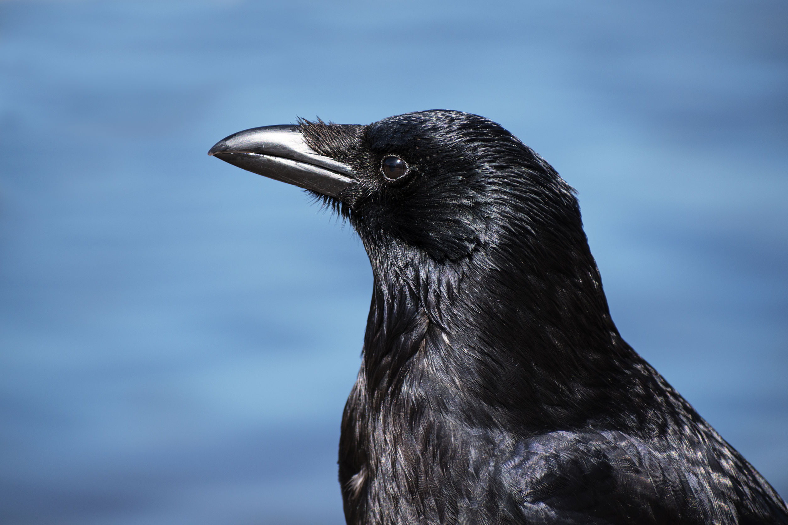
Carrion Crow (Corvus corone)
If mathematics is the language of the universe, might we find the ability to do math hard-wired in species?
Research in primates has demonstrated that even without training, humans and monkeys possess numerosity, the ability to assess the number of items in a set (2,3).
A paper in Current Biology from Wagener and colleagues provides evidence that crows are born with a subset of neurons that are “hard wired” to perceive the number of items in a set (4). This work provides yet more evidence supporting a hypothesis of an innate “number sense” that is provided by a specific group of “preprogrammed” neurons.
In this study, Wagener’s group measured the responses of single neurons in two “numerically naïve” crows to color dot arrays. They measured neurons in the endbrain region known as the niopallium caudolaterale (NCL), which is thought to be the avian analog of the primate prefrontal cortex. They found that 12% of the neurons in NCL specifically responded to numbers and that specific neurons responded to specific numbers of items with greater or lesser activity.
This is the first such study to investigate the idea of an innate “sense of number” in untrained vertebrates that are not primates, and as such it suggests that a hard-wired, innate “sense of number” is not a special feature of the complex cerebral cortex of the primate brain but is an adaptive property that evolved independently in the differently structured and evolved end brains of birds.
Many questions remain. Are there similarities in the actual neurons involved? What does learning do on a physiological level to these neurons: Increase their number, increase connections to them? What other vertebrates have similar innate mechanisms for assessing numbers of items? What about other members of the animal kingdom that need to have a sense of number for social or foraging behavior? How is it accomplished?
And finally, one last burning question, if birds are dinosaurs, does that mean that dinosaurs perished because they didn’t do their math homework? Asking for an eleven-year-old I know.
Real-time, up-to-the-minute access to information provides new opportunities for scientists to monitor cellular events in ever more meaningful ways. Real-time cytotoxicity and cell viability assay reagents now allow constant monitoring of cell health status without the need to lyse or remove aliquots from plates for measurement. With a real-time approach, data can be collected from cell cultures or microtissues at multiple time points after addition of a drug compound or other event, and the response to treatment continually observed.
The CellTox™ Green assay is a real-time assay that monitors cytotoxicity using a fluorescent DNA binding dye, which binds DNA released from cells upon loss of membrane integrity. The dye cannot enter intact, live cells and so fluorescence only occurs upon cell death, correlating with cytotoxicity. Here’s a quick overview showing how the assay works:
More Data Using Fewer Samples and Reagents The ability to continually monitor cytotoxicity in this way makes it easy to conduct more than one type of analysis on a single sample. Assays can be combined to determine not only the timing of cytotoxicity, but to also understand related events happening in the same cell population. As long as the readouts can be distinguished from one another multiple assays can be performed in the same well, providing more informative data while using less cells, plates and reagents.
Combining assays in this way can reveal critical information regarding mechanism of cell death. For example, assay combinations can be used to determine whether cells are dying from apoptosis or necrosis, or to distinguish nonproliferation from cell death. Combining CellTox Green with an endpoint luminescent caspase assay or a real-time apoptosis assay allows you to determine whether observed cytotoxic effects are due to apoptosis. Cytotoxic and anti-proliferative effects can be distinguished by combining the cytotoxicity assay with a luminescent or fluorescent cell viability assay.
Valued for ease of use and scalability, plate-based, bioluminescent cell viability assays are widely used to support research in biologics, oncology and drug discovery.
Cell viability assays are a bread-and-butter method for many researchers using cultured cells —everyday lab tools that are a part of many newsworthy papers, but rarely make news themselves.
Over time, cell viability assays have become easier to use and more “plug ‘n play”. Among modern assays, luminescent plate-reader based systems have been a favorite for several years because of their superior sensitivity, robustness, simple protocols and uncomplicated equipment requirements (all you need is a plate-reading luminometer). These qualities combine to allow easy scalability and adaptability from bench research to high throughput applications.
CellTiter-Glo® Luminescent Cell Viability Assay is an accepted go-to viability assay for many researchers. The assay measures ATP as an indicator of metabolically active cells. A quick search on Google Scholar returns 3,990 CellTiter-Glo results for 2017 and over 500 so far in January and February of 2018. A sampling of these recent publications gives a snapshot of some of the ways the CellTiter-Glo assay is used to support key areas of research today.
Does a treatment kill cells?
The obvious application of a cell viability assay is to understand whether cells are alive. In cancer research, the CellTiter-Glo assay is often used to confirm killing of tumor cells and to verify that normal cells survive. Therefore, these assays are a key part of the evaluation and screening of drug candidates and other therapies for cancer. Many papers reporting use of CellTiter-Glo are developing and evaluating the effectiveness of novel anti-cancer treatments. Continue reading “A Cell Viability Assay for Today”
Recently, I had the opportunity to attend a fascinating symposium held at Promega featuring conservationist Steward Brand, where he described some of the projects developed by his foundation, Revive & Restore.
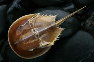
The organization’s mission is to apply emerging biotechnology techniques to endangered and extinct species with the intent to increase genetic diversity, provide disease resistance and facilitate adaptation to changing climates. Although the overall message of enhancing biodiversity through the application of new genetic technology was inspiring, the project that resonated most for me was related to the plight of horseshoe crabs.
Horseshoe crabs, often referred to as living fossils, include four extant species with origins dating back about 450 million years. Although they look like crabs, they belong to their own subphylum and are more closely related to spiders. When horseshoe crabs spawn, they leave their usual habitat on the ocean floor and migrate to shore in large numbers. As a result, they have been exploited for bait and fertilizer for decades.
Enter endotoxins, an indicator for bacterial contamination in biologicals, drugs and medical devices. U.S. Food & Drug Administration regulations dictate that finished products be tested for the presence of endotoxins. These pyrogenic compounds, found in the cell wall of Gram-negative bacteria, can cause fever and affect a wide range of biological activity, possibly leading to aseptic shock and death. The most common method for testing is the gel clot and Limulus Amebocyte Lysate (LAL) Test.
I first learned about the LAL test during graduate school, where it was presented as a ubiquitous and standard requirement for testing bacterial contamination in injectable drugs. I remember being fascinated that horseshoe crabs (Limulus sp.), contain a substance that could be used to detect endotoxins. Although the instructors mentioned the need to collect blood from horseshoe crabs in order to produce the test, the method or scale of this harvest wasn’t discussed, nor were the true costs of using this method of endotoxin testing.
The LAL test has served as a faster, more inexpensive endotoxin testing replacement for the rabbit pyrogens test for the past 35 years. Every year during mating season horseshoe crabs move to shallow water, where they are removed in huge numbers. (To get an idea of scale for the harvest and read a much more comprehensive investigation of the issue, check out this article in The Atlantic, which features an archive photo of Delaware Bay horseshoe crab harvest from 1928—for fertilizer, not pharmaceutical testing.)
After collection, the crabs end up in a lab where up to 30% of their blood is drained from a needle stuck in tissue around their heart. The LAL is extracted from the blood and can yield a product worth up to $15,000/quart. In order to avoid recollection, the crabs are returned to the ocean far from the shore where they were collected a few days before. Although it’s estimated that only 10-30% of these crabs die as a result of the process, there are indications that the horseshoe crab population and their ecosystems are impacted in other ways.
Researchers at the University of New Hampshire and Plymouth State University used accelerometers attached to recently bled female horseshoe crabs to test the hypothesis that harvesting for LAL was affecting their ability to spawn. While the research supported previous estimates with a death rate of 18%, females were found to be less likely to mate after being bled.
During his talk, Brand shared results from a study still in review that confirm the effect of over-harvesting Limulus on the survival of long distance migratory shorebirds. These birds synchronize their migration with horseshoe crab spawning, which provides a needed feast of eggs before the homestretch of their journey. Along with other ecosystem threats from climate change, the accelerated decline in the horseshoe crab population and dependency of migratory birds will likely to lead to a devastating ecological domino effect.
Fortunately, a synthetic alternative to LAL, recombinant factor C (rFC), has been available for nearly 20 years. Alas, there has been no significant shift by pharmaceutical companies away from the test based on horseshoe crab blood. rFC was patented and licensed to one company, Lonza, which Brand posited as one reason for the reluctance of drug companies to adopt its use.
Obviously, relying on one source for an essential testing reagent with no competition to temper cost is quite unattractive. But that argument has less bearing now that the patent is scheduled to expire in a few months, opening the door for additional manufacturers and creating an economic incentive for switching to the synthetic test.
Another reason may be that implementing a new test would require additional resources to validate the synthetic test for products that are already being tested with the LAL. Since the LAL has been specified in FDA guidance documents on endotoxin testing for decades, quality standards for existing products are based on the LAL, limiting momentum to change.
If both tests offered the same benefits, these arguments would make sense; however, research by one of the discoverers of rFC, Jeak Ling Ding of the National University of Singapore, shows that in many respects rFC is more efficacious than LAL. Since the raw material for the LAL test depends on an organism, there is seasonal variation in the components of the processed blood that must be taken into account. The reaction of the LAL also depends on a cascade of multiple compounds that can be affected by temperature, pH and proteins—leaving the test vulnerable to false positive results.
Although Eli Lilly is the only pharmaceutical company to date to use rFC in place of LAL, It seems the tide may be turning. According to Brand, others are interested in making the transition. It seems foolish not to, given the source for LAL shows signs of dwindling due to overexploitation. Perhaps pharmaceutical companies are beginning to see the value of a “slower/better” philosophy (the cornerstone of the Long Now Foundation, another brainchild of Brand’s), rather than “faster/cheaper.” I have certainly gained a new perspective on endotoxin testing and a deep appreciation for the work of Brand and his foundation.
Does your organization use the LAL test? What is preventing you from switching to the synthetic alternative? Let us know!
As a science writer, much of my day entails reviewing and revising marketing materials and technical literature about complex life science research products. I take for granted the understanding that I, my colleagues and our customers have of how these technologies work. This fact really struck me as I read an article about research to improve provider-patient communication in healthcare settings.
The researchers completed an analysis revealing that patient information materials had an average readability at a high school level, while the average patient reads at a fourth-grade level. These findings inspired the researchers to conduct a study in which they enlisted the help of elementary students to revise the content of the patient literature after giving them a short lesson on the material.
The resulting content did not provide more effective ways to communicate indications, pre- and post-op care, risks or procedures—that wasn’t really the point. Instead, the study underscores the important connection between patient literacy and health outcomes. More specifically, a lack of health literacy is correlated with poor outcomes and increased healthcare costs, prompting action from the US Department of Health & Human Services.
While healthcare information can be complex and full of specific medical terminology, I recognized that a lot of the technical and marketing information we create for our products at Promega has similar features. Wouldn’t it be interesting to find out how descriptions of some of our biggest technologies translate through the eyes and mouths of children?
After enlisting some help from my colleagues, I was able to catch a glimpse of how our complex technologies are understood by the little people in our lives. The parents and I explained a technology and then had our child provide a description or drawing of what they understood.
The mammalian brain is extremely complex. We know that it processes and stores information through synaptic connections within a complicated neural network. But how exactly do neurons communicate with each other? And how did this neural network come to exist? A recent paper published in Cell may provide some answers. It describes a previously unknown signaling pathway–with surprising origins–that transports RNA between neurons. Continue reading “A Virus-like Neural Pathway Hints at the Origins of the Mammalian Brain”
The Foundation for Food and Agriculture Research (FFAR) announced on November 30 that they are awarding $1M to a project based at the University of California, Davis, to study protein kinases of rice plants. The team is led by Dr. Pamela Ronald, a leading expert in plant genetics who has engineered disease- and flood-resistant rice. This project aims to address the growing agricultural problem of water scarcity by gaining a better understanding of the role kinases play in enabling drought-resistance. Promega will be supporting this research by providing NanoBRET™ products to help characterize kinase inhibitors.
Principal Investigator Pamela Ronald, Ph.D. Photo Credit: Deanne Fitzmaurice
The research team will begin by screening over 1,000 human kinase inhibitors to determine which ones do interact with the plant kinome and, if applicable, which kinase(s) they inhibit. Once the compound library has been established, the team will assess the inhibitors’ phenotypic effects on rice to identify kinases that, when inhibited, positively impact root growth and development. The long-term goal is to use these findings to engineer drought-resistant rice.
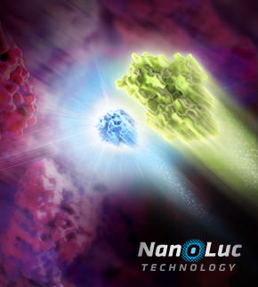
Researchers having been sharing plasmids ever since there were plasmids to share. Back when I was in the lab, if you read a paper and saw an interesting construct you wished to use, you could either make it yourself or you could “clone by phone”. One of my professors was excellent at phone cloning with labs around the world and had specific strategies and tactics for getting the plasmids he wanted. Addgene makes this so much easier to share your constructs from lab to lab. Promega supports the Addgene mission statement: Accelerate research and discovery by improving access to useful research materials and information. Many of our technology platforms like HaloTag® Fusion Protein, codon-optimized Firefly luciferase genes (e.g., luc2), and NanoLuc® Luciferase are present in the repository. We encourage people to go to Addgene to get new innovative tools. Afterall, isn’t science better when we share?
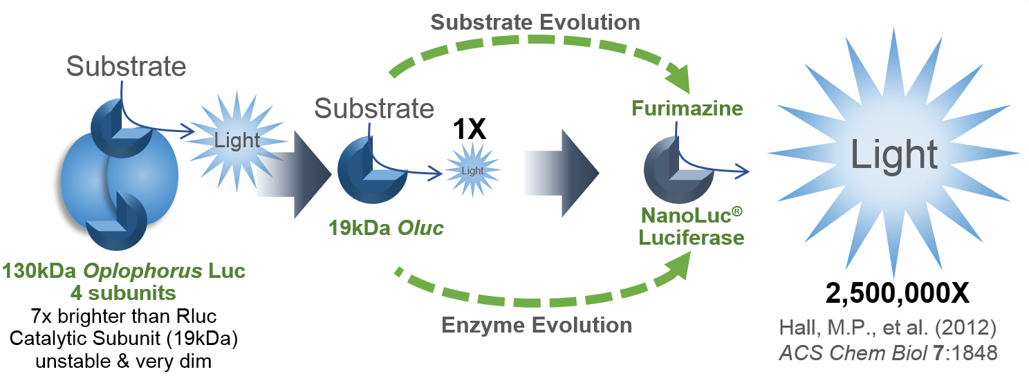
I’d like to focus on some tools in the Addgene collection based on NanoLuc® Luciferase (NLuc). The creation of NanoLuc® Luciferase and the optimal substrate furimazine is a good story (1). From a deep sea shrimp to a compact powerhouse of bioluminescence, NLuc is 100-fold brighter than our more common luciferases like firefly (FLuc) and Renilla (RLuc) luciferase. This is important not so much for how bright you can make a reaction but for how sensitive you can make a reaction. NLuc requires 100-fold less protein to produce the same amount of light from a Fluc or RLuc reaction. NLuc lets you work at physiological concentrations. NLuc is bright enough to detect endogenous tagged genes generated through the CRISPR/Cas9 knock-in. NLuc is very inviting for endogenous tagging as it is only 19kDa. An example is the CRISPaint-NLuc construct (Plasmid #67178) for use in the system outlined in Schmid-Burgk, J.L. et al (2).
“Glycobiology is the study of carbohydrates and their role in biology. Glycans, defined as ‘compounds consisting of a large number of monosaccharides linked glycosidically’ are present in all living cells; They coat cell membranes and are integral components of cell walls. They play diverse roles, including critical functions in cell signaling, molecular recognition, immunity and inflammation. They are the cell-surface molecules that define the ABO blood groups and must be taken into consideration to ensure successful blood transfusions.
The process by which a sugar moiety is attached to a biological compound is referred to as glycosylation. Protein glycosylation is a form of post-translational modification, which is important for many biological processes and often serves as an analog switch that modulates protein activity. The class of enzymes responsible for transferring the sugar moiety onto proteins is called a glycosyltransferase (GT).”
Illustration showing NanoLuc and firefly luciferase reporters.
The luciferase immunoprecipitation system (LIPS) assay is a liquid phase immunoassay allowing high-throughput serological screening of antigen-specific antibodies. The immunoassay involves quantitating serum antibodies by measuring luminescence emitted by the reporter enzyme Renilla luciferase (Rluc) fused to an antigen of interest. The Rluc-antigen fusion protein is recognized by antigen-specific antibodies, and antigen-antibody complexes are captured by protein A/G beads that recognize the Fc region of the IgG antibody (1).
XWe use cookies and similar technologies to make our website work, run analytics, improve our website, and show you personalized content and advertising. Some of these cookies are essential for our website to work. For others, we won’t set them unless you accept them. To learn more about our approach to Privacy we invite you to Read More
By clicking “Accept All”, you consent to the use of ALL the cookies. However you may visit Cookie Settings to provide a controlled consent.
We use cookies and similar technologies to make our website work, run analytics, improve our website, and show you personalized content and advertising. Some of these cookies are essential for our website to work. For others, we won’t set them unless you accept them. To find out more about cookies and how to manage cookies, read our Cookie Policy.
If you are located in the EEA, the United Kingdom, or Switzerland, you can change your settings at any time by clicking Manage Cookie Consent in the footer of our website.
Necessary cookies are absolutely essential for the website to function properly. These cookies ensure basic functionalities and security features of the website, anonymously.
Cookie
Duration
Description
cookielawinfo-checbox-analytics
11 months
This cookie is set by GDPR Cookie Consent plugin. The cookie is used to store the user consent for the cookies in the category "Analytics".
cookielawinfo-checbox-functional
11 months
The cookie is set by GDPR cookie consent to record the user consent for the cookies in the category "Functional".
cookielawinfo-checbox-others
11 months
This cookie is set by GDPR Cookie Consent plugin. The cookie is used to store the user consent for the cookies in the category "Other.
cookielawinfo-checkbox-advertisement
1 year
The cookie is set by GDPR cookie consent to record the user consent for the cookies in the category "Advertisement".
cookielawinfo-checkbox-necessary
11 months
This cookie is set by GDPR Cookie Consent plugin. The cookies is used to store the user consent for the cookies in the category "Necessary".
cookielawinfo-checkbox-performance
11 months
This cookie is set by GDPR Cookie Consent plugin. The cookie is used to store the user consent for the cookies in the category "Performance".
gdpr_status
6 months 2 days
This cookie is set by the provider Media.net. This cookie is used to check the status whether the user has accepted the cookie consent box. It also helps in not showing the cookie consent box upon re-entry to the website.
lang
This cookie is used to store the language preferences of a user to serve up content in that stored language the next time user visit the website.
viewed_cookie_policy
11 months
The cookie is set by the GDPR Cookie Consent plugin and is used to store whether or not user has consented to the use of cookies. It does not store any personal data.
Analytical cookies are used to understand how visitors interact with the website. These cookies help provide information on metrics the number of visitors, bounce rate, traffic source, etc.
Cookie
Duration
Description
SC_ANALYTICS_GLOBAL_COOKIE
10 years
This cookie is associated with Sitecore content and personalization. This cookie is used to identify the repeat visit from a single user. Sitecore will send a persistent session cookie to the web client.
vuid
2 years
This domain of this cookie is owned by Vimeo. This cookie is used by vimeo to collect tracking information. It sets a unique ID to embed videos to the website.
WMF-Last-Access
1 month 18 hours 24 minutes
This cookie is used to calculate unique devices accessing the website.
_ga
2 years
This cookie is installed by Google Analytics. The cookie is used to calculate visitor, session, campaign data and keep track of site usage for the site's analytics report. The cookies store information anonymously and assign a randomly generated number to identify unique visitors.
_gid
1 day
This cookie is installed by Google Analytics. The cookie is used to store information of how visitors use a website and helps in creating an analytics report of how the website is doing. The data collected including the number visitors, the source where they have come from, and the pages visted in an anonymous form.
Advertisement cookies are used to provide visitors with relevant ads and marketing campaigns. These cookies track visitors across websites and collect information to provide customized ads.
Cookie
Duration
Description
IDE
1 year 24 days
Used by Google DoubleClick and stores information about how the user uses the website and any other advertisement before visiting the website. This is used to present users with ads that are relevant to them according to the user profile.
test_cookie
15 minutes
This cookie is set by doubleclick.net. The purpose of the cookie is to determine if the user's browser supports cookies.
VISITOR_INFO1_LIVE
5 months 27 days
This cookie is set by Youtube. Used to track the information of the embedded YouTube videos on a website.
Performance cookies are used to understand and analyze the key performance indexes of the website which helps in delivering a better user experience for the visitors.
Cookie
Duration
Description
YSC
session
This cookies is set by Youtube and is used to track the views of embedded videos.
_gat_UA-62336821-1
1 minute
This is a pattern type cookie set by Google Analytics, where the pattern element on the name contains the unique identity number of the account or website it relates to. It appears to be a variation of the _gat cookie which is used to limit the amount of data recorded by Google on high traffic volume websites.