Light enters eyes and is transmitted to SCN and PHb.

Light: A Happy Pill for Dark Days?

Have you ever had a day where you feel exceptionally good? As intake on the world kind of good? You feel so much better than the previous couple of days that you stop to wonder why.

Then it dawns on you.

The sun is out. It’s been cloudy for the past week but now—SUNSHINE.

You go out to lunch or for a walk just to take in those rays. Sure, it feels warmer than your darkened office space, but it’s the light rather than warmth that’s making a difference.

You purposely don’t wear sunglasses and it feels like the light is coming in through your eyes and massaging that part of your brain that is your happy zone. Are you imagining it or is the sun really affecting how you feel?

In a study reported in the September 2018 issue of Cell we learn that this is not a figment of your or my imagination (1). There is, in fact, a type of retinal cell that transports sunlight directly to the part of our brains that affects mood.

Eyes and the Body’s Master Clock

Circadian rhythms are innate time-keeping functions found in all multicellular organisms. This subject of the 2017 Nobel prize in Physiology or Medicine, circadian rhythms are fueled by daily light-dark cycles and are critical to the function of neurologic, immune, musculoskeletal and cardiac tissues (2). Nearly every mammalian cell is affected by circadian rhythms.

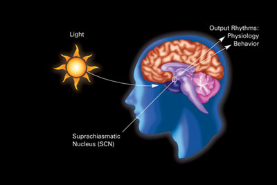
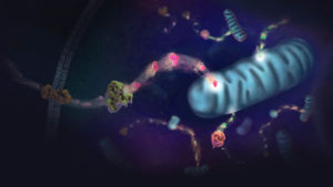
The human body has a circadian master clock, the suprachiasmatic nucleus or SCN. The SCN is a highly innervated tissue located in the hypothalamus (see image). It is connected directly to the retina by the optic nerve, and thus is influenced by external light and dark.

Light enters eyes and is transmitted to SCN and PHb.
Light enters the eyes and affects the SCN (physiologic effects), and as discussed in recent research, Fernandez et al. here, the perihabenular nucleus (behavioral effects). (Image in public domain.)

The retina of the eye is the light-gathering instrument for this organ. Historically, it’s been understood that the retina is composed of two cell types, rods and cones, that function in transmitting light and images to the optic nerve, which sends those signals to the brain.

Drawing of the retina with rods and cones, some nervous tissues.
Some parts of the retina. Light enters the eye (from left) and passes through to the rods and cones. Here a chemical change converts the light to nerve signals. Image-based on drawing by Ramón y Cajal, 1911 and licensed under Wikimedia commons.

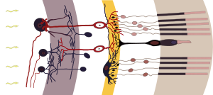
Studies by Hattar et al. in the early 2000s identified another cell found in the retina, the melanopsin-containing intrinsically photoactive retinal ganglion cells (ipRGCs) as the transmitter of circadian light signals (3). Through this direct connection to the SCN, the circadian master clock, the ipRGCs can influence a wide range of light-dependent functions independent of image processing (4).

Now Fernandez et al. have identified multiple types of ipRGCs. They showed that ipRGCs that mediate the effects of light on learning work via the SCN, while the pathway for light influencing emotions is different.

They discovered a new target of ipRGC cells, the perihabenular nucleus (PHb). The PHb is a newly recognized thalamic region of the brain. The authors showed that the connection between light and mood is regulated by ipRGCs through the PHb versus the SCN. They show that the PHb is integrated into other mood-regulating centers of the thalamic region.

In Conclusion

Daylight, and lack thereof, does affect both our mood and our ability to learn. In this 2018 report, we have learned that the pathways for these effects are distinct, and gain an understanding of a new thalamic region by which the light and mood actions occur. This information could influence the development of better drugs and/or therapies for major depressive disorders.

For those of us with seasonal affective disorder, the evidence is undeniable—lack of light can cause issues, from sleep-wake problems, to mood and learning issues.

And while we can’t create sunshine, a special lamp or lightbox may help to gain some full-spectrum light. To learn more about how to choose such a lamp and when to use it, see this Mayo clinic article for details.

References

  1. Fernandez, D.C. et al. (2018) Light affects mood and learning through distinct retinal pathways. Cell 175, 71–84.
  2. Ledford, H. and Callaway, E. (2017) Circadian clock scoops Nobel prize. Nature 550, 18.
  3. Hattar, S. et al. (2002) Melanopsin-containing retinal ganglion cells: architecture, projections, and intrinsic photosensitivity. Science 295, 1065–70.
  4. Hattar, S. et al. (2003) Melanopsin and rod-cone photoreceptive systems account for all major accessory visual functions in mice. Nature 424(6944)76–81.

How Prostate Cancer Cells Survive Glucose Deprivation

Illustration of energy metablism in cell.

Glucose is an energy metabolite necessary for cellular survival and growth whether or not the cell is part of a tumor. Not only do cancer cells switch from oxidative phosphorylation to aerobic glycolysis (the Warburg effect) to gain more glucose, a hallmark of cancer, but they also increase the amount of glucose taken up from the surrounding extracellular space. However, the lack of glucose can have a negative effect on cells, causing them to become apoptotic in the absence of this metabolite. Cancer cells have methods to get around the requirement for glucose, including upregulating glucose transporters to improve access to the energy metabolite. In this Redox Biology article, researchers describe how activating androgen receptor in response to a lack of glucose affects the amount of GLUT1 expressed on prostate cancer cells, making the cells resistant to glucose deprivation.

To set the stage, two prostate cancer cell lines, LNCaP, an androgen-sensitive cell line, and LNCaP-R, an androgen-insensitive cell line, were deprived of glucose. Both cell lines showed signs of cell death, but LNCaP-R cells died in greater numbers. To probe how LNCaP cells died, several inhibitors (a pan-caspase inhibitor, two necroptosis inhibitors and a ferroptosis inhibitor) were added but did not change the way the cells died. However, an autophagy inhibitor enhanced cell death, suggesting the cells were necrotic not apoptotic. Teasing apart if the necrosis of LNCaP cells was due to glucose availability or merely disrupted glycolysis, the glucose analog 2DG was added to the medium with glucose. The cells survived when treated with 2DG, suggesting it was the absence of glucose that induced necrosis. When LNCaP cells were cultivated in medium that replaced glucose with mannose or fructose, the cells survived, another point in favor of sugar depletion causing cell death.

Continue reading “How Prostate Cancer Cells Survive Glucose Deprivation”

How To Make Medicine on Mars

Today NASA’s InSight lander will touch down on Mars. InSight, which launched on May 5, is NASA’s first Mars landing since the Curiosity rover in 2012. The lander will begin a two-year mission to study Mars’ deep interior, gathering data that will help scientists understand the formation of rocky planets, including Earth.

NASA's InSight lander approaching Mars.
Image credit: NASA/JPL-Caltech

While every spacecraft that reaches Mars offers more knowledge of the Red Planet, a lot of the excitement is fueled by hopes that someday these missions will bring humans to Mars and enable us to start colonies there. While this goal seems very distant, tremendous progress is being made. Scientists around the globe are making incremental discoveries that will lead to the advances necessary to make colonization of Mars a reality.

I had the pleasure of meeting one team of scientists doing just this—eight high school students from iGEM Team Navarra BG. I met the team and their advisors at the 2018 iGEM Giant Jamboree, where they presented their synthetic biology project, BioGalaxy, as part of the iGEM competition. The problem they aimed to solve is key to helping humans stay on Mars for an extended period of time—how do you take everything you need when there isn’t enough room on the spacecraft? Continue reading “How To Make Medicine on Mars”

Nano, Nano: Tiny Lipid Particles with Big Therapeutic Potential

cell-transfection-viafect-luciferase-assay

Getting DNA or RNA into cells can be a tricky business, and a variety of transfection reagents have been developed over the years to make the process easier. Lipid-based reagents are especially popular because they combine efficient transfection with relatively low toxicity.

When it comes to transfection, it pays to think small. Human cells range in volume from 20–40 µm3 (sperm cells) to as large as 4 million µm3 (mature egg cells, or oocytes). For several decades, transfection reagents have targeted this size range. However, breakthrough research involves leaving the “micro” realm and entering a world that was once the domain only of science fiction: nanotechnology.

Continue reading “Nano, Nano: Tiny Lipid Particles with Big Therapeutic Potential”

A Tale of Two Toxins: the mechanisms of cell death in Clostridium difficile infections

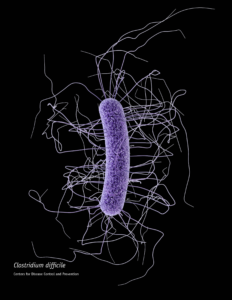
When someone is admitted to a hospital for an illness, the hope is that medical care and treatment will help them them feel better. However, nosocomial infections—infections acquired in a health-care setting—are becoming more prevalent and are associated with an increased mortality rate worldwide. This is largely due to the misuse of antibiotics, allowing some bacteria to become resistant. Furthermore, when an antibiotic wipes out the “good” bacteria that comprise the human microbiome, it leaves a patient vulnerable to opportunistic infections that take advantage of disruptions to the gut microbiota.

One such bacteria, Clostridium difficile, is of growing concern world-wide since it is resistant to many different antibiotics. When a patient is treated with an antibiotic, C. difficile can thrive in the intestinal tract without other bacteria populating the gut. C. difficile infection is the leading cause of antibiotic-associated diarrhea. While symptoms can be mild, aggressive infection can lead to pseudomembranous colitis—a severe inflammation of the colon which can be life-threatening.

C. difficile causes disease by releasing two large toxins, TcdA and TcdB. Understanding the role these toxins play in colonic disease is important for treatment strategies. However, most published research data only report the effects of the toxins independently. A 2016 study demonstrated a method of comparing the toxins side-by-side using the same time points and cell assays to investigate the role each toxin plays in the cell death that leads to disease of the colon. Continue reading “A Tale of Two Toxins: the mechanisms of cell death in Clostridium difficile infections”

MSI Analysis and the Application of Therapies Based on 2018 Nobel Immuno-Oncology Work

The 2018 Nobel Prize in Physiology and Medicine was awarded to James P. Allison of the United States and Tasuku Honjo of Japan for their work to identify pathways in the immune system that can be used to attack cancer cells (1). Although immunotherapy for cancer has been a goal for many decades, Dr. Allison and Dr. Honjo succeeded through their manipulation of “checkpoint inhibitor” pathways to target cancer cells.

Immune checkpoint inhibitor drugs have been effective in cancers such as aggressive metastatic melanoma, some lung cancers, kidney, bladder and head and neck cancers. These therapies have succeeded in pushing many aggressive cancers below detectable limits, though these cases are notably not relapse-free or necessarily “cured” (2,3).

One challenge in implementing immunotherapy in a cancer treatment regime is the need to understand the genetic makeup of the tumor. Certain tumors, with specific genetic features, are far more likely to respond to immune checkpoint therapy than others. For this reason, Microsatellite Instability (MSI) analysis has become an increasingly relevant tool in genetic and immuno-oncology research.

What is MSI Analysis?

Continue reading “MSI Analysis and the Application of Therapies Based on 2018 Nobel Immuno-Oncology Work”

What Could You Do with a Faster, More Consistent ADCC Reporter Bioassay?

Fc receptor-mediated antibody-dependent cell-mediated cytotoxicity (ADCC) is an important mechanism of action (MOA) by which antibodies target diseased cells for elimination. Traditional methods for measuring ADCC require primary donor peripheral blood mononuclear cells (PBMCs) or purified natural killer (NK) cells that express Fc receptors on the cell surface. Killing of target cells is an endpoint of this pathway activation and is used in classic ADCC bioassays.

PBMCs and NK cells are notoriously difficult to isolate and culture. Furthermore, cultured cells can be a source of variability.

There is a Better Way

Watch this video to learn why traditional ADCC assays can be problematic. You’ll also learn a solution. Find out how  to not only save time but also reduce assay variability.

For more details on the benefits of working with ADCC Reporter Bioassays go to the product page.

There you’ll see how standardized reagents in Promega ADCC Reporter Bioassays ensure better results and better consistency in an ADCC Reporter Bioassay that saves you time.

Overcoming Challenges When Scaling Antibody Production

Tradeoffs are a constant source of challenge in any research lab. To get faster results, you will probably need to use more resources (people, money, supplies). The powerful lasers used to do live cell imaging may well kill those cells in the process. Purifying DNA often leaves you to choose between purity and yield.

Robot performing autosampling

Working with biologics also involves a delicate balancing act. Producing compounds in biological models rather than by chemical synthesis offers many advantages, but it is not without certain challenges. One of those tradeoffs results from scaling up; the more plasmid that is produced, the greater probability of endotoxin contamination.

Continue reading “Overcoming Challenges When Scaling Antibody Production”

Quantitating Kinase-Inhibitor Interactions in Live Cells

Kinase target engagement is a new way to study kinase inhibitors for target selectivity, potency and residency. The NanoBRET™ TE Intracellular Kinase Assays enable you to quantitate kinase-inhibitor binding in live cells, making these assays an exciting new tool for kinase drug discovery research.

For today’s blog about NanoBRET™ TE Intracellular Kinase Assay, we feature spokesperson Dr. Matt Robers. Matt is part of Promega’s R & D department and is one of the developers of the NanoBRET™ TE Intracellular Kinase Assay.

Continue reading “Quantitating Kinase-Inhibitor Interactions in Live Cells”

From Drug Screening to Agriculture to Cardiac Development, A Dual-Luciferase Reporter Brings You the Story

Today’s blog was written by guest blogger Katarzyna Dubiel, marketing intern in Cellular Analysis and Proteomics. Last updated 02/12/2021

Reporter gene assays have been critical for the study of a wide-range of biological questions, from regulation of gene expression to cellular signaling. While reporter gene assays constitute a large group of technologies, here we highlight the diversity of new discoveries enabled by highly quantitative and easily measured bioluminescent luciferase-based reporter assays. Below are our top picks of exciting research discoveries involving the Dual-Luciferase Reporter Assay format using firefly and Renilla luciferases.

Continue reading “From Drug Screening to Agriculture to Cardiac Development, A Dual-Luciferase Reporter Brings You the Story”