MSI Analysis and the Application of Therapies Based on 2018 Nobel Immuno-Oncology Work

The 2018 Nobel Prize in Physiology and Medicine was awarded to James P. Allison of the United States and Tasuku Honjo of Japan for their work to identify pathways in the immune system that can be used to attack cancer cells (1). Although immunotherapy for cancer has been a goal for many decades, Dr. Allison and Dr. Honjo succeeded through their manipulation of “checkpoint inhibitor” pathways to target cancer cells.

Immune checkpoint inhibitor drugs have been effective in cancers such as aggressive metastatic melanoma, some lung cancers, kidney, bladder and head and neck cancers. These therapies have succeeded in pushing many aggressive cancers below detectable limits, though these cases are notably not relapse-free or necessarily “cured” (2,3).

One challenge in implementing immunotherapy in a cancer treatment regime is the need to understand the genetic makeup of the tumor. Certain tumors, with specific genetic features, are far more likely to respond to immune checkpoint therapy than others. For this reason, Microsatellite Instability (MSI) analysis has become an increasingly relevant tool in genetic and immuno-oncology research.

What is MSI Analysis?

Continue reading “MSI Analysis and the Application of Therapies Based on 2018 Nobel Immuno-Oncology Work”

Automated Approach for Multiomic Analysis

With the use of a suite of “-omics” technologies you can examine the way in which complex cellular processes work together across all molecular domains (i.e., proteomics, metabolomics, transcriptomics) in a single biological system. Several studies have been published across a wide range of fields illustrating the power of such a unified approach (1,2). Most studies however did not focus on the development of a high-throughput, unified sample preparation approach to complement high-throughput “omic” analytics.

A recent publication by Gutierrez and colleagues presents a simple high-throughput process (SPOT) that has been optimized to provide high-quality specimens for metabolomics, proteomics, and transcriptomics from a common cell culture sample (3). They demonstrate that this approach can process  16−24 samples from a cell pellet to a desalted sample ready for mass spectrometry analysis within 9 hours. They also demonstrated that the combined process did not sacrifice the quality of data when compared to individual sample preparation methods.

Literature Cited

1. Roume, H. (2013) Sequential Isolation of Metabolites, RNA, DNA, and Proteins from the Same Unique Sample. Methods Enzymol. 531, 219−236.
2. Lo, A. W. et al. (2017) ‘Omic’ Approaches to Study Uropathogenic Escherichia Coli Virulence. Trends Microbiol. 25, 729−740.
3. Gutierrez, D. et al. (2018)  An Integrated, High-Throughput Strategy for Multiomic Systems Level Analysis J. Proteome Res.

Are you looking for proteases to use in your research?
Explore our portfolio of proteases today.

Is MPS right for your forensics lab?

Today’s post was written by guest blogger Anupama Gopalakrishnan, Global Product Manager for the Genetic Identity group at Promega. 

Next-generation sequencing (NGS), or massively parallel sequencing (MPS), is a powerful tool for genomic research. This high-throughput technology is fast and accessible—you can acquire a robust data set from a single run. While NGS systems are widely used in evolutionary biology and genetics, there is a window of opportunity for adoption of this technology in the forensic sciences.

Currently, the gold standard is capillary electrophoresis (CE)-based technologies to analyze short tandem repeats (STR). These systems continue to evolve with increasing sensitivity, robustness and inhibitor tolerance by the introduction of probabilistic genotyping in data analysis—all with a combined goal of extracting maximum identity information from low quantity challenging samples. However, obtaining profiles from these samples and the interpretation of mixture samples continue to pose challenges.

MPS systems enable simultaneous analysis of forensically relevant genetic markers to improve efficiency, capacity and resolution—with the ability to generate results on nearly 10-fold more genetic loci than the current technology. What samples would truly benefit from MPS? Mixture samples, undoubtedly. The benefit of MPS is also exemplified in cases where the samples are highly degraded or the only samples available are teeth, bones and hairs without a follicle. By adding a sequencing component to the allele length component of CE technology, MPS resolves the current greatest challenges in forensic DNA analysis—namely identifying allele sharing between contributors and PCR artifacts, such as stutter. Additionally, single nucleotide polymorphisms in flanking sequence of the repeat sequence can identify additional alleles contributing to discrimination power. For example, sequencing of Y chromosome loci can help distinguish between mixed male samples from the same paternal lineage and therefore, provide valuable information in decoding mixtures that contain more than one male contributor. Also, since MPS technology is not limited by real-estate, all primers in a MPS system can target small loci maximizing the probability of obtaining a usable profile from degraded DNA typical of challenging samples.

Continue reading “Is MPS right for your forensics lab?”

From Drug Screening to Agriculture to Cardiac Development, A Dual-Luciferase Reporter Brings You the Story

Today’s blog was written by guest blogger Katarzyna Dubiel, marketing intern in Cellular Analysis and Proteomics. Last updated 02/12/2021

Reporter gene assays have been critical for the study of a wide-range of biological questions, from regulation of gene expression to cellular signaling. While reporter gene assays constitute a large group of technologies, here we highlight the diversity of new discoveries enabled by highly quantitative and easily measured bioluminescent luciferase-based reporter assays. Below are our top picks of exciting research discoveries involving the Dual-Luciferase Reporter Assay format using firefly and Renilla luciferases.

Continue reading “From Drug Screening to Agriculture to Cardiac Development, A Dual-Luciferase Reporter Brings You the Story”

Tetanus Neurotoxin: Potential Mechanism for Drug Delivery

Tetanus neurotoxin (TeNT), produced by Clostridium tetani, is one of the most potent neurotoxins in humans. TeNT causes tetanus, which is characterized by painful muscular contractions and spasms as well as seizure. TeNT is composed of a light chain and a heavy chain (TTH). The toxic properties of TeNT reside in the toxin light chain (L), but like complete TeNT, the TeNT heavy chain (TTH) and the C-terminal domain (TTC) alone can bind and enter into neurons.

Based on these properties, a recent publication (1) considered that TTC could be a promising vehicle to deliver drug cargos to neurons. To explore this possibility, they engineered fusion proteins containing various Tetanus neurotoxin fragments. They chose B-cell leukemia/lymphoma 2 protein (Bcl-2) as a partner protein, because Bcl-2 is one of the most potent anti-apoptotic proteins and has an appropriate size (26kDa) to act as a fusion partner.

They tested these fusion proteins in both cell-based and cell-free protein expression systems to determine whether the purified fusion products retained both anti-apoptotic and neuronal migration properties. One construct (Bcl2-hTTC) exhibited neuronal binding and prevented cell death of neuronal PC12 cells induced by serum and NGF deprivation, as evidenced by the inhibition of cytochrome C release from the mitochondria. For in vivo assays, Bcl2-hTTC was injected into the tongues of mice and was seen to selectively migrate to hypoglossal nuclei mouse brain stems.

  1. Watanbe, Y. et. al. (2018) Tetanus toxin fragments and Bcl-2 fusion proteins : cytoprotection and retrograde axonal migration. BMC Biotechnology 18, 39.

New Recombinant Asp-N Mass Spec Protease: Improved Format and Reduced Price

Asp-N is a endoproteinase hydrolyzes peptide bonds on the N-terminal side of aspartic residues. The native form is isolated from Pseudomonas fragi. The majority of vendors currently provide a commercial product that consists of 2µg of lyophilized material in a flat bottom vial, and sold for $175–200 US. Formatting such a small amount of material in flat bottom vial can lead to inconsistent resuspension of the protease. Inconsistent working concentrations will lead to non-reproducible data. The current high price also prohibits large-scale use.

Are you looking for proteases to use in your research?
Explore our portfolio of proteases today.

The new recombinant Asp-N protease is cloned from Stenotrophomonas maltophilia and expressed in E. coli. Recombinant Asp-N has similar amino acid cleavage specificity as compared to native Asp-N. Digestion of a yeast extract with native and recombinant Asp-N produces very similar results. Providing 10µg lyophilized material in V-shaped vial with a visible cake enables more consistent re-suspension resulting in reproducible data. Due to improved yields the list price is now approximately 40% less when compared to native enzyme.
Learn more about this new recombinant Asp-N protease.

Kicking Off Summer with Some Cookout Favorites

In the United States, the last Monday of May is Memorial Day, a national holiday in which we honor those who have given their lives in service to the country. For those of us living in Wisconsin, Memorial Day is also usually preceded by the first truly warm weekend of summer. So as families remember their loved ones, they gather together to create new memories in parks and backyards, around picnic tables or on grassy lawns–beginning the summer season with a cookout, picnics and bar-b-ques.

Here at Promega we love a good cookout too. So a few of us have cobbled together some of our favorite summer recipes to share with you. Do you have a favorite summer recipe? Share it in the comments below.  (Please note metric conversions are approximate and have not been tested.) Continue reading “Kicking Off Summer with Some Cookout Favorites”

HaloTag Application: Fluorescence Under Stress

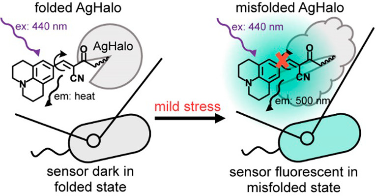
diagram illustrating the mechanism of the stress reporter

Cellular stress is associated with global misfolding and aggregation of the endogenous proteome. Monitoring stress-induced abnormalities remains one of the major technical challenges facing established sensors. Misfolded monomers induced by mild stresses, however, remain largely invisible with current sensors.

In a recent publication (1) Fares and colleagues describe a new sensor based upon a fluorescent molecular rotor that is conjugated to a Halo mutant (AgHalo). In non-stressed cells, the AgHalo sensor remains largely folded, and is fluorescent when misfolded. The fluorescent molecular rotor, when conjugated to purified AgHalo to form the proteome stress sensor, is able to report on urea-induced partially unfolded (misfolded) conformations with a higher fluorescent increase than the previously reported fluorophore-based sensors. Heat-induced misfolding is also effectively monitored by the fluorescence change of the sensor that is based on fluorescent molecular rotor, but not the solvatochromic fluorophore. The unique feature of the fluorescent molecular rotor makes the new generation of the AgHalo proteome sensor more sensitive to misfolded conformations that are primarily induced by mild proteome stress. Further, the new sensor exhibits a higher fluorescence signal when detecting soluble and insoluble protein aggregates that are induced by more severe proteome stress. These data collectively suggest that thermo-labile Halo conjugated with a fluorescent molecular rotor serves as a suitable sensor to detect a wide range of proteome stress conditions.

Literature Cited

Fares, M. et al. (2018) A Molecular Rotor-Based Halo-Tag Ligand Enables a Fluorogenic Proteome Stress Sensor to Detect Protein Misfolding in Mildly Stressed Proteome. Bioconjugate Chem 29, 215–24.

Learn more about protein analysis using HaloTag here.

Characterizing Multi-Subunit Protein Complexes Using Cell-Free Expression

artist's concept of a cell membrane

Multi-subunit protein complexes control membrane fusion events in eukaryotic cells (1). CORVET and HOPS are two such multi-subunit complexes, both containing the Sec1/Munc18 protein subunit VPS33A (2). Metazoans additionally possess VPS33B, which has considerable sequence similarity to VPS33A but does not integrate into CORVET or HOPS complexes and instead stably interacts with VIPAR. Recent research suggests that VPS33B and VIPAR comprise two subunits of a novel multi-subunit complex analogous in configuration to CORVET and HOPS (3).

In a recent publication (4), Hunter and colleagues, further characterized the VPS33B and VIPAR complex. Using co-immunoprecipitation and proximity-based ligation assay, they identified two novel VPS33B-interacting proteins, VPS53 and CCDC22.

In vitro binding experiments, VPS33B and GST-VIPAR were co-expressed in Escherichia coli and purified by GSH affinity. The VPS33B/GSTVIPAR complex was used as bait in pulldown experiments, with myc-CCDC22 and myc-VPS53 expressed by cell-free in vitro transcription/translation in wheat germ lysate. Myc-CCDC22 was very efficiently pulled down by VPS33B/GST-VIPAR, whereas myc-VPS53 was not .The interaction between VPS53 and the VPS33B-VIPAR complex was either indirect, requires other proteins contribute to the interaction, or requires a post-translational modification not conferred in the plant cell-free expression system (wheat germ). Pull-down experiments with individual subunits or expressing as complexes, was inefficient and did not result in binding to VPS33B/GST-VIPAR.

To further understand how VPS33B-VIPAR may interact with CCDC22, Hunter and colleagues attempted to refine the region of CCDC22 that interacts with VPS33B/GST-VIPAR by generating a series of truncated forms of CCDC22. However, none of five CCDC22 truncations were able to bind to VPS33B/GST-VIPAR. The hypothesis was that truncated forms of CCDC22 are unstable and unable to fold correctly in this assay system.

Additional experiments noted that the protein complex in HEK293T cells which contained VPS33B and VIPAR was considerably smaller than CORVET/HOPS, suggesting that, unlike VPS33A, VPS33B does not assemble into a large stable multi-subunit protein complex.


  1. D’Agostino, M. et. al. (2017) A tethering complex drives the terminal stage of SNARE-dependent membrane fusion. Nature 551, 634–638.
  2. Balderhaar, H. J. K. and Ungermann, C. (2013) CORVET and HOPS tethering complexes – coordinators of endosome and lysosome fusion. J. Cell Sci. 126, 1307–16.
  3. Spang, A. (2016) Membrane Tethering Complexes in the Endosomal System. Front. Cell Dev. Biol. 4, 35.
  4. Hunter, M.  et. al.  (2017) Proteomic and biochemical comparison of the cellular interaction partners of human VPS33A and VPS33B. [Internet bioRxiv http://dx.doi.org/10.1101/236695  Accessed 3/12/2018]