From Chromosomes to the Brain: How HaloTag Is Expanding What We Can See

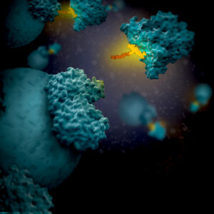
Artist's 3D concept of HaloTag with linker attaching to a target protein

Sometimes in the life sciences, itโ€™s really tempting to look at a process like mitosis and think, โ€œWow, we really understand that,โ€ and stop asking questions. In other cases, significant barriers like reliable expression of a non-endogenous reporter in an organ such as the brain is a challenge that seems impossible to overcome. However, those are precisely the moments when science and scientists need to push the boundaries of what we can see. In this post, I discuss two studies where researchers are literally seeing what could not be seen before by developing new methods for looking at their model systems.

In both papers discussed here, the HaloTagยฎ protein and ligands were key to developing new methods allowing scientists to visualize biological phenomena that had been previously inaccessible. HaloTagยฎ protein allows different functionalities to be linked onto a single genetic construct, rapidly and covalently under physiological conditions for experiments ranging from live-cell imaging to capture of protein interaction complexes (1). In the first study, HaloTag-CENP-A + JF635 ligand was the specific tool that let them directly see, in real time, that chromosome-attached microtubules pivot as the spindle elongates. In the second study, HaloTag was used to create a protocol for PET imaging of reporter gene systems in the brain.

Continue reading “From Chromosomes to the Brain: How HaloTag Is Expanding What We Can See”

Trends and Tools Transforming Drug Discovery: Five Takeaways from Discover Glo 2025

In biologics, cell therapy, and targeted protein degradation, the science is moving fastโ€”and so are the tools. From GPCR-targeted therapies to real-time CAR-T manufacturing tools, new techniques are reshaping how scientists approach drug development, live-cell imaging, and protein degradation.

The โ€œBringing Light to Scienceโ€ Discover Glo 2025 speaker series brought together researchers from across academia and industry to share real-world examples of how bioluminescent technologies are helping them advance their research. Now available on demand, these sessions offer fresh perspectives and actionable takeaways on the future of therapeutic development, cellular analysis and assay design.

Weโ€™ve distilled five key takeaways from the sessionsโ€”practical insights you can apply to your own work or use to stay current with where the field is heading.

Continue reading “Trends and Tools Transforming Drug Discovery: Five Takeaways from Discover Glo 2025”

Polyserine Targeting: A New Strategy Against Neurodegeneration

Neurodegenerative diseases like Alzheimerโ€™s are marked by the accumulation of misfolded proteins that wreak havoc on neurons. One of the most notorious culprits is tau, a structural protein that, in its diseased form, clumps together into aggregates that spread throughout the brain. These aggregates interfere with normal cellular processes, leading to memory loss, behavioral changes, and other devastating symptoms. Preventing tau aggregation is therefore a key strategy for slowing the progression of these symptoms.

What if we could recruit molecular โ€œhelpersโ€ to stop tau from accumulating?

Continue reading “Polyserine Targeting: A New Strategy Against Neurodegeneration”

Understanding Wnt Signaling Through ฮฒ-Catenin Localization in Live Cells

The Wnt/ฮฒ-catenin pathway, long studied in the context of developmental biology, has become increasingly recognized for its role in a wide range of human diseases. Its dysregulation has been implicated in cancer, fibrosis, immune modulation, and neurodegenerative conditionsโ€”making it a clinically actionable target across diverse therapeutic areas1. In this blog, we cover the fundamentals of Wnt/ฮฒ-catenin signaling, highlight ongoing research efforts to understand its role in disease, and show how combining live-cell imaging with luminescent assays complements functional studies.

Continue reading “Understanding Wnt Signaling Through ฮฒ-Catenin Localization in Live Cells”

Seeing Signals in a New Light: Far-Red Chemigenetic Biosensors Illuminate Kinase Activity

Cell signaling is a finely tuned process where both timing and spatial context play essential roles. Whether itโ€™s a hormone triggering a cellular response or a drug modulating a pathway, these processes unfold in dynamic, spatially organized ways. To study them, researchers rely on chemigenetic biosensorsโ€”genetically encoded tools that light up in response to molecular activity. However, traditional biosensors are constrained by several limitations: poor photostability under prolonged imaging, limited spectral flexibility for multiplexing, and insufficient spatial resolution for studying signaling events at subcellular scales.

Continue reading “Seeing Signals in a New Light: Far-Red Chemigenetic Biosensors Illuminate Kinase Activity”

Mapping the Mind: In Vivo Imaging of Synaptic Plasticity with HaloTagยฎ Ligands

The brain is constantly rewiring itself, fine-tuning connections that shape how we think, learn, and remember. But capturing those fleeting molecular changes as they happen โ€” at the level of individual synapses and across entire brain regions โ€” has long been a challenge in neuroscience. Now, thanks to recent advances in HaloTagยฎ dye technology, researchers can visualize protein dynamics in living brains with stunning clarity and specificity.

Continue reading “Mapping the Mind: In Vivo Imaging of Synaptic Plasticity with HaloTagยฎ Ligands”
