Mass Spectrometry Application: Antibody Quantitation for Preclinical PK studies

Isoform_Antibodies_LinkedIn
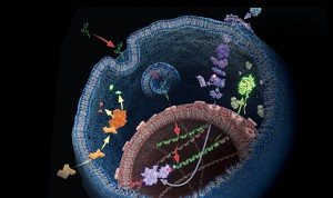
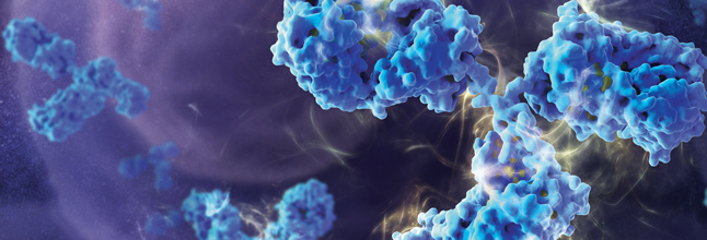
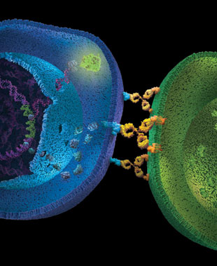
Therapeutic monoclonal antibodies (mAbs) represent the majority of therapeutics biologics now on the market, with more than 20 mAbs approved as drugs (1–3). During preclinical development of therapeutic antibodies, multiple variants of each antibody are assessed for pharmacokinetic (PK) characteristics across model systems such as rodents, beagles and  primates. Ligand-binding assays (LBA) are the standard technology used to perform the PK studies for mAb candidates (4). Ligand-binding assays (LBAs) are methods used  to detect and measure a macromolecular interaction between a ligand and a binding molecule. In LBAs, a therapeutic monoclonal antibody is considered to be the ligand, or analyte of interest, while the binding molecule is usually a target protein.

LBAs have certain well-documented limitations (5). Specific assay reagents are often not available early in a program. Interferences from endogenous proteins, antidrug antibodies, and soluble target ligands are potential complicating factors.

Liquid chromatography coupled to tandem mass spectrometry (LC–MS/MS)-based methods represent a viable and complementary addition to LBA for mAb quantification in biological matrixes. LC–MS/MS provides specificity, sensitivity, and multiplexing capability.

A recent reference (6) illustrates an automated method to perform LC–MS/MS-based quantitation, with IgG1 conserved peptides, a heavy isotope labeled mAb internal standard,and anti-human Fc enrichment. The method was applied to the pharmacokinetic study of a mAb dosed in cynomolgus monkey, and the results were compared with the immunoassay data. The interesting finding of the difference between ELISA and LC–MRM-MS data indicated that those two methods can provide complementary information regarding the drug’s PK profile.

Literature Cited

  1. Mao, T. et al. (2013) Top-Down Structural Analysis of an Intact Monoclonal Antibody by Electron Capture Dissociation-Fourier Transform Ion Cyclotron Resonance-Mass Spectrometry. Anal.Chem. 85, 4239–46.
  2. Weiner, L. M. et al. (2010) Monoclonal antibodies: versatile platforms for cancer immunotherapy. Nat. Rev. Immunol. 10, 317–27.
  3. Nelson, A. et al. (2010) Development trends for human monoclonal antibody therapeutics. Nat. Rev. Drug Discovery. 9, 767–74.
  4. DeSilva, B. et al. (2003) Recommendations for the Bioanalytical Method Validation of Ligand-Binding Assays to Support Pharmacokinetic Assessments of MacromoleculesPharm. Res. 20, 1885–00.
  5. Ezan, E.et al. (2009) Critical comparison of MS and immunoassays for the bioanalysis of therapeutic antibodiesBioanalysis 1, 1375–88.
  6. Zhang, Q. et al. (2014) Generic Automated Method for Liquid Chromatography–Multiple Reaction Monitoring Mass Spectrometry Based Monoclonal Antibody Quantitation for Preclinical Pharmacokinetic Studies. Anal.Chem. 86, 8776–84.

What Things Are You Thankful for in Science?

What are you thankful for in science?
As the social media lead for Promega, I keep my eye on trends in new media. I have personal accounts that I keep mostly to see what other people are doing. I try hangouts, social networking and other things so that I have an idea of developing practices outside of the biotechnology industry. One activity that has been popular over the last couple of years during the month of November in the United States is the Facebook post of “30 days of thanksgiving”.

I wondered what “thanksgiving” looks like to the research scientist. So I asked:

What are the things you are thankful for in science?

The answers have been as varied as the people I talked to ranging from little things like water bath floats to really big things, like the renewal of your research funding or achieving tenure.

Here are some of the answers from my informal inquiries:

“Tube floaties for water baths.”

—E.V., genomics product manager

“I was always thankful for Geiger counters.”

—K. G., science writer

“Thermal cyclers and Taq Polymerase. As an undergrad I watched someone sit with a timer and move their tubes between water baths at 3 different temperatures, opening tubes and adding polymerase at the end of each cycle. Modern PCR is SOOO much easier.”

—M.M., research scientist

“I am thankful for competent cells. I remember preparing the CaCl2 and doing slow centrifugation. Also thankful for serum-compatible transfection, rapid ligations and online journal access (no longer have to traipse over to the university library to get papers photocopied- uuurrrgggghhh).”

—R.D., technical services scientist

“How about T-vectors for cloning? I was no molecular biologist, but could make a T-vector work.”

—K.K., science writer

“I am thankful for open-access journals and the ability to read the full article without an institutional subscription.”

—S.K., science writer

“I am ever so thankful for ONLINE ORDERING! So awesome. Throw in online technical manuals, on-line support tools, on-line calculators – all are awesome!!”

—A.P., director, scientific courses

“I am thankful for automated sequencing- manual sequencing was laborious and hazardous!!!”

—R.G., technical services scientist

Do any of these resonate with you? What are you thankful for as a scientist? Let us know in the comments.

Use of Mass Spectrometry to Quantitate Food Allergens


Food allergies are becoming increasingly prevalent among children. Credit: James Gathany, CDC

Food allergies are increasing worldwide and becoming a public health issue, especially among children are concerned. Children have a higher prevalence of food allergies, with about 4–8%, compared to adults (1–5%).  Currently antibody-based methods such ELISA (enzyme-linked immunosorbent assay) are the primary method for food allergen analysis. In most cases antibodies are only available for single well-known allergens. Often those that are commercially available are poorly characterized resulting cross-reactivity that leads to false-positive results in diagnostic tests.

A recent publication (1) presented a review of an alternative technology based on mass spec (i.e., multiple reaction monitoring, MRM) that circumvents the drawbacks of antibody based methods. MRM allows precise quantitative determination of target proteins in complex samples with broad dynamic range.  MRM also provides quantification of different isoforms. It is noted that tryptic digestion followed by mass spec analysis, has already identified several unique peptides for different allergens, including those found in crustaceans, eggs, fish, peanuts, soy and wheat. In summary the challenge is now to select the appropriate tryptic signature peptide(s) for the respective allergen and to develop well characterized standards (i.e., isotope labeled standards) to ensure accurate quanititation.

Citation:

Koeberl, M et al. (2014) Next Generation of Food Allergen Quantification using Mass Spectrometric Systems J. Proteome Research  13, 3499–509.

Top 10 Innovator—Two Years Running

The ADCC Reporter Bioassays were named a Top 10 innovation by The Scientist Magazine.
The ADCC Reporter Bioassay systems were named a Top 10 innovation by The Scientist Magazine.
For the second year running a Promega technology has made The Scientist Magazine’s list of Top 10 Innovations. Last year it was the NanoLuc® luciferase technology; this year it is the ADCC Reporter Bioassay.

Antibody-dependent cell-mediated cytotoxicity (ADCC) is the main mechanism of action (MOA) of antibodies through which virus-infected or other diseased cells are targeted for destruction by components of the cell-mediated immune system. ADCC assays are often used to assess the effectiveness of monoclonal antibody therapies during the manufacture and development of biologic drugs. The bioluminescent ADCC Reporter Bioassays use an alternative readout at an earlier point in ADCC MOA pathway for the quantification of Fc effector function of antibody-based molecules: the activation of gene transcription through the NFAT (nuclear factor of activated T-cells) pathway in the effector cell.

The bioassay uses engineered Jurkat cells stably expressing the FcγRIIIa receptor, V158 (high affinity) variant, and an NFAT response element driving expression of firefly luciferase. The assay is ADCC MOA-based and features frozen, thaw-and-use effector cells and optimized reagents and protocol to perform a reporter-based ADCC bioassay in a single day. The ADCC Reporter Bioassay correlates with classic cytotoxic ADCC assays and is a suitable replacement for these cumbersome and highly variable assays.

The novel bioassay is linear, accurate, precise and stability indicating. Moreover, the bioassay shows good linear correlation between levels of glycosylation or fucosylation and ADCC activity. All of these features indicate the assay is suitable for use across biologic drug development programs.

Resources for the ADCC Reporter Bioassays:

Piecing the Puzzle Together: Using Multiple Assays to Better Understand What Is Happening with Your Cells

You often need several pieces of information to really understand what is happening within a cell or population of cells. If your cells are not proliferating, are they dying? Or, are you seeing cytostasis? If they are dying, what is the mechanism? Is it apoptosis or necrosis? If you are seeing apoptosis, what is the pathway: intrinsic or extrinsic?

If you are measuring expression of a reporter gene and you see a decrease in expression, is that decrease due to transfection inefficiencies, cytotoxicity, or true down regulation of your reporter gene?

To investigate these multiple parameters, you can run assays in parallel, but that requires more sample, and sample isn’t always abundant.

Multiplexing assays allows you to obtain information about multiple parameters or events (e.g., reporter gene expression and cell viability; caspase-3 activity and cell viability) from a single sample. Multiplexing saves sample, saves time and gives you a more complete picture of the biology that is happening with your experimental sample.

What information do you need about your cells to complete the picture?

Multiplexing assay reagents to measure biomarkers in the same sample has often been considered an application only accomplished with antibodies or dyes and sophisticated detection instrumentation. However, Promega has developed microwell plate based assays for cells in culture that allow multiplexed detection of biomarkers in the same sample well using standard multimode multiwell plate readers. Continue reading “Piecing the Puzzle Together: Using Multiple Assays to Better Understand What Is Happening with Your Cells”

Is This What a Scientist Looks Like?

scientists-at-workI am the mother of a six-year-old girl who loves to get magazines in the mail. For several years my daughter has received an enjoyed popular kids’ science/international culture magazine. The stories are short and simple, and this magazine usually does a good job of presenting factual information in easy-to-digest forms. Each magazine comes with a set of animal cards, which we have diligently collected.

However, the latest issue that came to our mailbox really got me thinking. The final pages featured artwork by the young readers. I love the idea of featuring the work of the readers.  Usually, my daughter loves seeing what other children her age from around the world draw and take pictures of, and sometimes we have some pretty interesting discussions about the work.

This time though we didn’t spend much time talking about the art work. She wasn’t particularly interested, and I wasn’t sure I what I thought. But I may have missed a teachable moment. The theme for the pages was a Halloween-minded “spooky science”, and all of the pictures were of “mad scientists” alone at work doing presumably nefarious things in their laboratories. Of the eight drawings pictured, six of them pictured scientists that were human, and five of the humans were male. All of them were pale-skinned. The sole female scientist, whose lab featured a certificate with the words “monster maker”, was drawn by a girl. The ages of the children submitting the work ranged from 9 to 14. Continue reading “Is This What a Scientist Looks Like?”

Convenient, Non-Radioactive Detection of Isoaspartate

Structure of the PCMT1 protein. Based on PyMOL rendering of PDB 1i1n. Licensed under creative commons http://creativecommons.org/licenses/by-sa/3.0/deed.en

The ISOQUANT® Isoaspartate Detection Kit is intended for quantitative detection of isoaspartic acid residues in proteins and peptides, which can result from the gradual, nonenzymatic deamidation of asparagine or rearrangement of aspartic acid residues.

The ISOQUANT® Kit is designed to provide information regarding the global formation of isoaspartic acid residues at Asn and Asp sites, not at each site separately.

The deamidation of asparagine residues and rearrangement of aspartic acid residues is characterized by the formation of a succinimide intermediate that resolves to form a mixture of isoaspartic acid (typically 70–85%) and aspartic acid.
The rate and extent of isoaspartic acid formation can vary widely among proteins, depending on the amino acid sequence and size of the target protein. Deamidation of Asn residues has been observed most frequently at Asn-Gly and Asn-Ser sites within proteins.

The ISOQUANT® Isoaspartate Detection Kit uses the enzyme Protein Isoaspartyl ethyltransferase (PIMT) to specifically detect the presence of isoaspartic acid residues in a target protein. PIMT catalyzes the transfer of a methyl group from S-adenosyl-L-methionine (SAM) to isoaspartic acid. Spontaneous decomposition of this methylated intermediate results in the release of methanol and reformation of the succinimide.

References:

Wang, W. et al. (2012) Quantification and characterization of antibody deamidation by peptide mapping with mass spectrometry. Int. J. Mass. Spec. 312, 107–13.

Grappin, P. et al. (2011) New proteomic developments to analyze protein isomerization and their biological significance in plants. J. Proteomics, 74, 1475–82.

Yang, H. and Zubarev, R.A. (2010) Mass spectrometric analysis of asparagine deamidation and aspartate isomerization in polypeptides. Electrophoresis 31, 1764–71.

Sinha, S. et al. (2009) Effect of protein structure on deamidation rate in the Fc fragment of an IgG1 monoclonal antibody. Protein Sci. 18, 1573–84.

Back in My Day We Trudged Up Hill to the Lab…

Tube racks stacked in the refrigeratorRemember the bubble getter? Siliconizing sequencing gel glass plates? Carrying out sequencing reactions in strip tubes? Diagramming, by hand, your cloning scheme and calculating the cut sizes with a hand-held calculator? Marking plates for plaque lifts with india ink?

This video is for all of you who were in the lab when life was “one gene, one graduate student”. What other oldie but goodies can you think of? Leave a comment or tweet @promega #backinmyday

[youtube=http://www.youtube.com/watch?v=voNepWllrMM]

From Zombies to Hanta Virus to Wizards in Training: Summertime Reading

iStock_000016760626XSmallJust the other day, the local library near Promega held its Friends of the Library Summer Book Sale, and several of the Promega editors trekked to the stacks to take advantage of bargain books. Many of us were looking for summer reading treasures.

Summer reading has always been a big tradition for me.  When I was younger I spent much of my summer breaks sitting under the shade of a big tree or, later, an airplane hangar with a good book.  When I was in college on the summer Glee Club tour, I remember an English Literature major who always boarded the bus with a shopping bag full of Harlequin romances—her summer reading. My daughter, now a “school-ager” herself has come home with own summer reading list, although many of the assignments were ebooks: times have changed a little.

As I was thinking about summer reading, I wondered, is there any pattern to what scientists take on vacation with them for their leisure reading hours? So I conducted an informal poll of science types hanging around Promega to see what they were planning for summer vacation reading. The results were as varied as the scientists whom I polled, but if you are stuck for a summer reading idea, perhaps one of these titles will appeal to you.

Do you have any summer reading suggestions for us? Share them on Twitter (@Promega), Facebook  or Google+

Science-related fiction Continue reading “From Zombies to Hanta Virus to Wizards in Training: Summertime Reading”

Epigenetics and Exercise

Turning on some genes

If, like me, you sometimes need more motivation to exercise consistently—even though you know that it is good for you—you may be interested in the findings of a paper published recently in PLOS Genetics. The paper showed that consistent exercise over a 6-month period caused potentially beneficial changes in gene expression. In short, regular exercise caused expression of some “good” genes, and repression of “bad” ones, and these changes appeared to be controlled by epigenetic mechanisms.

Epigenetic changes are modifications to DNA that affect gene expression but don’t alter the underlying sequence. Perhaps the best understood example of an epigenetic change is DNA methylation—where methyl groups bind to the DNA at specific sites and alter expression, often by preventing transcription. Epigenetic changes have been shown to occur throughout all stages of development and in response to environmental factors such as diet, toxin exposure, or stress. The study of epigenetics is revealing more and more about how the information stored in our DNA is expressed in different tissues at different times and under different environmental circumstances. Continue reading “Epigenetics and Exercise”