Targeting Glioblastoma Cells by Packaging a Lentiviral Vector Inside a Zika Virus Coat

A recent article published in Cancers demonstrates a new method for targeting glial cells using a lentiviral packaging system that incorporated Zika virus envelope proteins. By using the reporter gene firefly luciferase, researchers demonstrated that a pseudotyped virus could infect cultured glioblastoma cells.

Introduction

Space-fill drawing of the outside of one Zika virus particle, and a cross-section through another as it interacts with a cell. The two main proteins of the viral envelope, the envelope proteins and membrane proteins, are shown in red and purple respectively. The lipid membrane of the envelope is shown in light lavender.The capsid proteins, in orange, are shown interacting with the RNA genome, in yellow, at the center of the virus. The cell-surface receptor proteins are in green, the cytoskeleton in blue, and blood plasma proteins in gold. Drawn by David Goodsell.
Space-fill drawing of the outside of one Zika virus particle, and a cross-section through another as it interacts with a cell. The two main proteins of the viral envelope, the envelope proteins and membrane proteins, are shown in red and purple respectively. The lipid membrane of the envelope is shown in light lavender. The capsid proteins, in orange, are shown interacting with the RNA genome, in yellow, at the center of the virus. The cell-surface receptor proteins are in green, the cytoskeleton in blue, and blood plasma proteins in gold. Drawn and copyright owned by David Goodsell.

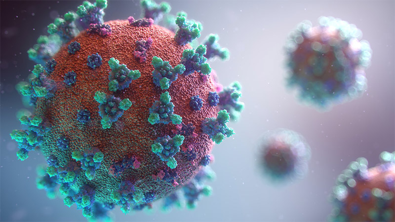
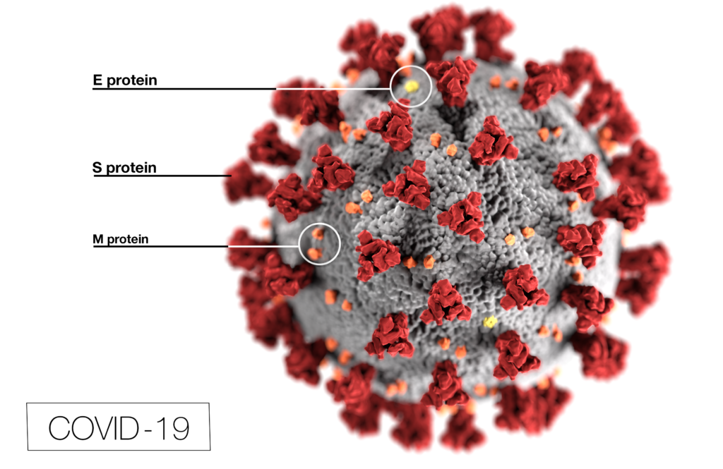
Viruses enjoy a fearsome reputation. SARS-CoV-2 is only the latest infectious agent that has garnered attention by becoming a worldwide pandemic. Even the viral name suggests that SARS-CoV-2 was not the first of its type [SARS-CoV is the virus behind the severe acute respiratory syndrome (SARS) that spread worldwide in the early 2000s]. There are many different families of viruses (e.g., coronavirus for SARS-CoV-2 or lentiviruses for HIV-1) and each show a preference to the cell types they want to infect. By investigating the life cycle of viruses to better understand their mechanisms, researchers can discover new opportunities that may be exploited.

In 2015 and 2016, the virus that concerned health authorities was Zika virus (ZIKV). While this virus generally caused mild disease, the babies of women who were infected during pregnancy were at increased risk for microcephaly and other brain defects. These defects were traced back to Zika virus infecting nerve tissue, specifically, glial cells. This discovery provided an opportunity to explore how Zika virus might affect the brain tumor, glioblastoma multiforme (GMB), especially the glioblastoma stem cells (GSCs) that resist conventional treatment and contribute to the poor prognosis for GMB. Studies suggested that Zika virus infection prolonged survival in animal glioma models and selectively killed GSC with minimal effects on normal cells. In fact, the molecules used by ZIKV to enter cells were predominantly found on tumors, not normal cells. Knowing that the ZIKV envelope proteins prM and E provide the target specificity for glial cells, Kretchmer et al. wanted to explore if ZIKV envelope proteins substituted in lentivirus packaging systems would be able to enter glioblastoma cells.

Continue reading “Targeting Glioblastoma Cells by Packaging a Lentiviral Vector Inside a Zika Virus Coat”

From Viral Outbreak to Vaccine Development: Our Top 10 Most Viewed Blog Posts of 2020

This illustration, created at the Centers for Disease Control and Prevention (CDC), reveals ultrastructural morphology exhibited by coronaviruses. Photo Credit:  Alissa Eckert, MS; Dan Higgins, MAM CDC It is one is used in several of our top 10 most viewed blogs of 2020
Illustration from CDC; Photo Credit: Alissa Eckert, MS; Dan Higgins, MAM

When you look at our top 10 most viewed blog posts of 2020, there’s no surprise that all relate to COVID-19. We have come a long way since the beginning of the year, thanks to tireless scientists and researchers around the globe. They have led the way in COVID-19 research, treatment, and testing. Let’s take a closer look at this top 10 list:

10. Tips to Maintain Physical Distance in the Lab 

The spread of COVID-19 forced us to adapt and adjust to new ways in life, in work, and for this blog post, in the lab. In response to the pandemic, some labs shut down completely. Others have stayed open, especially those involving coronavirus research. This post provides 10 helpful distancing tips for researchers to stay safe and productive while working in the lab.  

9. Investigation of Remdesivir as a Possible Treatment for SARS-2-CoV (2019 nCoV) 

Scientists have worked hard to determine possible treatment for COVID-19. This blog post focuses on Remdesivir (RDV or GS-5734), an encouraging treatment used for the first case in the United States. It provides an in-depth look at numerous studies and clinical trials on Remdesivir as treatment for COVID-19. One key finding is that RDV needed to be administered either before or shortly after infection to limit lung damage. 

Continue reading “From Viral Outbreak to Vaccine Development: Our Top 10 Most Viewed Blog Posts of 2020”

The Path Brightens for Vaccine Researchers: Luminescent Reporter Viruses Detect Neutralizing Antibodies

Developing a vaccine that is safe, effective, easily manufactured and distributed is a daunting task. Yet, that is exactly what is needed in response to the COVID-19 pandemic.

Computer generated 3D image of coronavirus

Vaccine development, safety and efficacy testing take time. The mumps vaccine is thought to be the quickest infectious disease vaccine ever produced, and its development required four years from sample collection to licensing (2). However, there are many reasons to anticipate quicker development for a COVID-19 vaccine: Researchers are collaborating in unprecedented ways, and most COVID-19 scientific publications are free for all to access and often available as preprints. As of August 11, 2020, researchers around the globe have more than 165 vaccine candidates in development, 30 of which are in some phase of human clinical trials (1). The range of vaccine formulations available to scientists has expanded to include RNA and DNA vaccines, replication-defective adenovirus vaccines, inactivated or killed vaccines and subunit protein vaccines. Equally important is that vaccine developers and researchers have greater access to powerful molecular biology tools like bioluminescent reporters that enable quicker testing and development.

Continue reading “The Path Brightens for Vaccine Researchers: Luminescent Reporter Viruses Detect Neutralizing Antibodies”

Choices for Measuring Luciferase-Tagged Reporter Pseudotyped Viral Particles in Coronavirus Research

Coronavirus (CoV) researchers are working quickly to understand the entry of SARS-CoV-2 into cells. The Spike or S proteins on the surface of a CoV is trimer. The monomer is composed of an S1 and S2 domain. The division of S1 and S2 happens in the virus producing cell through a furin cleavage site between the two domains. The trimer binds to cell surface proteins. In the case of the SARS-CoV, the receptor is angiotensin converting enzyme 2. (ACE2). The MERS-CoV utilizes the cell-surface dipeptidyl peptidase IV protein. SARS-CoV-2 uses ACE2 as well. Internalized S protein goes though a second cleavage by a host cell protease, near the S1/S2 cleavage site called S2′, which leads to a drastic change in conformation thought to facilitate membrane fusion and entry of the virus into the cell (1).  

CDC / Alissa Eckert, MS; Dan Higgins, MAMS

Rather than work directly with the virus, researchers have chosen to make pseudotyped viral particles. Pseudotyped viral particles contain the envelope proteins of a well-known parent virus (e.g., vesicular stomatitis virus) with the native host cell binding protein (e.g., glycoprotein G) exchanged for the host cell binding protein (S protein) of the virus under investigation. The pseudotyped viral particle typically carries a reporter plasmid, most commonly firefly luciferase (FLuc), with the necessary genetic elements to be packaged in the particle. 

To create the pseudotyped viral particle, plasmids or RNA alone are transfected into cells and the pseudotyped viruses work their way through the endoplasmic reticulum and golgi to bud from the cells into the culture medium. The pseudoviruses are used to study the process of viral entry via the exchanged protein from the virus of interest. Entry is monitored through assay of the reporter. The reporter could be a luciferase or a fluorescent protein.   

Continue reading “Choices for Measuring Luciferase-Tagged Reporter Pseudotyped Viral Particles in Coronavirus Research”