mRNA Vaccines for COVID-19: The Promise and Pitfalls

Updated 8/25/2021

Multiple battles are being fought in the war against the SARS-CoV-2 coronavirus that causes COVID-19. Currently, there are nearly 5,000 clinical trials listed in the World Health Organization (WHO) database, either underway or in the recruiting stage, for vaccines and antiviral drugs. The Moderna mRNA vaccine and Janssen vaccine have received emergency use authorization (EUA) from the Food and Drug Administration (FDA); the Pfizer-BioNTech Vaccine (marketed as Comiraty) received FDA approval in August 2021.

mrna vaccines and coronavirus covid-19

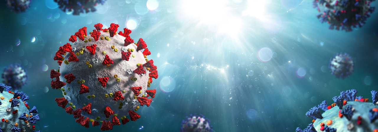
Both the Moderna vaccine and Comiraty are mRNA-based, as opposed to most conventional vaccines against established diseases that are protein-based. Typically, the key ingredient in viral vaccines is either part of an inactivated virus, or one or more expressed proteins (antigens) that are a part of the virus. These protein antigens are responsible for eliciting an immune response that will fight future infection by the actual virus. Another approach is to use a replication-deficient viral vector (such as adenovirus) to deliver the gene encoding the antigen into human cells. This method was used for the coronavirus vaccine developed by Oxford University in collaboration with AstraZeneca; phase 3 interim data were announced on the heels of the Pfizer/BioNTech and Moderna announcements. All three vaccines target the SARS-CoV-2 spike protein, because it is the key that unlocks a path of entry into the host cell.

Continue reading “mRNA Vaccines for COVID-19: The Promise and Pitfalls”

Optimized Protein Expression: Flexi Rabbit Reticulocyte Lysate

A protein chain being produced from a ribosome.

mRNAs commonly exhibit differing salt requirements for optimal translation. Small variations in salt concentration can lead to dramatic differences in translation efficiency. The Flexi® Rabbit Reticulocyte Lysate System allows translation reactions to be optimized for a wide range of parameters, including
Mg2+ and K+ concentrations and the choice of adding DTT. To help optimize Mg2+ for a specific message, the endogenous Mg2+ concentration of each lysate batch is stated in the product information included with this product.

The following references utilize the features of Flexi Rabbit Reticulocyte Lysate System to investigate certain parameters of translation:

Vallejos, M. et al. (2010)The 5′-untranslated region of the mouse mammary tumor virus mRNA exhibits cap-independent translation initiation. Nucl Acids Res. 38, 618–32. Identification of internal ribosomal ribosomal entry site in the 5’ untranslated region of the mouse mammary tumor virus mRNA.

Spriggs, K. et al. (2009) The human insulin receptor mRNA contains a functional internal ribosome entry segment. Nucl. Acids. Res. 17, 5881–93. Identification of a functional internal ribosome entry site in the human insulin receptor mRNA.

Powell, M. et al. (2008) Characterization of the termination-reinitiation strategy employed in the expression of influenza B virus BM2 protein. RNA 14, 2394–06. Analysis of the mRNA signals involved in the expression of influenza B virus BM2 protein.

Sato, V. et al. (2007) Measles virus N protein inhibits host translation by binding to eIF3-p40. J. Vir. 81, 11569–76. Charaterized the effect of the measles virus N protein binding to the translation initiation factor eIF3-p40 on the expression of various proteins in rabbit reticulocyte lysate.

Hirao, K. et al. (2006) EDEM3, a soluble EDEM homolog, enhances glycoprotein endoplasmic reticulum-associated degradation and mannose trimming. J. Biol. Chem. 281, 9650–58. The EDEM3 protein was expressed in the presence of canine microsomal membranes to establish that co-translational translocation occurs into the endoplasmic reticulum.

Shenvi, C. et al. (2005) Accessibility of 18S rRNA in human 40S subunits and 80S ribosomes at physiological magnesium ion concentrations–implications for the study of ribosome dynamics. RNA 11, 1898–08. Characterization of ribosome dynamics under different ionic conditions.

Nair, A. et al. (2005) Regulation of luteinizing hormone receptor expression: evidence of translational suppression in vitro by a hormonally regulated mRNA-binding protein and its endogenous association with luteinizing hormone receptor mRNA in the ovary. J. Biol. Chem. 280, 42809–16. Examined the affect of luteinizing hormone receptor mRNA binding protein on transltional suppression of luteinizing hormone receptor RNA.