Understanding how a compound or drug affects cellular pathways often requires measuring kinetic changes over an extended period of time—from several hours to days. Live-cell kinetic cell-based assays that measure cell viability, cytotoxicity, apoptosis and other cellular pathways are great for collecting real-time data. You don’t necessarily need expensive equipment to run these types of assays. In the videos below, Dr. Sarah Mahan, a research scientist at Promega, demonstrates how you can easily get great 24-hour or multi-day kinetic data using a GloMax® Microplate Reader.
Continue reading “How to Get Real-Time Kinetic Data With GloMax® Microplate Readers”glomax
How Promega Helped Our Lab Scale Up Drug Discovery for Bloodborne Pathogens
This blog was written by Sebastien Smick, Research Technician in Dr. Jacquin Niles’ laboratory at Massachusetts Institute of Technology (MIT)
Our lab is heavily focused on the basic biology and drug discovery of the human bloodborne pathogen Plasmodium falciparum, which causes malaria. We use the CRISPR/Cas9 system, paired with a TetR protein fused to a native translational repressor alongside a Renilla luciferase reporter gene, to conditionally knock down genes of interest to create modified parasites. We can then test all kinds of compounds as potential drug scaffolds against these gene-edited parasites. Our most recent endeavor, one made possible by Promega, is a medium-low throughput robotic screening pipeline which compares conditionally-activated or-repressed parasites against our dose-response drug libraries in a 384-well format. This process has been developed over the past few years and is a major upgrade for our lab in terms of data production. Our researchers are working very hard to generate new modified parasites to test. Our robots and plate readers rarely get a day’s rest!
Small Changes With Large Consequences: The Role of Genetic Variance in Disease Development
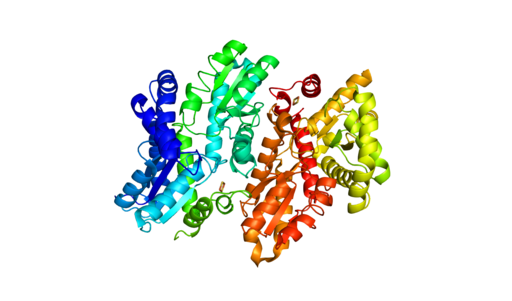
Understanding how disease states arise from genetic variants is important for understanding disease resistance and progression. What can complicate our understanding of disease development is when two people have the same genetic variant, but only one has the disease. To investigate what might be happening with ferrochelatase (FECH) variant alleles that result in erythropoietic protoporphyria (EPP), scientists used next-generation sequencing (NGS) along with RNA analysis and DNA methylation testing to assess the FECH locus in 72 individuals from 24 unrelated families with EPP.
What is FECH and its relationship to EPP?
FECH is the gene for ferrochelatase, the last enzyme in the pathway that synthesizes heme. The inherited metabolic disorder, EPP, is caused when the activity of FECH is reduced to less than a third of normal levels thus, increasing the levels of protoporphyrin (PPIX) without metal in erythrocytes. The consequences of the low-metal PPIX include severe phototoxic skin reactions and hepatic injury due to PPIX accumulation in the liver.
How does FECH expression affect EPP?
The EPP disease state is not simply the lack of two functional FECH genes. Disease occurs with a hypomorphic allele, mutations in FECH that reduce its function, in trans to a null FECH allele. Researchers focused on three common variants called the GTC haplotype that are associated with expression quantitative trait loci (eQTL) that reduce FECH activity. Interestingly, these three variants have been found in trans, but researchers wanted to learn if there were individuals who were homozygous for the GTC allele and how EPP manifested for them.
Continue reading “Small Changes With Large Consequences: The Role of Genetic Variance in Disease Development”Designing a Reporter Construct for Analyzing Gene Regulation
Bioluminescent reporter assays are an excellent choice for analyzing gene regulation because they provide higher sensitivity, wider dynamic range and better signal-to-background ratios compared to colorimetric or fluorescent assays. In a typical genetic reporter assay, cells are transfected with a vector that contains the sequence of interest cloned upstream of a reporter gene, and the reporter activity is used to determine how the target sequence influences gene expression under experimental conditions. A second control reporter encoded on the same or a different plasmid is an essential internal control. The secondary reporter is used to normalize the data and compensate for variability caused by differences in cell number, lysis efficiency, cell viability, transfection efficiency, temperature, and measurement time.
For genetic reporter assays, using a secondary control vector with a weak promoter like PGK or TK to ensures that the control does not interfere with activation of your primary reporter vector. Transfection of high amounts of the control plasmid or putting the control reporter under control of a strong promoter like CMV or SV40 often leads to transcriptional squelching or other interference with the experimental promoter (i.e., trans effects). Reporter assays can also be used to quantitatively evaluate microRNA activity by inserting miRNA target sites downstream or 3´ of the reporter gene. For example, the pmirGLO Dual-Luciferase miRNA Target Expression Vector is based on dual-luciferase technology, with firefly luciferase as the primary reporter to monitor mRNA regulation and Renilla luciferase as a control reporter for normalization.
Here in Technical Services we often talk with researchers who are just starting their project and looking for advice on designing their genetic reporter vector. They have questions like:
- How much of the upstream promoter region should be included in the vector?
- How many copies of a response element will be needed to provide a good response?
- Does the location of the element or surrounding sequence alter gene regulation?