Like the recipe book for life, every living creature has DNA. DNA contains genes, which contain instructions for making proteins. There are many types of important proteins that impact the way our body functions. Transcription factors (TFs) are a special protein that controls what other proteins are made by directly interacting with DNA to turn genes “on” or “off.”
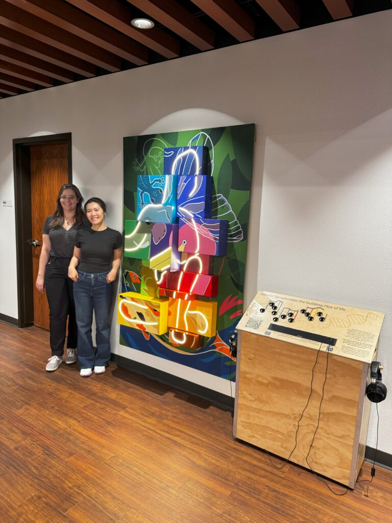
The newest art installation at our Biopharmaceutical Technology Center Institute (BTCI) brings this concept to life. “Genetic Symphonies: Building Hox of Life” uses a human skeleton to showcase how TFs turns on Hox genes by flipping the switches in the correct order. Hox proteins are a special TF that function during growth and development—and all mammals have them. There are 13 groups of Hox TFs (Hox1-Hox13) and unlike other proteins, Hox TFs must be made in a certain order for proper development to occur, starting with Hox1 and ending with Hox13.
In this interactive exhibit, the user is a TF and must turn on Hox genes by flipping the switches in the correct order on a control podium. Every switch (Hox gene) you flip will be accompanied by light and sound (Hox proteins), representing the production of Hox TF proteins. If you successfully turn on all 13 light switches in the correct order, then the entire skeleton will be lit up, orchestrating your own developmental symphony.
Cyanobacteria, microscopic photosynthetic bacteria, have been quietly shaping our planet for billions of years. Responsible for producing the oxygen we breathe, these tiny organisms play a critical role in the global carbon cycle and are now stepping into the spotlight for another reason: their potential to both understand and potentially combat climate change.
Baia di Levente. Marine, volcanic seeps in Italy where UTEX 3221 and UTEX 3222 were discovered.Image credit: Adobe Stock.
Recently, researchers discovered two new strains of cyanobacteria, UTEX 3221 and UTEX 3222, thriving in a marine volcanic seep off the coast of Italy. While cyanobacteria are virtually everywhere there is water and light—from calm freshwater ponds to extreme environments like Yellowstone’s hot springs—this particular habitat is remarkable for its naturally high CO₂ levels and acidic conditions. For these newly identified strains, a geochemical setting like marine volcanic seeps have likely driven the evolution of unique traits that could make them valuable for carbon sequestration and industrial applications.
For decades, the concept of “undruggable” targets has presented one of the most significant challenges in drug discovery. At our recent virtual event, Illuminating New Frontiers: Cracking the Undruggable Code, leading researchers and industry experts gathered to showcase cutting-edge technologies and fresh perspectives that are expanding the boundaries of therapeutic development. Over three engaging days, participants explored groundbreaking advances in targeting RAS signaling, leveraging protein degradation and induced proximity strategies, and exploring RNA as a therapeutic target.
Luminescent live-cell assays are powerful tools for cellular biology research. They offer both qualitative and quantitative insights into processes such as gene expression, cell viability, metabolic activity, protein and small molecule interactions, and targeted protein degradation. But what if you could go beyond the numbers and actually see what is happening in your cells? With luminescent imaging, you have the opportunity to uncover more dynamic data by visualizing what happens with your cells in real time.
Why Luminescent Imaging?
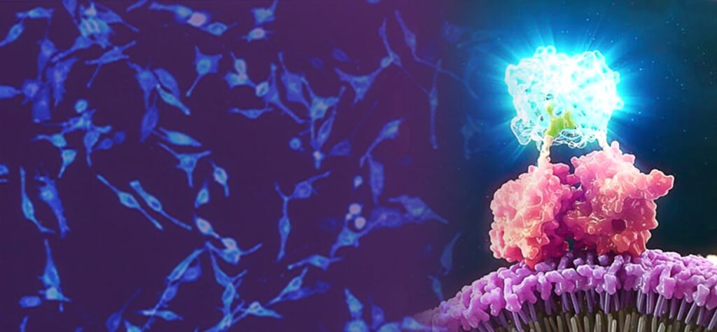
Bioluminescent reporters such as NanoLuc® luciferase are well-suited for bioluminescent imaging. NanoLuc, the smallest engineered luciferase available, is up to 150X brighter than firefly or Renilla luciferase, so exposure times can be shorter than they would be with other luminescent reporters. Its small size also makes it less likely to perturb normal biology or protein function.
Another benefit of bioluminescence for imaging is the inherent stability and sustainability of the bioluminescent signal, which does not require external excitation like fluorescent tags. This allows direct visualization of protein dynamics in living cells without the need for repeated sample excitation. The lack of external excitation also reduces the risk of phototoxicity and photobleaching, common issues that can adversely affect cell viability and signal integrity over time.
Applications Across Cellular Research
Luminescent imaging complements traditional luminescence assays by adding spatial and temporal dimensions. With luminescent live-cell imaging, researchers can visualize NanoLuc® Luciferase assays to gain a deeper understanding of the real-time cellular processes occurring in each experiment. Applications include:
Determining which cells provide signal
Analyzing mixed cell populations
Identifying rare events
Monitoring protein:protein interactions
Identifying protein localization and translocation
Tracking protein degradation and stability over time
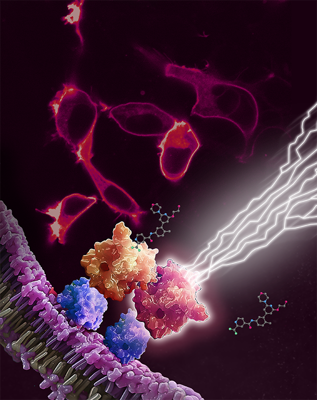
Selectively targeting proteins for removal from the cell—instead of inhibiting protein activity—is a newer approach with therapeutic potential. In this method, the protein is targeted for degradation using the cell’s natural ubiquitin proteasome system (UPS). The degradation process is initiated by compounds such as molecular glues and proteolysis targeting chimeras (PROTACs) linking the target protein to an E3 ligase. Once this linkage occurs, the cell’s UPS does the rest.
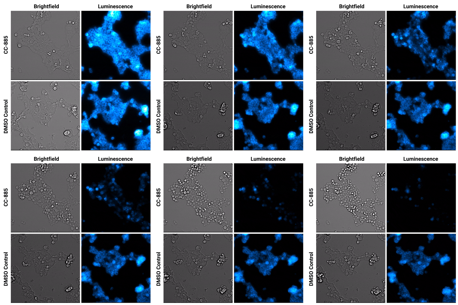
Luminescent substrates with increased signal stability, such as the Nano-Glo® Extended Live Cell Substrate, enable researchers to image targeted protein degradation in their cells in real time. In the example shown below, Nano-Glo® Vivazine™ Live Cell Substrate was used to image degradation of the GSPT1 protein by the CC-885 degrader over 5 hours.
Targeted protein degradation over time. HEK293 cells expressing endogenous HiBiT-tagged GSPT1 and stably expressing LgBiT were treated with CC-885 degrader or DMSO control treatment. Assayed with Nano-Glo® Vivazine™ Live Cell Substrate and imaged over 5 hours using GloMax® Galaxy Bioluminescence Imager.
Combining Luminescent and Fluorescent Imaging to Detect Protein:Small Molecule Interactions
Using bioluminescence resonance energy transfer (BRET)-based assays such as NanoBRET® assays allows you to detect protein:protein interactions by measuring energy transfer from a bioluminescent protein donor to a fluorescent protein acceptor. These assays can be used to monitor changes in protein interactions over time, making them a useful tool for small-molecule screening.
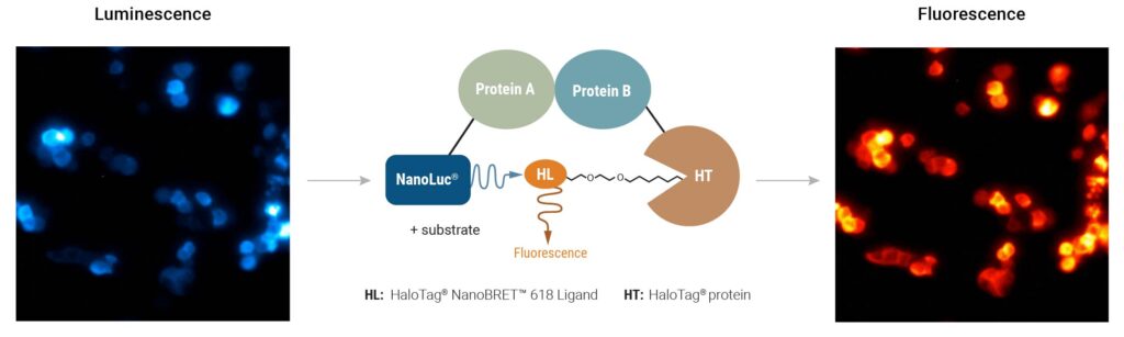
The schematic below illustrates how the NanoBRET® NanoGlo® Detection Systems can be used to visualize target engagement. The cells on the left are expressing a NanoLuc® fusion protein, resulting in a luminescent signal. Adding a fluorescent small tracer (center) results in energy transfer and a fluorescent signal (right). Using an imaging platform that has luminescence and fluorescence imaging capabilities will let you see this energy transfer in action.
Detecting protein:small molecule interactions with NanoBRET® NanoGlo® Detection Systems. HCT116 cells expressing a PRMT5–NanoLuc® fusion were supplemented with a fluorescent small molecule tracer (center panel). Before tracer addition, luminescent signal indicates energy is present on the donor protein (left; 3-minute exposures for 15 minutes). Binding of fluorescent tracer results in energy transfer and fluorescent signal (right; 3-minute exposures for 60 minutes). Images were captured on the GloMax® Galaxy Bioluminescence Imager.
Bringing the Power of Luminescent Imaging to Your Lab
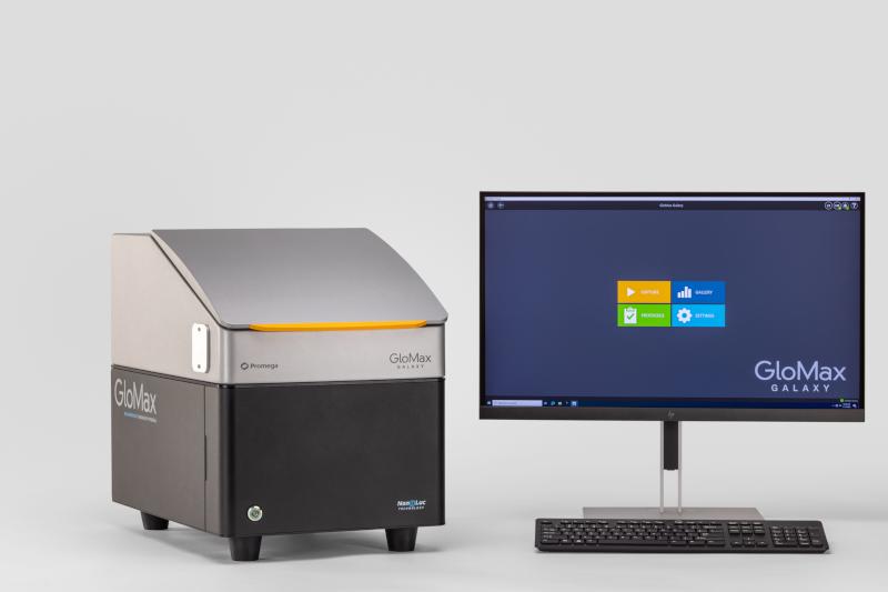
The GloMax® Galaxy Bioluminescence Imager was built to visualize the NanoLuc®-based assays we engineered, in a benchtop instrument accessible to any lab. The Galaxy is a fully equipped microscope with luminescence, fluorescence, and brightfield imaging capabilities, giving researchers a way to see functional and dynamic cellular events across a cell population.
Conclusion
Luminescent imaging can enrich what we learn from live-cell assays and offers an unprecedented view into the dynamics of cellular processes. From monitoring drug responses to visualizing protein interactions, this technology delivers insights that go beyond the capabilities of traditional assays.
Whether you’re studying cancer biology, drug development or cellular signaling, luminescent imaging can help you uncover what’s hidden in your data and see your research in a whole new light.
GloMax® Galaxy Luminescent Imager, NanoBRET® Nano-Glo® Detection Systems and Nano-Glo® Vivazine live Cell Substrate are for Research Use Only. Not for Use in Diagnostic Procedures.
Age-related macular degeneration (AMD) is a common eye disease that can result in progressive loss of vision. While AMD typically affects older adults, a specific rare type of AMD called Malattia Leventinese/Doyne honeycomb retinal dystrophy (ML/DHRD) can appear as early as the teenage years. Although ML/DHRD is rare, its study may provide insights into broader mechanisms of retinal degeneration, which could benefit millions affected by AMD.
While the genetic cause of ML/DHRD is known, there have been no small molecule inhibitors identified that reduce the production of the disease-causing protein. However, researchers from the University of Texas Southwestern Medical Center and the University of Minnesota recently published a paper that describes a small-molecule inhibitor that addresses the primary pathology of ML/DHRD. In the paper, titled “GSK3 inhibition reduces ECM production and prevents age-related macular degeneration-like pathology,” the team used CRISPR-engineered cell lines to study production of the disease-causing protein in response to treatment with inhibitors. The work was supported by the Promega Academic Access Program, which helped defray the costs of needed reagents. Their results point to future strategies for developing therapeutics at the currently incurable disease.
In the permafrost of Siberia, a remarkable discovery has been made—a mummified juvenile sabretooth cat, Homotherium latidens, frozen in time for over 35,000 years. This discovery, made along the Badyarikha River in the Indigirka River Basin of Yakutia, Russia, offers an exciting glimpse into a species that has no modern analog (a living equivalent of something extinct) (1). For paleontologists and evolutionary biologists, it provides an unprecedented look at an ancient predator that roamed the Earth during the Ice Age. So, how is this cub mummy truly fascinating scientists?
A Rare Find
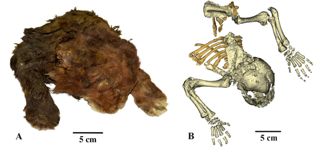
The frozen mummy of Homotherium latidens: (A) external appearance; (B) skeleton, CT-scan, dorsal view (1).
The permafrost of Siberia is a treasure trove of Ice Age fossils, but the discovery of a mummified Homotherium cub stands out for its rarity and significance. While bones can tell us a lot about the history of an extinct species, mummies—where the animal’s soft tissues, such as fur, skin and sometimes internal organs, are preserved—offer far more detailed information. ‘Mummies’ refer to animals (or humans) that have been preserved with their soft tissues intact, often through natural or intentional processes like drying or embalming. This preservation allows scientists to gain insights into the organism’s diet, health, development and adaptations—details that bones alone can’t reveal!
With Thanksgiving just around the corner, kitchens across the country will soon be alive with the sights, smells and sounds of cooking. But what if we dove deeper into those recipe books and looked beyond the instructions? You might find you’re more than just a cook; you’re quite the chemist!
Inspired by the popular show Lessons in Chemistry, an adaptation of Bonnie Garmus’s bestselling novel, cooking is presented as applied science, encouraging viewers to think critically about their cooking and the chemical reactions that create the flavors and textures they love. In this blog, we’ll explore the chemistry behind Thanksgiving cooking, revealing how different techniques bring out the best in each dish.
The Eppendorf Family Exchange program offers a unique opportunity for Promega families to participate in an international exchange initiative designed to bring our global community closer together. Through this program, children of Eppendorf and Promega employees immerse themselves in new cultures, gain language skills, and forge lifelong friendships.
The program began in 2019 during the 40th anniversary of Promega when we received the generous gift of an exchange program from a friend in the industry: Eppendorf. Every year, ten children ages 14-18 have the opportunity to participate in this enriching exchange, experiencing daily life from a new cultural perspective in a different country for two to four weeks. In return, Promega families will host a child from an Eppendorf family.
Preparing samples, conducting test series with cell cultures, or writing laboratory reports. Laboratory tasks cover a broad range of activities. Technical assistants support researchers in performing and evaluating experiments or carrying out laboratory tests in the medical field. A lab without them? Hard to imagine. However, it is not just scientific and technical understanding that is important. “Certain soft skills are necessary to be successful in your job. This also applies to the scientific field,” says Anette Leue, Head of Digital Marketing & Communications at Promega GmbH. “The focus is often on technical skills, while personal development is neglected. This inspired us to come up with our ‘Develop Yourself with Promega’ program.”
What is Develop Yourself with Promega?
“Develop Yourself with Promega” is a training series for laboratory personnel, focusing on personal development. It covers topics such as “How do I present my results in an interesting and structured way?” or “What do I need to make my lab more sustainable?” The aim is to expand professional competencies through soft-skill training. “At the beginning, we conducted a survey with our partner, the Life Science Learning Lab (in German Glaesernes Labor) in Berlin, among technical assistants to find out which topics are important to them,” Leue continues. These insights became the starting point for the first four trainings:
Green your lab: How can my lab become more sustainable?
Presentation training: A few steps to a good presentation
Post-translational modifications of proteins are critical for proper protein function. Modifications such as phosphorylation/dephosphorylation can act as switches that activate or inactivate proteins in signaling cascades. The addition of specific sugars to membrane proteins on cells are critical for recognition, interaction with the extracellular matrix and other activities. While we know volumes about some types of protein modifications, ADP-ribosylation on aspartate and glutamate residues has been more difficult to study because of the chemical instability of these ester-linked modifications.
Matić Lab (Eduardo José Longarini and Ivan Matić) recently published a study that explored mono-ADP-ribosylation (ADPr) on aspartate and glutamate residues by the protein PARP1 and its potential reversal by PARG. PARP1 and PARG signaling are central to DNA repair and apoptosis pathways, making them potentially powerful therapeutic targets in cancer or neurodegenerative diseases in which DNA repair processes are often disrupted.
XWe use cookies and similar technologies to make our website work, run analytics, improve our website, and show you personalized content and advertising. Some of these cookies are essential for our website to work. For others, we won’t set them unless you accept them. To learn more about our approach to Privacy we invite you to Read More
By clicking “Accept All”, you consent to the use of ALL the cookies. However you may visit Cookie Settings to provide a controlled consent.
We use cookies and similar technologies to make our website work, run analytics, improve our website, and show you personalized content and advertising. Some of these cookies are essential for our website to work. For others, we won’t set them unless you accept them. To find out more about cookies and how to manage cookies, read our Cookie Policy.
If you are located in the EEA, the United Kingdom, or Switzerland, you can change your settings at any time by clicking Manage Cookie Consent in the footer of our website.
Necessary cookies are absolutely essential for the website to function properly. These cookies ensure basic functionalities and security features of the website, anonymously.
Cookie
Duration
Description
cookielawinfo-checbox-analytics
11 months
This cookie is set by GDPR Cookie Consent plugin. The cookie is used to store the user consent for the cookies in the category "Analytics".
cookielawinfo-checbox-functional
11 months
The cookie is set by GDPR cookie consent to record the user consent for the cookies in the category "Functional".
cookielawinfo-checbox-others
11 months
This cookie is set by GDPR Cookie Consent plugin. The cookie is used to store the user consent for the cookies in the category "Other.
cookielawinfo-checkbox-advertisement
1 year
The cookie is set by GDPR cookie consent to record the user consent for the cookies in the category "Advertisement".
cookielawinfo-checkbox-necessary
11 months
This cookie is set by GDPR Cookie Consent plugin. The cookies is used to store the user consent for the cookies in the category "Necessary".
cookielawinfo-checkbox-performance
11 months
This cookie is set by GDPR Cookie Consent plugin. The cookie is used to store the user consent for the cookies in the category "Performance".
gdpr_status
6 months 2 days
This cookie is set by the provider Media.net. This cookie is used to check the status whether the user has accepted the cookie consent box. It also helps in not showing the cookie consent box upon re-entry to the website.
lang
This cookie is used to store the language preferences of a user to serve up content in that stored language the next time user visit the website.
viewed_cookie_policy
11 months
The cookie is set by the GDPR Cookie Consent plugin and is used to store whether or not user has consented to the use of cookies. It does not store any personal data.
Analytical cookies are used to understand how visitors interact with the website. These cookies help provide information on metrics the number of visitors, bounce rate, traffic source, etc.
Cookie
Duration
Description
SC_ANALYTICS_GLOBAL_COOKIE
10 years
This cookie is associated with Sitecore content and personalization. This cookie is used to identify the repeat visit from a single user. Sitecore will send a persistent session cookie to the web client.
vuid
2 years
This domain of this cookie is owned by Vimeo. This cookie is used by vimeo to collect tracking information. It sets a unique ID to embed videos to the website.
WMF-Last-Access
1 month 18 hours 24 minutes
This cookie is used to calculate unique devices accessing the website.
_ga
2 years
This cookie is installed by Google Analytics. The cookie is used to calculate visitor, session, campaign data and keep track of site usage for the site's analytics report. The cookies store information anonymously and assign a randomly generated number to identify unique visitors.
_gid
1 day
This cookie is installed by Google Analytics. The cookie is used to store information of how visitors use a website and helps in creating an analytics report of how the website is doing. The data collected including the number visitors, the source where they have come from, and the pages visted in an anonymous form.
Advertisement cookies are used to provide visitors with relevant ads and marketing campaigns. These cookies track visitors across websites and collect information to provide customized ads.
Cookie
Duration
Description
IDE
1 year 24 days
Used by Google DoubleClick and stores information about how the user uses the website and any other advertisement before visiting the website. This is used to present users with ads that are relevant to them according to the user profile.
test_cookie
15 minutes
This cookie is set by doubleclick.net. The purpose of the cookie is to determine if the user's browser supports cookies.
VISITOR_INFO1_LIVE
5 months 27 days
This cookie is set by Youtube. Used to track the information of the embedded YouTube videos on a website.
Performance cookies are used to understand and analyze the key performance indexes of the website which helps in delivering a better user experience for the visitors.
Cookie
Duration
Description
YSC
session
This cookies is set by Youtube and is used to track the views of embedded videos.
_gat_UA-62336821-1
1 minute
This is a pattern type cookie set by Google Analytics, where the pattern element on the name contains the unique identity number of the account or website it relates to. It appears to be a variation of the _gat cookie which is used to limit the amount of data recorded by Google on high traffic volume websites.