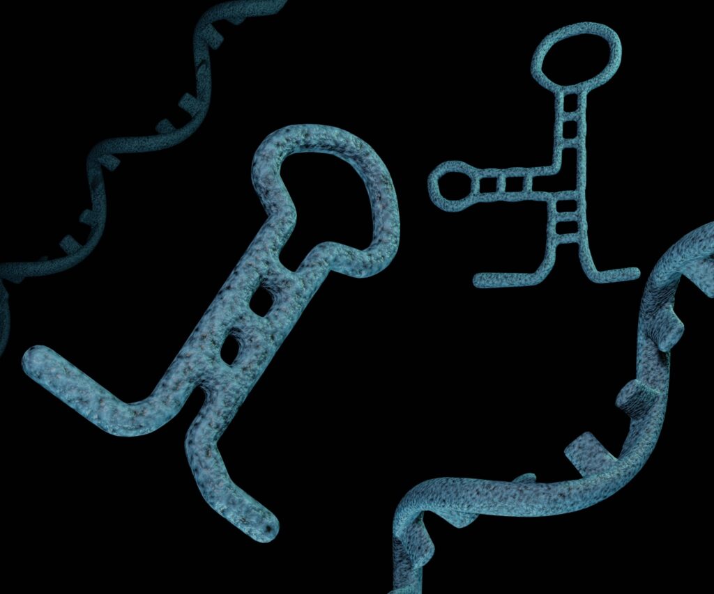
Discovered in 1983 and initially dismissed as ‘cellular dust,’ exosomes have since emerged as pivotal players in biomedical research due to their roles in intercellular communication, potential as drug delivery vectors and as biomarkers for various diseases. These small extracellular vesicles, measuring 30–150nm, are crucial for transferring proteins, lipids, and nucleic acids — including microRNA (miRNA), mRNA, and non-coding RNA– between cells (1). miRNAs are particularly critical as they regulate gene expression and offer insights into the cellular mechanisms underlying diseases like cancer, enhancing the value of exosomes in cancer research.
Beyond exosomes importance in understanding intracellular communication and organ cross-talk, exosomes can also alter the functions of recipient cells based on their cargo. This capability makes them extremely valuable in providing insights into alterations in cellular communication, tumor microenvironments, metastasis and immune evasion.
Despite their growing importance, the research into exosomes faces challenges such as inconsistent isolation methodologies, varied nomenclature, and the lack of standardized analysis strategies, which can limit the understanding of their full potential in cancer progression (1).
Some Current Research Method Limitations
Studying exosomes is critical for understanding their role in the progression of cancer. Common research methods include differential ultracentrifugation, size exclusion chromatography and immunoaffinity capture; each of these methods, however, have their own strengths and limitations:
- Differential Ultracentrifugation (dUC): Widely regarded as the ‘gold standard’ in exosome isolation, dUC uses high-speed centrifugation to separate exosomes from other cellular components based on density. Although effective, this method is time-consuming and labor-intensive, requiring specialized equipment that can be a significant investment. Moreover, dUC can co-isolate other extracellular vesicles similar in size and density, which may contaminate the exosome samples and affect the purity of the results (1).
- Size Exclusion Chromatography (SEC): SEC offers a gentle alternative to dUC that maintains the integrity and functionality of exosomes. This method separates particles based on their size as they pass through a gel filtration column. While SEC does not require large sample volumes and is less harsh on the vesicles, it has limitations in distinguishing exosomes from other vesicles of similar size, such as macrovesicles. Additionally, the throughput of SEC is generally limited, which can be a bottleneck when processing multiple samples (1).
- Immunoaffinity Capture: This method enhances the specificity of exosome isolation by using antibodies that target exosomal surface proteins. Immunoaffinity capture can be particularly useful for isolating specific subsets of exosomes from heterogeneous mixtures; however, the method relies heavily on the availability and specificity of antibodies, which can vary. Non-specific binding by antibodies can lead to the isolation of non-exosomal vesicles, compromising the purity of the sample. Furthermore, the selected antibodies might only recognize certain exosomal markers, potentially missing other exosome populations that do not express these markers (1).
Innovative Tools for Improving Exosome Isolation
Given the limitations of traditional methods in exosome isolation and miRNA analysis, the development of more effective techniques has become paramount. This is why in July 2023, Promega and INOVIQ announced a strategic partnership that combines INOVIQ’s EXO-NET® exosome capture technology with Promega’s nucleic acid purification systems. This collaboration aims to revolutionize cancer diagnostics by enhancing both manual and high-throughput methods for exosome isolation and miRNA analysis.

INOVIQ’s EXO-NET® technology automates exosome isolation from various samples, ensuring high purity essential for downstream applications like biomarker discovery. The EXO-NET tool also uniquely captures exosomes based on size and surface proteins, ensuring the isolated exosomes are reflective of their cells of origin, providing valuable insights into cellular functions and disease states.
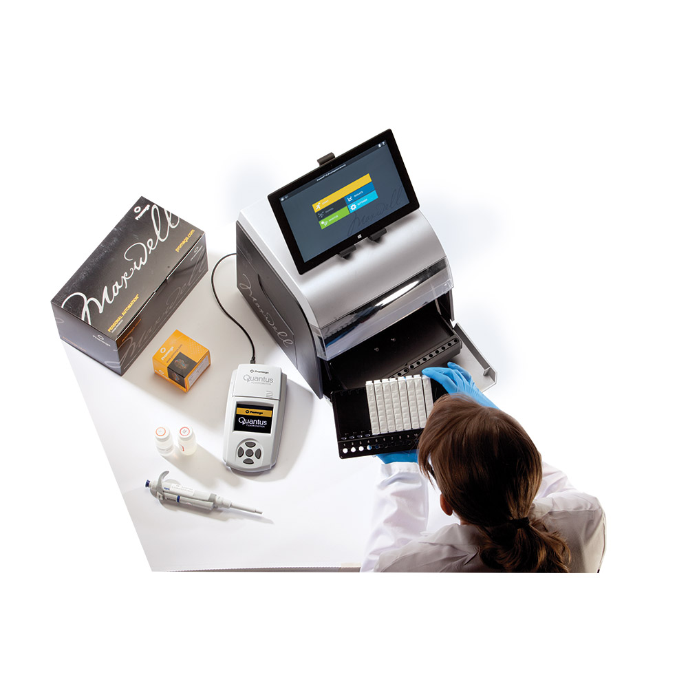
Complementing this, Promega’s Maxwell® HT systems offer efficient and automated nucleic acid extraction, including miRNA, from the isolated exosomes. This synergy allows for the rapid processing of numerous samples, essential for scaling studies and accelerating research without compromising quality. Ultimately, the enhanced sensitivity and specificity of these tools can provide researchers with more reliable and nuanced insights into the mechanisms of cancer.
Looking Ahead
The partnership between INOVIQ and Promega not only promises to improve the precision of cancer research, but also opens new avenues for biomarker discovery. By providing tools that improve the efficiency and accuracy of exosome-based diagnostics, this partnership will play a pivotal role in cancer research and development of new diagnostic modalities.
For more information on this joint marketing agreement, please visit Promega’s Press Release.
Reference
Latest posts by Shannon Sindermann (see all)
- Extreme Makeover, Epidemic Edition: Can Ants Modify Their Nests for Survival? - October 10, 2024
- Taking the Plunge: How the Seine Became Olympic-Ready - August 27, 2024
- What do Exosomes have to do with Cancer Research? - May 30, 2024
