What Can We Do to Make Your Work Easier? Addressing the Challenges of Working with Primary Antibodies

Scientific progress is iterative. Small advances and bits and pieces of knowledge are accumulated over time with each advance depending on the body of work that has preceded it. Being able to replicate the work from one step to another in this march of science is critical, and it is not easy. We all recognize that a well-documented procedure goes a long way toward enabling reproducibility, but the procedure is not the only hurdle to reproducibility, and it’s not even the main one.
Artistic Image of Hibit Tag

Many of the reagents researchers use are derived from or produced by living organisms. A decade ago, in 2015, an analysis of preclinical research estimated that irreproducibility costs the U.S. around $28 billion a year. When the authors traced the causes, the largest single category wasn’t study design or data analysis. It was biological reagents and reference materials (1).

When Bill Linton first developed the idea of a company that supports scientists in the 1970s, his question was: “What can we do to make your work easier?” The answer he kept getting was the same: researchers wished they could buy reliable restriction enzymes instead of purifying their own for every experiment. It would reduce variability and enable them to focus on the interesting biological questions rather than the requisite protein isolations needed to ask those questions.

Biotechnology and life science research have come a long way since then. We now take reliable, commercially available restriction enzymes for granted. Still, Promega keeps asking Bill’s original question: What can we do to make your work easier? The answers to that question have led to ground-breaking luciferase reporter technology, add-mix-measure assays for everything from cell viability to P450 activity to NADP/H detection, and standardized thaw-and-use primary cell lines for bioassays.

Today’s Difficult Reagent: Primary Antibodies

Among the most difficult reagents to work with and standardize are antibodies. If you are generating polyclonal primary antibodies that are not commercially available to study your target protein, you first need to find an animal with clean pre-immune serum. Usually that serum is evaluated by Western blot, which itself is an inherently fussy technique. And polyclonal antibodies are just that—polyclonal. What is generated after exposure to your antigen is a mixture of antibodies, and the composition of the mixture can change over time as the animal’s immune response matures. This gives you batch-to-batch variability and can make reproducibility more difficult. Commercially produced polyclonal antibodies are subject to the same exact issues. In August 2026, a news article in Nature reported that a metascientist had released a database of more than 18,000 questionable validation images across the catalogs of 15 antibody suppliers. All reflect the antibodies may not be performing like the scientists expect (Garisto 2026). Additionally, production of antibodies requires animal resources. High-quality, application validated antibodies are expensive as well. Using primary antibodies to study proteins is a significant pain point for the bench scientist.

It’s Not Just the Antibodies. It’s the Western Blots Too.

Tagged proteins or high-abundance exogenous expression of a target protein will not always reflect the endogenous protein behavior, localization or function. Large tags can interfere with folding and function; over expression of a protein can produce false protein interactions. Your results could be showing you artifacts resulting from a situation in your cells that is simply not biologically relevant.

If you are studying your target proteins (and other molecules) using Western blotting, post-translational modifications such as phosphorylation, ubiquitination, and cleavage shifts can complicate the interpretation of your results. Multiple bands on a blot can be hard to understand—is the protein cleaved? Is there cross-reactivity with your antibody to another protein? Is your sample degraded?

Continue reading “What Can We Do to Make Your Work Easier? Addressing the Challenges of Working with Primary Antibodies”

Charged Molecular Glues: The Molecular Trojan Horse That Slipped Past Biology’s Toughest Bouncer 

The cell membrane is notoriously selective about what it lets in. Charged molecules? Mostly rejected at the door. That’s a problem, because some of the most promising drug targets sit behind that barrier, and reaching them requires chemistry the membrane won’t tolerate. 

Continue reading “Charged Molecular Glues: The Molecular Trojan Horse That Slipped Past Biology’s Toughest Bouncer “

A Historic Milestone for PROTAC Research: What Vepdegestrant’s FDA Decision Means for Drug Discovery

Protein degrader research has yielded its first approved therapeutic for specific breast cancer patients: Vepdegestrant received FDA approval on May 1, 2026 (1). Vepdegestrant is an oral PROteolysis TArgeting Chimera (PROTAC) that targets the estrogen receptor for degradation in breast cancer patients with ESR1-mutated ER+/HER2– advanced breast cancer (2) produced by Arvinas, Inc. in collaboration with Pfizer Inc.

A Different Kind of Drug Development

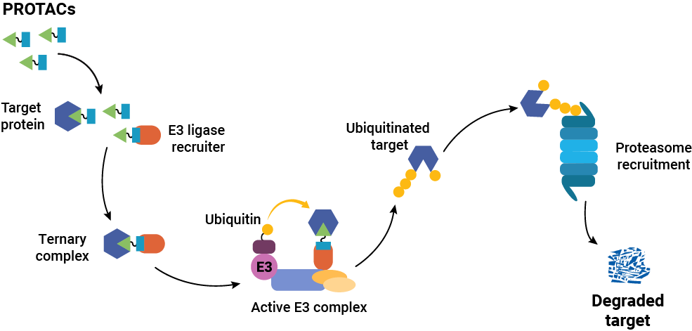
Targeted protein degraders (or PROTACs) have opened new possibilities in drug discovery research. Instead of inhibiting protein function or interaction, degraders cause the removal of the target protein itself. Traditional small molecule drugs work by binding a protein to inhibit it or block function, and they must remain bound to work. That means that the target protein should be well-characterized in terms of binding and activity sites, and the drug must bind specifically only to the target protein. In contrast, degraders only need to bind long enough to recruit cellular protein degradation machinery to the target protein, and the method does not rely on an accessible and specific binding site on the target protein. Once degradation occurs, the degrader is released and can engage with the next target.

The approval of vepdegestrant is a landmark moment for the entire TPD and induced proximity field, demonstrating that it is possible to rationally design molecules whose pharmacology is categorically distinct from traditional drugs, relying on a catalytic rather than occupancy-driven mechanism of action.  More importantly, this translates to meaningful clinical outcomes in patients. —Dr. Kristin Riching, Promega R&D Scientist

The first peptide-based PROTAC was described in 2001 in the laboratories of Craig Crews and Ray Deshaies (3), but translating the concept into orally bioavailable, clinically viable molecules took nearly two decades, using tools that did not exist when the field began. More than 40 PROTAC degraders have now entered clinical trials (4), with vepdegestrant the most advanced, supported by Phase 3 data from the VERITAC-2 trial demonstrating statistically significant improvement in progression-free survival in ESR1-mutant patients. That progress required solving a measurement problem as much as a chemistry one: how do you quantify target protein degradation at endogenous levels, with enough sensitivity and throughput to drive a screening campaign? CRISPR-engineered protein tagging combined with the small bioluminescent reporter tag, HiBiT solved that problem, providing a sensitive, HTS-compatible readout of endogenous target levels without relying on laborious, artifact-prone western blots. Critically, HiBiT also enabled researchers to watch target protein degradation unfold in real time in living cells.

“Seeing it happen in real time, frankly, may have been what convinced many people that the modality had genuine merit.”
—Dr. Kristin Riching

Developing a PROTAC is not like developing a traditional inhibitor. Success requires successful completion of a complex cascade of cellular events: the molecule must enter the cell, engage the target protein and the E3 ligase simultaneously, form a productive ternary complex in the right geometry, trigger ubiquitination, and drive proteasomal degradation, all while competing with cellular noise that can blunt each step. “PROTACs are large molecules, so they are often not very permeable,” Riching explains. “They also need to simultaneously engage both the target and the E3 ligase machinery, but they need to do so in a productive geometry that leads to ubiquitination, which is not easily predicted. In cells, many compounding factors can limit activity, making it difficult to identify which parameters most need improvement. Event-driven modalities like PROTACs rely on robust tools to tease apart each mechanistic step to aid SAR optimization.”

Getting that data means adopting a screening framework built around mechanistic understanding of the full degradation cascade from the earliest stages of optimization, while preserving the native biology and the stoichiometric relationships that govern degradation efficiency. It also means going beyond endpoint measurements. Knowing whether a target is degraded is a starting point; knowing how fast, how completely, and how durably it degrades is what distinguishes a development candidate from a dead end. Riching’s research has shown that different proteins in the same family can respond to the same PROTAC with dramatically different kinetic profiles (5,6), which is a distinction that endpoint assays cannot capture, and one that can determine which compounds are worth advancing.

What’s Next after Vepdegestrant?

The approval of Vepdegestrant validates more than just a single drug, it validates the PROTAC drug category and the tools and methods that enabled it. For researchers working on next-generation degraders, the signal is clear: the modality works. Now the question is how far we can push it.

Riching points to E3 ligase diversity as the field’s most pressing unresolved problem. “The greatest challenge will be expanding beyond the two E3 ligases — CRBN and VHL — that have driven most PROTAC progress to date,” she says. “We don’t yet fully understand the scope of targets accessible through these ligases, but it stands to reason that additional ligases will be necessary to unlock a larger portion of the degradable proteome. Their broad distribution also limits opportunity for tissue-selective targeting. Developing the tools and chemistry to recruit a wider repertoire of E3 ligases remains one of the most important unsolved problems the field faces.”

Beyond ligase diversity, the field is expanding its conception of what a degrader can be. Molecular glues, LYTACs, and other induced proximity strategies are broadening the range of accessible targets — including extracellular and membrane-bound proteins that sit outside the reach of classical PROTACs. Each new modality brings its own characterization challenges, and the same principle holds: understanding mechanism at the cellular level, early and rigorously, is what separates the compounds worth advancing from those that look promising in a tube.

The approval of vepdegestrant is a landmark. But researchers working in this space know it is a beginning as much as it is a culmination — proof that the approach is sound, and a starting line for everything that follows.


Read more about Innovative Imaging Solutions for Targeted Protein Degradation on our website.


Literature Cited

  1. Arvinas, Inc. Arvinas Announces FDA Approval of VEPPANU (vepdegestrant) for the Treatment of ESR1m, ER+/HER2– Advanced Breast Cancer Accessed: May 5, 2026 
  1.  Arvinas, Inc. (2025) Arvinas Announces FDA Acceptance of the New Drug Application for Vepdegestrant for the Treatment of ESR1m, ER+/HER2– Advanced Breast Cancer. August 8. Accessed: April 27, 2026.  
  1. Sakamoto, K.M. et al. (2001) Protacs: Chimeric Molecules that Target Proteins to the Skp1-Cullin-F Box Complex for Ubiquitination and Degradation. Proc. Natl. Acad. Science USA 98, 8554-–9. Accessed: May 4, 2026. 
  1. Chen, S. (2026) Protein Degraders (PROTACS & Molecular Glues) in 2026: The Emergining Challenge to Traditional Drug Development Accessed: May 5, 2026 
  1. Riching, K.M et al. (2018) Quantitative Live-Cell Kinetic Degradation and Mechanistic Profiling of PROTAC Mode of Action. ACS Chem. Biol. 13, 2758–70. Accessed: May 4, 2026 
  1. Riching, K.M. et al.  (2022) The Importance of Cellular Degradation Kinetics for Understanding Mechanisms in Targeted Protein Degradation. Chem. Soc. Rev. 51, 6210–6221. 

This article was written with AI assistance.

Targeted Protein Degradation: How Chemoproteomics and Induced Proximity Are Shaping Drug Discovery

Earlier this fall, more than 90 researchers from academia and industry gathered at the Promega Madison campus for the 4th TPD & Induced Proximity Symposium. The event focused on the rapidly advancing field of targeted protein degradation (TPD) and the broader concept of induced proximity—therapeutic strategies that bring two or more proteins into proximity to trigger a specific biological effect. 

This 4th year reflected of the symposium a maturing and diversifying field with chemoproteomics and proteomescale mapping redefining what it means to be “druggable,” while AI and high throughput biology are connecting molecular design to cellular function. Yet the mission remains unchanged—using molecular approaches that leverage the cellular machinery to make progress against targets once deemed “undruggable.” 

Continue reading “Targeted Protein Degradation: How Chemoproteomics and Induced Proximity Are Shaping Drug Discovery”

A New View of Protein Degradation with HiBiT and Live Cell Imaging

Targeted protein degradation (TPD) is an emerging drug discovery strategy that offers an entirely different approach to tackle disease-relevant proteins, including classic “undruggable” targets. Instead of inhibiting protein function, small molecules like PROTACs and molecular glues co-opt the cell’s own ubiquitin-proteasome system to eliminate specific proteins altogether. But as this targeted approach gains traction, it also challenges existing methods for validating compound activity.  

How do you confirm that degradation is happening in a biologically-relevant system? Can you validate protein degradation in real-time?  

Continue reading “A New View of Protein Degradation with HiBiT and Live Cell Imaging”

Unlocking the Power of Live-Cell Kinetics in Degrader Development

In targeted protein degradation (TPD), timing is everything. Understanding not just whether a degrader works—but how fast, how thoroughly and how sustainably—can dramatically influence early discovery decisions. Dr. Kristin Riching (Promega) dove into the real-time world of degradation kinetics in the webinar: Degradation in Motion: How Live-Cell Kinetics Drive Degrader Optimization, sharing how dynamic data provides a clearer view of degrader performance than traditional endpoint assays.

Whether you’re exploring your first PROTAC or optimizing a molecular glue series, the expertise offered in Dr. Riching’s presentation gives you actionable insights that will help you connect kinetic data to better therapeutic design.

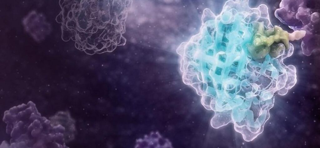
3D visualization of a protein structure within a live-cell environment, highlighting the interaction site relevant to targeted protein degradation, set against a dark cellular background to emphasize kinetic dynamics.
Continue reading “Unlocking the Power of Live-Cell Kinetics in Degrader Development”

Cracking the Undruggable Code: Top 10 Key Takeaways

For decades, the concept of “undruggable” targets has presented one of the most significant challenges in drug discovery. At our recent virtual event, Illuminating New Frontiers: Cracking the Undruggable Code, leading researchers and industry experts gathered to showcase cutting-edge technologies and fresh perspectives that are expanding the boundaries of therapeutic development. Over three engaging days, participants explored groundbreaking advances in targeting RAS signaling, leveraging protein degradation and induced proximity strategies, and exploring RNA as a therapeutic target.

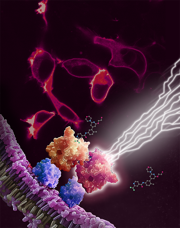
Target engagement of RAF dimer inhibitor LXH254 at RAF kinases, in complex with KRAS (blue). RAF inhibitor LXH254 engages BRAF or CRAF protomers (orange), but spares ARAF (red). Unoccupied ARAF is competent trigger downstream mitogenic signaling (lightning bolts). Red cells in the background are fluorescently labeled RAS proteins, expressed in live cells.
Continue reading “Cracking the Undruggable Code: Top 10 Key Takeaways”

Third Annual Targeted Protein Degradation Symposium: Embracing the Excitement of Discovery

The third annual Targeted Protein Degradation (TPD) Symposium just wrapped up last month. It was kicked off with Poncho Meisenheimer, VP of Research and Development at Promega, likening the gathering of researchers to “kids in a biology candy store.” This playful analogy captured the vibrant energy and sense of exploration among the attendees, who convened to delve into the future possibilities of proximity-induced degradation. Poncho left attendees with three key questions to consider throughout the symposium:

  1. How can we focus on quantitative measures of cellular events in relevant models?
  2. How do we generate results that serve both human and AI models?
  3. How do we best embrace the excitement of discovery?

Nearly 150 participants from both industry and academia attended the two-day symposium. It was held on September 11th and 12th at Promega’s R&D hub, the Kornberg Center, in Madison, Wisconsin. The event, now in its third year, provided a familiar environment where collaborations flourished, and many attendees rekindled connections forged through previous interactions or partnerships in the field.

Continue reading “Third Annual Targeted Protein Degradation Symposium: Embracing the Excitement of Discovery”

Cancer Moonshot: Solving Tough Problems

At the American Association for Cancer Research meeting in April 2016, then Vice President of the United States, Joe Biden, revealed the Cancer Moonshot℠ initiative— a program with the goals of accelerating scientific discovery in cancer research, fostering greater collaboration among researchers, and improving the sharing of data (1,2). The Cancer Moonshot is part of the 21st Century Cures Act, which earmarked $1.8 billion for cancer-related initiatives over 7 years.  The National Cancer Institute (NCI) and the Cancer Moonshot program have supported over 70 programs and consortia, and more than 250 research projects.  According to the NCI, the initiative from 2017 to 2021 resulted in over 2,000 publications, 49 clinical trials and more than 30 patent filings. Additionally, the launch of trials.cancer.gov has made information about all cancer research trials accessible to anyone who needs it (3).

“We will build a future where the word ‘cancer’ loses its power.”

First Lady, Dr. Jill Biden

In February 2022, the Biden White House announced a plan to “supercharge the Cancer Moonshot as an essential effort of the Biden-Harris administration” (4).  Biden noted in his address that, in the 25 years following the Nixon administration’s enactment of the National Cancer Act in 1971, significant strides were made in understanding cancer. It is now recognized not as a single disease, but as a collection comprising over 200 distinct diseases. This period also saw the development of new therapies and enhancements in diagnosis. However, despite a reduction in the cancer death rate by more than 25% over the past 25 years, cancer continues to be the second leading cause of death in the United States [4].

The Cancer Moonshot is a holistic attempt to improve access to information, support and patient experiences, while fostering the development of new therapeutics and research approaches to studying cancer. In this article, we will focus on research, diagnostics and drug discovery developments.

Solving for Undruggable Targets

KRAS , a member of the RAS family, has long been described as “undruggable” in large part because it is a small protein with a smooth surface that does not present many places for small molecule drugs to bind. The KRAS protein acts like an off/on switch depending upon whether it has GDP or GTP bound.  KRAS mutations are associated with many cancers including colorectal cancer (CRC), non-small cell lung cancer (NSCLC), and pancreatic ductal adenocarcinoma (PDAC). The G12 position in the protein is the most commonly mutated; G12C accounts for 13% of the mutations at this site, and is the predominant substitution found in NSCLC, while G12D is prevalent in PDAC (5).

Continue reading “Cancer Moonshot: Solving Tough Problems”

Insights from our Second Annual Targeted Protein Degradation Symposium

In the opening remarks of our second annual Targeted Protein Degradation Symposium, Tom Livelli, VP of Life Sciences Products & Services at Promega, posed a question to the attendees: “What do you want to be able to do today that you can’t?” This aspirational question set the tone for an event where building connections to advance the study and application of proximity-induced degradation took center stage.

More than 90 attendees from academia and industry gathered September 20–21 for the two-day symposium, which was hosted in our inspirational Kornberg Center—the R&D heart of Promega. Through engaging talks, a poster session, “Learn n’ Burn” challenges and social gatherings, participants had the opportunity to reinforce existing collaborations and to connect with others who are making an impact in the field of targeted protein degradation.

Continue reading “Insights from our Second Annual Targeted Protein Degradation Symposium”