NanoLuc® Luciferase: A Good Thing for Small Packages

influenza viruses

A paper published on October 2 in the Journal of Virology describes an exciting development in the world of influenza research—the construction of a luciferase reporter virus that does not affect virulence and can be used to track development and spread of infection in mice.

Insertion of luciferase reporter genes into viruses has been accomplished before for several viruses, but has not been successful for influenza. Construction of influenza reporter viruses is complicated because the viral genome is small and all the viral genes are critical for infection. Therefore, replacement of an existing gene with a reporter gene or insertion of additional reporter sequences without affecting the virus’s ability to replicate and cause infection has proven difficult. To be successful, a reporter gene needs to be small enough to insert into the viral genome without eliminating any other vital functionality.

Continue reading “NanoLuc® Luciferase: A Good Thing for Small Packages”
Trypsin/Lys-C Mix: Alternative for standard trypsin protein digestions

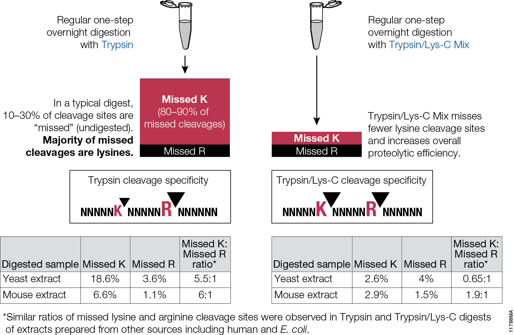
Trypsin/Lys-C Mix, Mass Spec Grade, is a mixture of Trypsin Gold, Mass Spectrometry Grade, and rLys-C, Mass Spec Grade. The Trypsin/Lys-C Mix is designed to improve digestion of proteins or protein mixtures in solution.It is a little known fact that trypsin cleaves at lysine residues with lesser efficiency than at arginine residues. Inefficient proteolysis at lysine residues is the major cause of missed (undigested) cleavages in trypsin digests.

11788MA


Supplementing trypsin with Lys-C enables cleavage at lysines with excepetional efficiency and specificity. Following the conventional trypsin digestion protocol (i.e., overnight incubation at nondenaturing conditions, reduction,alkylation, 25:1 protein:protease ratio [w/w], mix and incubate overnight at 37°C.) Replacing trypsin with Trypsin/Lys-C Mix in this conventional protocol leads to multiple benefits for protein analysis including more accurate mass spectrometry-based protein quantitation and improved protein mass spectrometry analytical reproducibility.

Are you looking for proteases to use in your research?
Explore our portfolio of proteases today.

Safety First (and Sensitivity too!): Diamond™ Nucleic Acid Dye

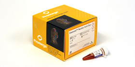
product photo for diamond™ nucleic acid dye

Gel electrophoresis and gel staining are common lab tasks that you may not think too much about.  It’s a fairly routine part of your day…purify DNA or RNA, check it on a gel.  As you probably know, interchelating agents like ethidium bromide can be used to visualize your nucleic acids on a gel for relatively low cost. The problem with ethidium bromide is that it’s highly mutagenic, making it less than ideal to work with and disposal of ethidium bromide can be quite costly. There are other commercially available alternatives to ethidium bromide that use fluorescent-based dyes to detect nucleic acids in gels. Some of these are touted to be safer than ethidium bromide; others are marketed as more sensitive.  If you are going to switch from an interchelating agent to something safer, you certainly don’t want to lose out on sensitivity.

To make your gel staining safer, more convenient, and more cost-effective, we’ve developed the Diamond™ Nucleic Acid Dye. This dye is not detectably genotoxic or cytotoxic at the 1:10,000 dilution recommended for gel staining, as determined by the Ames MPF™ Assay, is more sensitive than competing fluorescent-type “safe” dyes, and, in its concentrated form, is room-temperature stable for 90 days (1, 2).   If you are looking to switch to a safer, more sensitive way to stain your polyacrylamide or agarose gels to visualize your DNA or RNA, you may want to give the Diamond™ Nucleic Acid Dye a try.

  1.  Schagat, T. and Hendricksen, A. Diamond™ Nucleic Acid Dye is a Safe and Economical Alternative to Ethidium Bromide. [Internet] July 2013; tpub 125. [cited: 2013, July, 29].
  2. Truman, A., Hook, B. and Hendricksen, A. Diamond™ Nucleic Acid Dye: A Sensitive Alternative to SYBR® Dyes. [Internet] June 2013; tpub 121. [cited: 2013, July, 29].

Convenient, Non-Radioactive Detection of Isoaspartate

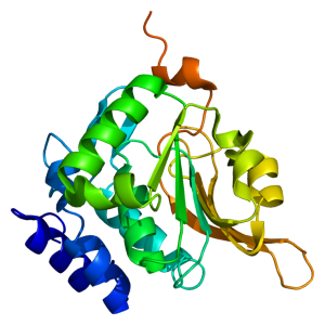
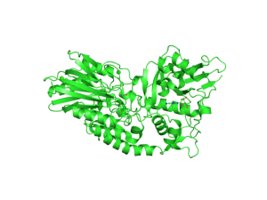
Structure of the PCMT1 protein. Based on PyMOL rendering of PDB 1i1n. Licensed under creative commons http://creativecommons.org/licenses/by-sa/3.0/deed.en

The ISOQUANT® Isoaspartate Detection Kit is intended for quantitative detection of isoaspartic acid residues in proteins and peptides, which can result from the gradual, nonenzymatic deamidation of asparagine or rearrangement of aspartic acid residues.

The ISOQUANT® Kit is designed to provide information regarding the global formation of isoaspartic acid residues at Asn and Asp sites, not at each site separately.

The deamidation of asparagine residues and rearrangement of aspartic acid residues is characterized by the formation of a succinimide intermediate that resolves to form a mixture of isoaspartic acid (typically 70–85%) and aspartic acid.
The rate and extent of isoaspartic acid formation can vary widely among proteins, depending on the amino acid sequence and size of the target protein. Deamidation of Asn residues has been observed most frequently at Asn-Gly and Asn-Ser sites within proteins.

The ISOQUANT® Isoaspartate Detection Kit uses the enzyme Protein Isoaspartyl ethyltransferase (PIMT) to specifically detect the presence of isoaspartic acid residues in a target protein. PIMT catalyzes the transfer of a methyl group from S-adenosyl-L-methionine (SAM) to isoaspartic acid. Spontaneous decomposition of this methylated intermediate results in the release of methanol and reformation of the succinimide.

References:

Wang, W. et al. (2012) Quantification and characterization of antibody deamidation by peptide mapping with mass spectrometry. Int. J. Mass. Spec. 312, 107–13.

Grappin, P. et al. (2011) New proteomic developments to analyze protein isomerization and their biological significance in plants. J. Proteomics, 74, 1475–82.

Yang, H. and Zubarev, R.A. (2010) Mass spectrometric analysis of asparagine deamidation and aspartate isomerization in polypeptides. Electrophoresis 31, 1764–71.

Sinha, S. et al. (2009) Effect of protein structure on deamidation rate in the Fc fragment of an IgG1 monoclonal antibody. Protein Sci. 18, 1573–84.

Basic Biology Matters

crop image2Every scientific paper is the story of a journey from an initial hypothesis to a final conclusion. It may take months or years and consists of many steps taken carefully one at a time. The experiments are repeated, the controls verified, the negative and positive results analyzed until the story finally makes sense. Sometimes the end of the story confirms the hypothesis, sometimes it is a surprise. A paper published last week in Cell describes a study where a team of researchers investigating one problem in basic biology (how one component of a signaling complex works), found an unexpected and potentially significant application in a different field (cancer research).

The paper, published in the June 6 issue of Cell, describes a previously unknown interaction between two cellular proteins—the transcription factor HIF1A and the cyclin-dependent kinase CDK8—in the regulation of genes associated with cellular survival under low-oxygen conditions. An accompanying press release describes how the discovery of a role for CDK8 in this process may have implications for cancer research, as CDK8 may be a potential target for drugs to combat “hypoxic” tumors. Continue reading “Basic Biology Matters”

Rabbit Reticulocyte Lysate Translation Systems: Tools for the analysis of translational regulation

TEM of Norovirus particles. Photo Credit: Charles D. Humphrey, Centers for Disease Control and Prevention

Rabbit Reticulocyte Lysate Translation Systems are used in the identification of mRNA species, the characterization of their protein products and the investigation of transcriptional and translational control. Rabbit Reticulocyte Lysate is prepared from New Zealand white rabbits. After the reticulocytes are lysed, the extract is treated with micrococcal nuclease to destroy endogenous mRNA and thus reduce background translation to a minimum.

Untreated Lysate is prepared from New Zealand white rabbits in the same manner as treated lysates with the exception that it is not treated with micrococcal nuclease. Unlike a coupled system that initiates transcription/translation from DNA, the RNA-based rabbit reticulocyte can be used for the direct investigation of transcriptional/translational control and the replication of RNA-based viruses.


References

Characterization of translation regulation (i.e., UTRs, Capping, IRES)

  1. Nguyen, H-L .et al. (2013) Expression of a novel mRNA transcript for human microsomal epoxide hydrolase is regulated by short reading frames within it 5’ –untranslated region. RNA. 19, 752–66.
  2. Wei, J. et al. (2013) The stringency of start codon selection in the filamentous fungus Neurospora crass. J. Biol. Chem. 288, 9549–62.
  3. Paek Ki-Y. et al. (2012) Cap-Dependent translation without base-by-base scanning of an messenger ribonucleic acid. Nucl. Acid. Res. 40, 7541–51.
  4. Se, and NH. Su.W. et al. (2011) Translation, stability, and resistance to decapping of mRNA containing caps substituted in the triphosphate with BH3. RNA 17, 978–88.
  5. Anderson, D. et al. (2011) Nucleoside modifications in RNA limit activation of 2’-5’ oligoadenylate synthetase and increase resistance to cleavage by RNase L. Nucl. Acid. Res. 39, 9329-38.

RNA virus Characterization

  1. Vashist, S. et al. (2012) Identification of RNA-protein interaction networks involved in the Norovirus life cycle. J. Vir. 86, 11977–90.
  2. Soto-Rifo, R. et al. (2012) Different effects of the TAR structure on HIV-1 and HIV-2 genomics RNA translation. Nucl. Acids. Res. 40, 2653–67.
  3. Poyry, T. et al. (2011) Mechanisms governing the selection of translation initiation sites on Foot-and-Mouth Disease Virus RNA. J.Vir. 85, 10178–88.
  4. Cheng, E. et al. (2011) Characterization of the interaction between Hantavirus nucleopcapsid protein and ribosomal protein S19. J. Biol. Chem. 286, 11814–24.
  5. Vera-Otarola, J. et al. (2011) The Andes Hantavirus NSs Protein is expressed from the Viral mRMA by a leaky scanning mechanism. J. Vir. 86, 2176–87.

Compound Screening Using Cell-Free Protein Expression Systems

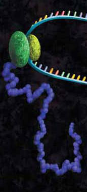
A protein chain being produced from a ribosome.
Both prokaryotic and eukaryotic cell-free protein expression systems have found great utility in efforts to screen organic compounds for inhibition of the basic cellular functions of transcription and translation, common targets for antibiotic compounds.

Cell-free systems can provide some advantages over cell-based systems for screening purposes. Cell-free systems allow exact manipulation of compound concentrations. This is an important parameter when evaluating the potential potency of the lead compound.

There is no need for cellular uptake to evaluate the effect of the compounds. While uptake evaluation is important for determining the eventual efficacy of the drug, it can unnecessarily eliminate valuable lead compounds in an initial screen. The interpretation of results in living cells is complicated by the large number of intertwined biochemical pathways and the ever-changing landscape of the growing cell. Cell-free systems allow the dissection of effects in a static system for simpler interpretation of results and the ability to specifically monitor individual processes such as transcription or translation. Individual targets not normally present, or found at low concentrations, can be added in controlled amounts.

The following references illustrate this application:

The Price for Convenience May Not Be That Pricey After All

Hour glass

I was having a discussion with my mother just the other day about cleaning products (lively topic, I know). She showed me her newest time saver…prediluted bleach. Huh, I thought. I guess that does save a bit of time, but I couldn’t resist telling her that she was paying triple the price for a whole lot of water. She said, without pause, that it was worth it to her to not have to splash fully concentrated bleach around. A convenience worth paying for, in her words.

I don’t know why this struck me as odd. I pay for convenience all the time as I get older. When I started running gels back in college, I wouldn’t have dreamed of buying a precast gel, but several years into my lab life I found myself running more than 15 gels a week, so precast was really a convenient alternative. When I was a grad student, I poured all of my own plates (and most of the plates for older students, too!). Fast forward a few years, and I running upwards of 300 microbial selective cultures per week. The switch to prepoured plates was a no brainer.

When put in the context of what our time is worth, would you rather be thawing and mixing loading dyes, buffers, stains, reagents, etc., or are you better of grabbing a premixed, room-temp stable dye or ladder/loading dye mix off the shelf and getting on with your research? I think most scientists would agree that these small conveniences allow you to free up a little more time to do the important work you should be doing.

I’m curious…what time savers or convenience items do you find that make your day a little easier in the lab?

Screening for Inhibitors of CD73 (5´-ectonucleotidase) Using a Metabolite Assay

CD73

CD73 also known as 5´-Ectonucleotidase (NT5E) is a membrane-anchored protein that acts at the outer surface of the cell to convert AMP to adenosine and free phosphate. CD73 activity is associated with immunosuppression and prometastatic effects, including angiogenesis. CD73 is highly expressed on the surfaces of many types of cancer cells and other immunosuppressive cells (1). A recent study by Quezada and colleagues showed that the high concentration of adenosine produced by the CD73-catalyzed reaction on glioblastoma multiforme cells, which are characterized by extreme chemoresistance, triggered adenosine signaling and in turn, the multi-drug resistance (MDR) phenotype of these cells (2).

Because of the roles of adenosine in immunosuppression, angiogenesis and MDR phenotypes, CD73 (NT5E) is an attractive therapeutic target. However, the current methods of assaying for the ectonucleotidase activity, HPLC and a malachite green assay, are cumbersome and not suited to high-throughput screening. The HPLC assay is expensive and difficult to automate and miniaturize (3). The malachite green assay is sensitive to phosphate found in media, buffers and other solutions used in the compound-screening environment.

To address the problem of developing a reliable high-throughput screening assay for CD73, Sachsenmeier and colleagues (3) looked to a luminescent ATP-detection reagent.

Continue reading “Screening for Inhibitors of CD73 (5´-ectonucleotidase) Using a Metabolite Assay”

A New Edge in Bisulfite Conversion

methyledge_featureproduct_280x140

Aberrant methylation events have significant impacts in terms of incidence of cancer and development disregulation. Researchers studying DNA methylation are often working with DNA from “difficult” tissues such as formalin-fixed, paraffin embedded tissues, which characteristically yield DNA that is more fragmented than that purified from fresh tissue. Traditional methods for bisulfite conversion involve a long protocol, harsh chemicals, and generally yield highly fragmented DNA. The DNA fragmentation may significantly impact the utility of the converted DNA in downstream applications such as bisulfite-specific PCR or bisulfite sequencing.

An ideal bisulfite conversion system enables complete conversion of a DNA sample in a short period of time, provides high yield of DNA, minimally fragments the DNA, works on a wide range of input DNA amounts (from a wide variety of sample types), and, while we’re at it, is easy to use and to store. Whew! That’s quite the list.

Continue reading “A New Edge in Bisulfite Conversion”